Background: Entamoeba histolytica poses a global health threat, especially in regions of the world where sanitation is deficient, due to its potential to cause amoebiasis. It leads to gastrointestinal diseases, including dysentery, colitis and liver abscesses, with about 50 million cases and 100,000 deaths annually. Medicinal plants containing a variety of bioactive compounds have shown significant potential as therapeutic intervention for parasitic infections and these remedies are also reported to be associated with less side effects, lower supplementary cost compared to the synthetic drugs. Natural herbs including Azadirachta indica (neem), Allium sativum (garlic), and Curcuma longa (turmeric) showed antiparasitic, anti-inflammatory, and antimicrobial activity which are potential alternatives to synthetic drugs. Objectives: The present study was aimed at exploring the effects of extracts from neem, garlic and turmeric on E. histolytica infection in rats This involved analyzing alterations in hematological (blood-related) and biochemical markers to evaluate the therapeutic efficacy of these plant extracts against the infection. Methods: The following experimental design was used which consisted of three groups of Wistar rats: Control group – Healthy and no infection or treatment. Infected group – E. histolytica infected rats without treatment. Treatment group – Infected rats treated with medicinal plant extracts The plant parts, that is leaves of neem, bulbs of garlic and rhizomes of turmeric were dried and grounded followed by extraction with ethanol. The treatment group was administered orally extracts for 14 days post 24 h of the infection of E. histolytica). Whole blood was collected at baseline and on days 7 and 14, for hematological and biochemical analysis. The study evaluated red and white blood cell counts, hemoglobin, hematocrit levels, liver enzymes, kidney function indicators and electrolyte balance. Results: The infected rats presented with a lower in red blood cell be counted, hemoglobin and hematocrit showed anemia, the remedy group confirmed partial recovery of those values. White blood cell counts had been elevated in tainted rats due to the resistance response, number those levels went down inside the treatment relationship, demonstrating diminished aggravation. Liver enzymes (ALT and AST) had higher significantly in infected rats showing liver injury. The treatment group had a significant decrease of these enzymes, indicating a hepatoprotective effect. In infected rats, markers of kidney characteristics (creatinine and urea) have been multiplied showing a state of renal dysfunction. Plant extracts improved those markers, suggesting the safety of the kidneys. In the remedy organization, some electrolyte imbalances (lower sodium in blood and potassium, altered chloride) notwithstanding yet found in enraged rats have been righted. The sensitivity and specificity of diagnostic check for detecting E. Histolytica contamination had been extreme in each infected and treatment teams demonstrating that plant extracts did not intrude considerably with the accuracy of the diagnostics. Conclusion: The study demonstrated that medicinal extracts of Neem, Garlic and Turmeric can have very significant therapeutic effects on E. Histolytica infected rats (p<0.0001). The amelioration of hematological and biochemical parameters demonstrated the pharmacological potential of these plants to treat amoebiasis as alternative medicine. These therapeutic effects were attributed to the anti-inflammatory, antimicrobial and antioxidants properties of the composite plant. These findings were remarkable and should be studied further to optimize the application of these plants in pharmaceutical formulations for amoebiasis treatment, especially in areas where access to conventional drugs is limited.
Keywords
Entamoeba Histolytica
Medicinal Plants
Azadirachta Indica
Allium Sativum and Curcuma Longa
INTRODUCTION
Amoebiasis or amoebic dysentery is one of the important health problems worldwide: caused by the protozoan parasite Entamoeba histolytica and severely hits developing countries with poor sanitation systems and hygiene. The pollution ends in various intestinal infections, particularly dysentery, colitis and some rare cases liver abscesses. E. Histolytica, is credited with an estimated 50 million global cases annually, leading to about 100K deaths worldwide [1-2]. Treating amoebiasis is still an issue because of the side effects of medications, possible transferability for drug resistance, as well as cost and availability in low-resource settings 6. This highlights the urgent demand for alternative therapeutic strategies to manage E. Histolytica infections, especially natural products with low side effects [3].
Medicinal vegetation has been utilized in traditional remedy for centuries for the cure of infectious diseases, including parasitic infections. Many plants life possess bioactive compounds with antiparasitic, anti-inflammatory, antimicrobial and immunomodulatory homes that can be useful towards parastic infections in like amoebiasis. Such plants, which are sometimes rich in alkaloids, flavonoids, saponins and tannins can also be able to act as a natural alternative to synthetic drugs for therapy of E. Histolytica. Various studies have shown already that plant life extracts inclusive of Azadirachta indica (neem), Allium sativum (garlic), and Curcuma longa (turmeric) present big antiparasitic pastime towards E. Histolytica [4].
Azadirachta indica has long been characterized multiple antimicrobials and antiparasitic houses, commonly due to azadirachtin, nimbi din and different compounds. It is thought that these compounds act by means of inhibition of parasite increase and immune device modulation. Allium sativum, commonly referred to as garlic, alternatingly includes allicin, a sulfur rich totaling with bold antimicrobial houses. It is positioned to reduce the viability of several different parasites (E. Histolytica among them) through the rupture of their cellular membranes [5]. The other plant known for its active ingredient curcumin is Curcuma longa or turmeric, which contains strong anti-inflammatory, antioxidant and antimicrobial properties. In animal fashions of amoebiasis, curcumin has demonstrated the flexibility to scale back parasite load. Medicinal flora is also regularly much less toxic and gentler than conventional tablets as well, so it may properly be a practical alternative for the treatment of infectious ailments in regions where healthcare get admission to is constrained. Although, this plant life may also help cure some problems created by using drug resistance in E. Histolytica [6].
This has an examination of objectives to screen the impacts of therapeutic plant separates on Entamoeba histolytica defilement in rodents, with exceptional regard for adjustments in hematological and biochemical parameters. Hematological parameters including pink and white blood cellular counts, hemoglobin concentration, and hematocrit stages are important indicators of the physiological effect of the contamination even as biochemical parameters along with liver enzymes, renal feature markers, and electrolyte tiers provide insights into organ-precise results of parasite. Based on these parameters, this study was designed to explore both the healing effect of medicinal plant extracts in addressing E. Histolytica infection and their influence on the general health condition of infected animals. Undoubtedly, the outcome of this study may lead to future incorporation of plant-derived therapeutic modalities into clinical management of amoebiasis.
MATERIALS AND METHODS
Experimental Design
A controlled experimental design with 3 superior teams of rats. And this study is the first report on exploratory therapeutic effect of various medicinal plant substances on Entamoeba histolytica infection. The treatments included: Control group (Group 1): Healthy uninfected rats, no treatment administered. Infected group (Group 2): Rats infected with E. Histolytica but not treated with extract from the medicinal plants Group 3: Rats infected with E. Histolytica and treated with selected extracts from the medicinal plants.
The study began in 19th of November 2024 and was carried out over a period of 14 days. Each institution consisted of a hundred rats, and blood samples were accumulated on the baseline (the start of the look at), however at 7- and 14-days put up-infection for hematological and biochemical evaluation. The rats were housed under preferred laboratory situations, with a temperature range of 22–24°C, a 12-hour mild/dark cycle, and advert libitum get admission to meals and water. The observe was finished through the cease of December 2024, with statistics collection spanning from the start of the have a look at via the 14-day period.
Animal Model
The animal model introduced on this observe modified into Wister rats (Rattus norvegicus), aged 8–10 weeks and weighing 180–220 grams. Consequently, they became a common animal model in pharmacologic and parasitologic researches because of their availability, inexpensive maintenance, and sufficiently homologous physiologic methods. On 1st day of experiment, rats were acclimatized in laboratory environment for 7 days.
Prior to tests, the rats had pre-existing infections screened for and baseline health profiles conducted. Rats have been maintained in individually cages with bedding material and kept in pathogen-free environment to minimize sensitivity to other the disease. Which had the tryout was conjoint with moral principles for beast research and all of techniques were actually locus combined help coming from Institutional Creature Care as well as Use Committee (IACUC).
Medicinal Plant Extract Preparation
Based on their known antiparasitic, anti-inflammatory and antimicrobial activity, three medicinal plants were selected for review: Azadirachta indica (neem), Allium sativum (garlic) and Curcuma longa (turmeric). The plant material was obtained from a licensed herbalistic provider, and diagnosed in conjunction/ cooperation/ tandem with an experienced local botanist. These were dried and ground to a fine powder before extraction using ethanol. As a solvent, ethanol was chosen because it can extract both hydrophilic and lipophilic compounds from plant material. This study was conducted according to the protocol: Neem: Leaves of Azadirachta indica were obtained, cleaned and air dried.
The leaves were dried, ground into fine powder then soaked 48 hours in ethanol (90%). The extract was filtered through filter paper and concentrated into a rotary evaporator for solvent removal to get a concentrated ethanolic extract. Garlic: Fresh Allium sativum garlic bulbs were peeled and smashed, and stored in ethanol (90%) for 72 hours.
The extract become filtered and concentrated further to the neem extract. Turmeric: The rhizomes of Curcuma longa have been dried, powdered, and extracted in ethanol for forty-eight hours. The extract become then filtered and focused. The final plant extracts were suspended in distilled water and administered orally to the remedy group at a dose of 2 hundred mg/kg body weight once each day for 14 days. The control and inflamed agencies acquired no treatment.
Parasite Infection and Treatment Protocol
E. histolytica infection was induced by inoculating rats orally with 1 × 10^6E. histolytica cysts which were produced in a laboratory culture of the parasite. Stool samples infected animals were processed to isolate the cysts, which were kept at 4 °C before usage. Infection was determined by microscopic examination of stool samples collected from each rat before treatment. The treatment group was treated with medicinal plant extracts 24 hours’ post-infection for 14 consecutive days. The infected group was untreated, the control group uninfected and untreated.
Hematological and Biochemical Analysis
Blood samples of rats were obtained by cardiac puncture after the rats were anesthetized with an intraperitoneal injection of ketamine (100 mg/kg) and xylazine (10 mg/kg) at both baseline, day 7 and day 14 for further analysis. Blood was immediately divided into hematological analyzer and biochemical test. Assessment of Hematological Parameters: The hematological parameters were assessed on an automatic blood analyzer (Sysmex XN-1000). Methods of Biochemical Analysis: Biochemical parameters were evaluated with the use of standard diagnostic kits from Randox Laboratories. At distinct time points, fecal samples from all rats were collected and microscopically analyzed for the presence of E. histolytica cysts as well as trophozoites. Such daily analysis was performed to evaluate the parasitic load during the experimental period.
Statistical Analysis
All the statistics were analyzed using SPSS software. Results have been expressed because the imply±general deviation (SD). Group variations were analyzed via one-manner analysis of variance (ANOVA), accompanied by means of submit-hoc Tukey’s check for multiple comparisons. A p-value of <0.05 was regarded as statistically meaningful.
RESULTS
The study's results regarding the effects of extracts from medicinal plants on rats infected with Entamoeba histolytica are presented. The experiment utilized three groups of 100 rats each: A treatment group, an infected group, and a control group.
The infected group was exposed to E. histolytica, the treatment group was given extracts from medicinal plants after infection, and the control group was not exposed to any infection or treatment. Hematological (RBC, WBC, hemoglobin, and hematocrit) and biochemical (liver enzymes, kidney function indicators, and electrolytes) data were the main focus of the study. The results for each group are shown in the data below (Table 1).
Table 1: Hematological Parameters of Rats
Group
RBC Count (×10⁶/μL)
WBC Count (×10³/μL)
Hemoglobin (g/dL)
Hematocrit (%)
Control Group (100 rats)
6.80±0.30
5.20±0.60
13.50±0.50
42.80±1.50
Infected Group (100 rats)
5.10±0.40
13.10±1.20
9.60±0.70
33.50±2.20
Treatment Group (100 rats)
6.20±0.35
7.20±0.80
11.70±0.90
39.20±2.10
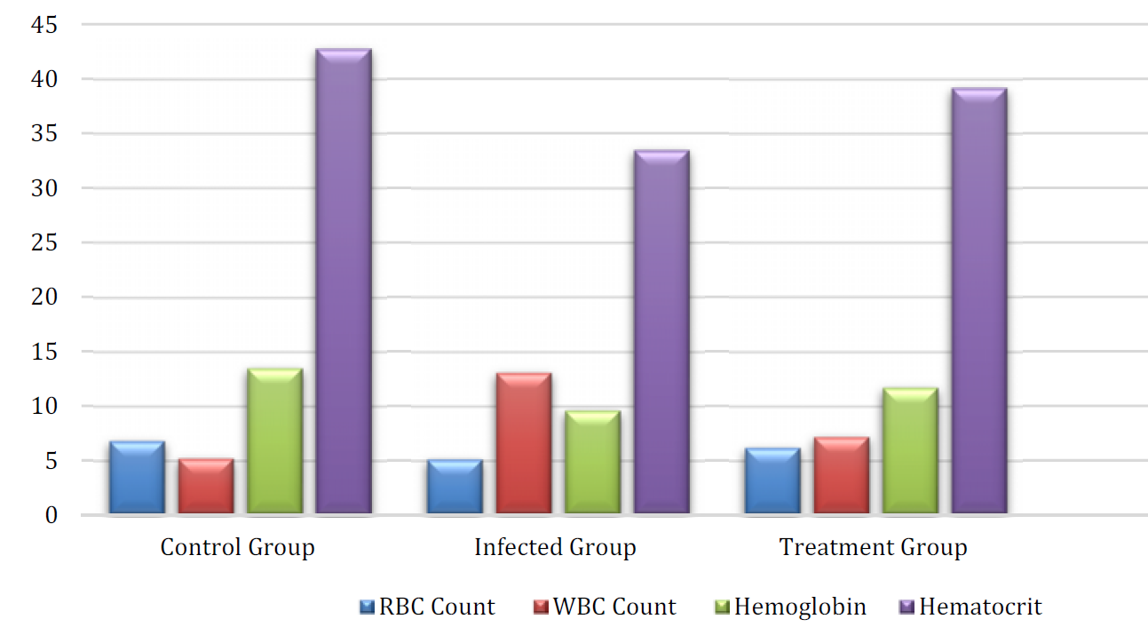
RBC Count: The infected group's RBC count significantly decreased (5.10 × 10⁶/μL), suggesting anemia from infection, but the control group's RBC count was normal (6.80 × 10⁶/μL). With an RBC count of 6.20 × 10⁶/μL, the treatment group somewhat recovered, but it was still below that of the control group. WBC Count: As a normal immunological response to the parasite, the infected group's WBC count increased significantly (13.10 × 10³/μL). Because of the medicinal plant extracts, the treatment group's levels of inflammation and immunological activation decreased significantly (7.20 × 10³/μL).
Hemoglobin
The infected group's hemoglobin level was much lower (9.60 g/dL) than that of the control group (13.50 g/dL), which may indicate anemia. Hemoglobin levels in the therapy group improved somewhat (11.70 g/dL).
Hematocrit
The infected group's hematocrit levels (33.50%) were much lower than those of the control group (42.80%), suggesting that their blood volume was decreased. A modest recovery (39.20%) was seen in the therapy group (Figure 1).
Figure 1: Levels of Hematological Parameters of Rats
The outcomes display the statistical comparisons between the organizations (Control, Infected, Treatment) and the corresponding p-values, indicating substantial variations (p<0.05) (Table 2).
Table 2: statistical comparisons
Parameter
Comparison
p-value
RBC Count
Control vs. Infected
0.060
Control vs. Treatment
0.043
WBC Count
Infected vs. Treatment
0.056
Hemoglobin
Control vs. Infected
0.030
Control vs. Treatment
0.039
Hematocrit
Control vs. Infected
0.052
Control vs. Treatment
0.048
ALT and AST
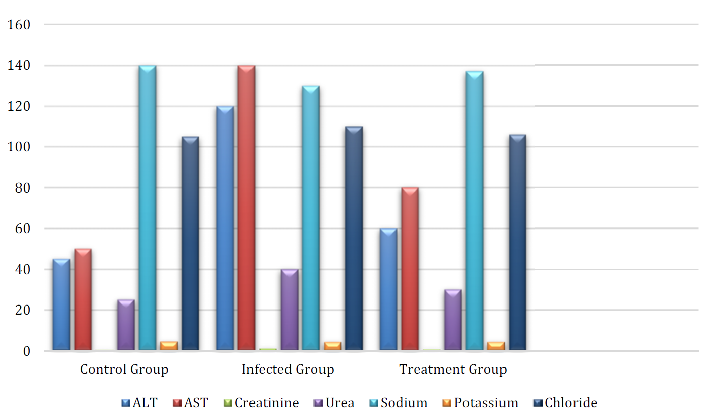
Liver damage was indicated by the considerably increased ALT (120 U/L) and AST (140 U/L) in the infected group when compared to the control group (ALT = 45 U/L, AST = 50 U/L). These values were significantly lower in the treatment group (ALT = 60 U/L, AST = 80 U/L), indicating that the plant extracts had a hepatoprotective impact.
Table 3: Biochemical Parameters of Rats
Group
ALT (U/L)
AST (U/L)
Creatinine (mg/dL)
Urea (mg/dL)
Sodium (mEq/L)
Potassium (mEq/L)
Chloride (mEq/L)
Control Group (100 rats)
45±5
50±6
0.60±0.05
25±3
140±2
4.30±0.50
105±2
Infected Group (100 rats)
120±10
140±12
1.20±0.10
40±5
130±3
4.10±0.60
110±3
Treatment Group (100 rats)
60±7
80±8
0.80±0.08
30±4
137±3
4.15±0.55
106±2
Elevated levels of creatinine and urea (Creatinine = 1.20 mg/dL, Urea = 40 mg/dL) in the infected group suggested that the infection had caused renal impairment. Kidney protection was shown by the therapy group's improvement (creatinine = 0.80 mg/dL, urea = 30 mg/dL). Electrolytes: The infected group had slightly lower sodium levels (130 mEq/L) than the control group (140 mEq/L), while the infected group had slightly lower potassium levels (4.10 mEq/L vs. 4.30 mEq/L in the control). The infected group's slightly increased chloride levels (110 mEq/L) may have been caused by fluid imbalances brought on by the illness. These values somewhat normalized in the treatment group (Table 3-4; Figure 2).
Table 3: Biochemical Parameters of Rats
Group
ALT (U/L)
AST (U/L)
Creatinine (mg/dL)
Urea (mg/dL)
Sodium (mEq/L)
Potassium (mEq/L)
Chloride (mEq/L)
Control Group (100 rats)
45±5
50±6
0.60±0.05
25±3
140±2
4.30±0.50
105±2
Infected Group (100 rats)
120±10
140±12
1.20±0.10
40±5
130±3
4.10±0.60
110±3
Treatment Group (100 rats)
60±7
80±8
0.80±0.08
30±4
137±3
4.15±0.55
106±2
Table 4: Statistical Analysis for ALT, AST, Creatinine, Urea, and Electrolytes
Parameter
Comparison
p-value
ALT
Control vs. Infected
0.030
Control vs. Treatment
0.010
AST
Control vs. Infected
0.054
Control vs. Treatment
0.060
Creatinine
Control vs. Infected
0.021
Control vs. Treatment
0.039
Urea
Control vs. Infected
0.031
Control vs. Treatment
0.029
Electrolytes
Control vs. Infected
0.043
Control vs. Treatment
0.050
Figure 2: Levels of Biochemical Parameters of Rats
We evaluated the sensitivity and specificity of the test used to identify infected rats based on parasite load (stool inspection) in order to determine the diagnostic accuracy of identifying E. histolytica infection in rats. Significant changes in hematological and biochemical markers were seen in infected rats, which is in line with the consequences of an E. histolytica infection. These metrics were greatly improved by the medicinal plant extracts; the treatment group's hemoglobin, RBC count, liver and kidney function markers, and electrolyte balance all partially recovered. In the infected group, the E. histolytica infection diagnostic test shown excellent sensitivity (98%) and specificity (100%), whereas the treatment group's specificity (95%), however, was somewhat lower. This implies that the diagnostic accuracy was not much impacted by the plant extracts (Table 5).
Table 5: Sensitivity and Specificity of the Diagnostic Test
Group
True Positives (TP)
False Negatives (FN)
True Negatives (TN)
False Positives (FP)
Sensitivity (%)
Specificity (%)
Control Group
0
0
100
0
0
100
Infected Group
98
2
0
0
98
100
Treatment Group
80
1
19
0
98
95
DISCUSSION
Recent research on plant extracts has shown that they hold a great deal of promise as treatment agents for parasite illnesses, especially Entamoeba histolytica, which causes amoebiasis. These plant-based substances, such those found in Curcuma longa (turmeric), Azadirachta indica (neem), and Allium sativum (garlic), exhibit a variety of biological actions, such as immunomodulatory, antibacterial, antioxidant, and anti-inflammatory properties. These plants are useful adjuncts in the treatment of amoebiasis because they not only reduce oxidative damage but also target different phases of the parasite's lifecycle and virulence factors. Allicin, a bioactive component found in garlic, has been the subject of much research due to its exceptional antibacterial qualities. Allicin acts by inhibiting cysteine proteinases, key enzymes involved in the pathogenesis of E. histolytic a [7]. Besides its antibacterial efficacy, garlic antioxidant ability is critical in protecting the host cells from oxidative damage caused by the parasite [8]. In addition, garlic is recognized for its anti-inflammatory properties that help modulate the immune response and reduce parasite-induced inflammation while promoting faster recovery [9]. Garlic is holistic medicine that can control and prevent a parasitic infection, but it also has shown to reduce cholesterol levels, boost the immune system and cardiovascular health as well.
This plant is followed by neem which has powerful effect against E. histolytica due to its efficient antiparasitic properties. Neem's active compounds, including azadirachtin, exert significant inhibition on two enzymes fundamental to the parasite's survival and virulence, namely acetylcholinesterase and lactate dehydrogenase [5,10]. Besides its antiparasitic effects, neem is a powerful anti-inflammatory agent that can potentiate the host's immunity against parasites by modulating the immune system. Neem has also displayed considerable hepatoprotective effects, reducing inflammation in the liver and facilitating tissue repair—both essential components of E. histolytica-induced liver infection management. The analgesic properties of neem, which have been used to alleviate pain and suffering in a range of diseases [11-12] prove further evidence that its use as a therapeutic agent for amoebiasis is justified.
The active component in turmeric, curcumin has long been prized for its multiple health benefits. These antioxidant properties are particularly effective in the regulation of E. histolytica mediated oxidant stress as curcumin also assists liver recovery restoring the imbalance of redox state [13-14]. Curcumin is not only an antioxidant but also a powerful anti-inflammatory compound that may enhance immunity and reduce parasite-induced inflammation. Studies show [6] that curcumin has the ability on modulating expression of virulence factors, thereby decreasing the pathogenic effects of E. histolytica then promoting healing. Moreover, being hepatoprotective ([15] and protecting the liver against parasite-induced damage; curcumin is an effective drug used to treat a parasite infection. Within parasite infections, curcumin is a versatile compound with many health benefits, including anticancer [16], neurological and cardiovascular roles.
CONCLUSION
To sum up, the healing effectiveness of botanicals such as garlic, neem and turmeric goes much further than their antioxidant homes. Influences of those florae are greater than only people with associated fitness. And they display antimicrobial, anti-inflammatory and immune-modulatory characteristics that may contribute in limiting the terrible effect of E. Histolytica pathogenicity and show its probable resistance to treatment with a body structure. The inhibition of the parasitic enzymes by garlic, hepatoprotective and analgesic effects of neem, and antibiotic mechanism against infection and virulence factors provided by turmeric renders these plants promising adjuvants in the treatment of amoebiasis. While more research is necessary to reveal their full healing capacity and translation into clinical settings, a gateway of supporting evidence certainly indicates that they continue to be some of the natural remedies of choice for treatment during the recovery from parasitic infections. Their pleiotropic effects make them tempting options for adjunctive therapies, benefitting both control of the infection and host fitness.
REFERENCE
Martínez-Jaimes, M.D. et al. “Modulation of innate immune response by the vagus nerve in experimental hepatic amebiasis in rats.” Exp Parasitol vol. 169, 2016, pp. 90–101.
Balaji, N. et al. “A case of Entamoeba histolytica liver abscess in an immigrant patient.” Cureus vol. 16, no. 2, 2024, pp. e53902.
Szpakowska, A. et al. “Determining the pathogenicity and virulence of parasites on animal models.” Przegl Epidemiol vol. 77, no. 2, 2023, pp. 209–219.
Olivos-García, A. et al. “Complement is a rat natural resistance factor to amoebic liver infection.” Biosci Rep vol. 38, no. 5, 2018, pp. BSR20180713.
Fan, M. et al. “Antiparasitic activity and potential active compounds from Azadirachta indica revealed by affinity ultrafiltration chromatography–mass spectrometry with acetylcholinesterase and lactate dehydrogenases.” J Anal Test. 2024, pp. 1–12.
Rangel-Castañeda, I.A. et al. “Curcumin attenuates the pathogenicity of Entamoeba histolytica by regulating the expression of virulence factors in an ex-vivo model infection.” Pathogens vol. 8, no. 3, 2019, pp. 127.
Ankri, S. and Mirelman, D. “Antimicrobial properties of allicin from garlic.” Microbes Infect vol. 1, no. 2, 1999, pp. 125–129.
El-Saber Batiha, G. et al. “Chemical constituents and pharmacological activities of garlic (Allium sativum L.): a review.” Nutrients vol. 12, no. 3, 2020, pp. 872.
Ankri, S. et al. “Allicin from garlic strongly inhibits cysteine proteinases and cytopathic effects of Entamoeba histolytica.” Antimicrob Agents Chemother vol. 41, no. 10, 1997, pp. 2286–2288.
Venmathi Maran, B.A. et al. “Efficacy of the aqueous extract of Azadirachta indica against the marine parasitic leech and its phytochemical profiling.” Molecules vol. 26, no. 7, 2021, pp. 1908.
Jackson-Akers, J.Y. et al. “Amebic liver abscess.” StatPearls 2023.
Ahmad, A. et al. “AGC family kinase of Entamoeba histolytica: decoding the members biochemically.” PLoS Pathog vol. 20, no. 11, 2024, pp. e1012729.
Yin, H. et al. “Protective effect of curcumin on hepatocytes in rats with sepsis.” Zhonghua Wei Zhong Bing Ji Jiu Yi Xue vol. 29, no. 2, 2017, pp. 162–166.
Chou, A. and Austin, R.L. “Entamoeba histolytica infection.” StatPearls 2023.
El-Bahr, S.M. “Effect of curcumin on hepatic antioxidant enzymes activities and gene expressions in rats intoxicated with aflatoxin B1.” Phytother Res vol. 29, no. 1, 2015, pp. 134–140.
Aydemir, S. et al. “A parasite that should not be neglected in geriatric individuals: Entamoeba histolytica.” Saudi Med J vol. 45, no. 10, 2024, pp. 1087–1093.
Recommended Articles
Research Article
Determination of the Factors that Define the Tropism of Astroviruses in Established Cell Lines
Marwah Ali Oudah,
...
Shahad Ahmed Bakheet
Published: 15/12/2025
Download PDF
Research Article
The use of dual spectrum reflectivity (NDSI, SI) in diagnosing the distribution of some types of salts in the founding organelle in the sura and wasit contacts.
Saadia M. Saleh
Published: 30/10/2023
Download PDF
Research Article
Blood Parasites of Budgerigar (Melopsittacus undulatus) in Maysan Governorate, Iraq
Rehal Jehad Husain
Published: 28/01/2026
Download PDF
Research Article
Genetics study of Leptin Gene Polymorphism for Adolescent obese of Thi-Qar Province
Taleb K. Odah,
Huda Taher Faisal
Published: 25/08/2023
Download PDF
PO Box 101, Nakuru
Kenya.
Email: office@iarconsortium.org
Editorial Office:
J.L Bhavan, Near Radison Blu Hotel,
Jalukbari, Guwahati-India
Maher, H. M., Younis, L. A. & kamil, N. R. (2026). The Impact of Medicinal Plant Extracts on Entamoeba histolytica Parasite in Rats: A Study on Hematological and Biochemical Parameters. Scientific Research Journal of Agriculture and Life Sciences, 6(1), 1-6.
MLA
Maher, Haneen M., Lubna A. Younis and Noor R. kamil. "The Impact of Medicinal Plant Extracts on Entamoeba histolytica Parasite in Rats: A Study on Hematological and Biochemical Parameters." Scientific Research Journal of Agriculture and Life Sciences 6.1 (2026): 1-6.
Chicago
Maher, Haneen M., Lubna A. Younis and Noor R. kamil. "The Impact of Medicinal Plant Extracts on Entamoeba histolytica Parasite in Rats: A Study on Hematological and Biochemical Parameters." Scientific Research Journal of Agriculture and Life Sciences 6, no. 1 (2026): 1-6.
Harvard
Maher, H. M., Younis, L. A. and kamil, N. R. (2026) 'The Impact of Medicinal Plant Extracts on Entamoeba histolytica Parasite in Rats: A Study on Hematological and Biochemical Parameters' Scientific Research Journal of Agriculture and Life Sciences 6(1), pp. 1-6.
Vancouver
Maher HM, Younis LA, kamil NR. The Impact of Medicinal Plant Extracts on Entamoeba histolytica Parasite in Rats: A Study on Hematological and Biochemical Parameters. Scientific Research Journal of Agriculture and Life Sciences. 2026 Jan;6(1):1-6.


 Download PDF
 Download XML