Coronaviruses are a diverse group of viruses infecting many different animals and they can cause mild to severe respiratory infections in humans. In 2002 and 2012, respectively, two highly pathogenic coronaviruses with zoonotic origin, Severe Acute Respiratory Syndrome Coronavirus (SARS-CoV) and Middle East Respiratory Syndrome Coronavirus (MERS-CoV), emerged in humans and caused fatal respiratory illness, making emerging coronaviruses a new public health concern in the twenty-first century. Severe Acute Respiratory Syndrome Coronavirus 2 (SARS-CoV-2) is a highly transmissible and pathogenic coronavirus that emerged in late 2019 and has caused a pandemic of acute respiratory disease, named ‘coronavirus disease 2019’ (COVID-19), which threatens human health and public safety. The disease is transmitted by inhalation or contact with infected droplets and the incubation period ranges from 2 to 14 days. The symptoms are usually fever, cough, sore throat, breathlessness, fatigue, malaise among others. The disease is mild in most people; in some (usually the elderly and those with comorbidities), it may progress to pneumonia, acute Respiratory Distress Syndrome (ARDS) and multi organ dysfunction. Many people are asymptomatic. The case fatality rate is estimated to range from 2 to 3%. Diagnosis is by demonstration of the virus in respiratory secretions by special molecular tests. Common laboratory findings include normal/ low white cell counts with elevated C-Reactive Protein (CRP). In this Review, we describe the basic virology of SARS-CoV-2, current knowledge of clinical, epidemiological and pathological features of COVID-19 and treatment approaches for SARS-CoV-2 infection.
Keywords
Pandemic
2019-nCOV
SARS-CoV-2
COVID-19
Bat-SL-CoV
RT-PCR
INTRODUCTION
History of Coronavirus
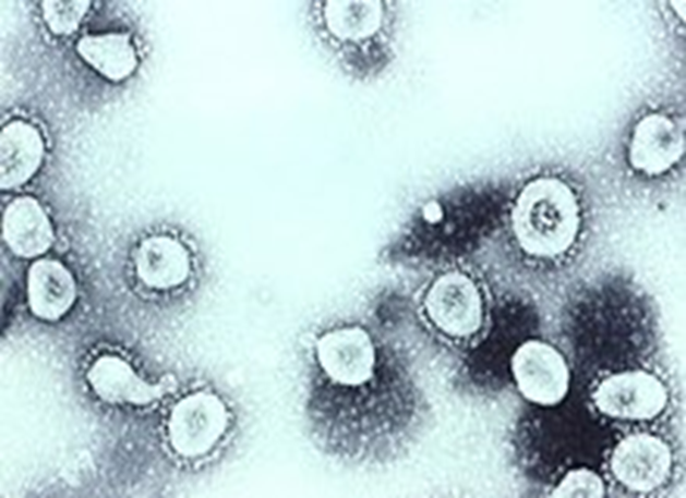
Coronaviruses were first discovered in the 1930s when an acute respiratory infection of domesticated chickens was shown to be caused by Infectious Bronchitis Virus (IBV). In 1931, Arthur Schalk and M.C. Hawn described new respiratory infection of chickens in North Dakota. The infection of new-born chicks was characterized by gasping and listlessness. The chicks' mortality rate was 40–90%. Fred Beaudette and Charles Hudson six years later successfully isolated and cultivated the infectious bronchitis virus [1]. In the 1940s, two more animal coronaviruses i.e., Mouse Hepatitis Virus (MHV) and Transmissible Gastro Enteritis Virus (TGEV) were isolated. It was not realized at the time that these three different viruses were related. Human coronaviruses were discovered in the 1960s. They were isolated using two different methods in United Kingdom and United States. E.C. Kendall, Malcom Byone and David Tyrrell working at the Common Cold Unit of the British Medical Research Council isolated from a boy a novel common cold virus B814. The virus was unable to cultivate using standard techniques which had successfully cultivated rhinoviruses, adenoviruses and other known common cold viruses. In 1965, Tyrrell and Byone successfully cultivated the novel virus serially by passing it through organ culture of human embryonictrachea. The new cultivating method was introduced to the lab by Bertil Hoorn. The isolated virus when intranasally inoculated into volunteers caused a cold and was inactivated by ether which indicates that it had a lipid envelope. Around the same time, Dorothy Hamre and John Proc know at the University of Chicago isolated a novel cold virus 229E from medical students, which they grew in kidney tissue culture. The novel virus 229 E, like the virus strain B814, when inoculated into volunteers caused a cold and was inactivated by ether. The two novel strains B814 and 229E were subsequently imaged by electron microscopy in 1967 by Scottish virologist Almeida at St. Thomas Hospital in London. Almeida through electron microscopy was able to show that B814 and 229E were morphologically related by their distinctive club-like spikes and were morphologically related to IBV [2]. A research group at the National Institute of Health the same year was able to isolate another member of this new group of viruses using organ culture and named the virus strain OC43 (OC means organ culture). Like B814, 229E and IBV, the novel cold virus OC43 had distinctive club-like spikes when observed under electron microscope. The IBV-like novel cold viruses were also morphologically related to the mouse hepatitis virus. This new group of IBV-like viruses came in existence, known as Coronaviruses. The coronavirus strain B814 was lost. It is not known that which human coronavirus was present. Other human coronaviruses have been identified, including SARS-CoV in 2003, HCoV NL63 in 2004, HCoV HKU1 in 2005, MERS-CoV in 2012 and SARS-CoV-2 in 2019. There also been a large number of animal coronaviruses identified since the 1960s (Figure 1).
Figure 1: Transmission Electron Micrograph of Organ Cultured Coronavirus OC43
Origin of Coronavirus
Many human coronaviruses have their origin in bats. The human coronavirus NL63 shared a common ancestor with a bat coronavirus (ARCoV.2) between 1190 and 1449 CE. The human coronavirus 229E shared a common ancestor with a bat coronavirus (GhanaGrp1 Bt CoV) between 1686 and 1800 CE. More recently, alpaca coronavirus and human coronavirus 229E diverged before 1960. MERS-CoV emerged in humans from bats through the intermediate host of camels. MERS-CoV, related to several bat coronavirus species, appears to have diverged from SARS-CoV in 1986. A possible path of evolution of SARS coronavirus and keen bat coronaviruses is that SARS-related coronaviruses coevolved in bats for a long time [3]. The ancestors of SARS-CoV first infected leaf-nose bats of the genus Hipposideridae; subsequently, they spread to horseshoe bats in the species Rhinolophidae, then to Asian palm civets and finally to humans. Unlike other strains such as beta coronaviruses, bovine coronavirus of the species Beta coronavirus 1 and subgenus Embe-covirus was originated in rodents and not in bats. In the 1790s, equine coronavirus diverged from the bovine coronavirus after a cross-species jump. Later in the 1890s, human coronavirus OC43 diverged from bovine coronavirus after another cross-species spillover event [4]. It is speculated that the flu pandemic of 1890 may have been caused by this spillover event, not by the influenza virus, because of related timing, neurological symptoms and unknown causative agent of the pandemic. Besides causing respiratory infections, human coronavirus OC43 is also suspected of playing a role in neurological diseases. In the 1950s, the human coronavirus OC43 began to diverge into its present genotypes. Phylogenetically, mouse hepatitis virus (Murine coronavirus), which infects the mouse's liver and central nervous system is related to human coronavirus OC43 and bovine coronavirus [5].
Microbiology of Coronavirus
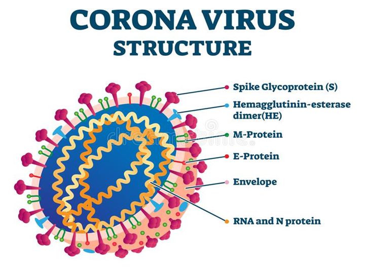
Coronaviruses are large, roughly spherical particles with bulbous surface projections. The average diameter of the virus particles is around 125 nm. The diameter of the envelope is 85 nm and the spikes are 20 nm long. The envelope of the virus in electron micrographs appears as a distinct pair of electron-dense shells (shells that are relatively opaque to the electron beam used to scan the virus particle). The viral envelope consists of a lipid bilayer, in which the Membrane (M), Envelope (E) and Spike (S) structural proteins are anchored. The ratio of E:S:M in the lipid bilayer is approximately 1:20:300. On average a coronavirus particle has 74 surface spikes. A subset of coronaviruses (specifically the members of betacoronavirussubgroup. A) Also have a shorter spike-like surface protein called Hema-glutinin Esterase (HE). The coronavirus surface spikes are homotrimers of S protein, which is composed of S1 and S2 subunit. The homotrimeric S protein is a class I fusion protein which mediates the receptor binding and membrane fusion between the virus and host cell. The S1 subunit forms the head of the spike and has the receptor binding domain.
The S2 subunit forms the stem which anchors the spike in the viral envelope and on protease activation enables fusion. The E and M proteins are important in forming the viral envelope and maintaining its structural shape. Inside the envelope, there is the nucleocapsid, which is formed from multiple copies of the Nucleocapsid (N) protein binds to the positive sense single stranded RNA genome in a continuous beads on a string type conformation. The lipid bilayer envelope, membrane proteins and nucleo-capsid protect the virus when it is outside the host cell (Figure 2).
Figure 2: Structure of Corona Virus
Epidemiology of Coronavirus
The COVID-19 epidemic expanded in early December from Wuhan, China’s 7th most populous city, throughout China and was then exported to a growing number of countries. The first confirmed case of COVID-19 outside China was diagnosed on 13th January 2020 in Bangkok (Thailand). On 2nd of March 2020, 67 territories outside mainland China had reported 8565 confirmed cases of COVID-19 with 132 deaths, as well as significant community transmission occurring in several countries worldwide, including Iran and Italy and it was declared as global pandemic by the WHO on 11th of March 2020. The number of confirmed cases is constantly increasing worldwide and after Asian and European regions, a steep increase in cases is currently (31 March 2020) being observed in low-income countries. It is problematic to quantify the exact size of this pandemic as it would necessary to count all cases including severe cases, symptomatic cases and mild ones [6]. Unfortunately, to date, there is not a global and standard response to the pandemic and each country is facing the crisis based on their own possibilities, expertise and hypotheses. Thus, there are different criteria for testing, hospitalization and estimating of cases making it difficult to calculate the number of people affected by epidemic. Based on the data, the estimated case fatality ratio among medically attended patients is approximately 2%. By 19th July 2020, based on the WHO reports, we have globally 1,40,07,791 confirmed cases and 5,97,105 deaths, distributed as follows: Western Pacific Region 1.7 billion cases and 3649 deaths, European Region 26,84,223 cases and 6,01,934 deaths, South East Asia Region 1,61,441 cases and 1,44,664 deaths, Eastern Mediterranean Region 6,76,508 cases and 4,18,955 deaths, Region of the Americas 75,93,083 cases and 6,01,934 deaths and in the Africa region 2,00,000 cases and 5,600 deaths [7].
Risk Factors
Age, race/ethnicity, gender, some medical conditions (HIV, AIDS), use of certain medications(corticosteroids), poverty and crowding, certain occupations, pregnancy, chronic kidney diseases, chronic obstructive pulmonary disease, weakened immune system because of organ transplant, obesity, serious heart conditions such as heart failure or coronary artery disease, sickle cell anemia, diabetes, moderate to severe asthma, disease that affect blood vessels and blood flow to the brain, cystic fibrosis, high blood pressure, dementia, liver diseases, smoking and thalassemia [8].
Modes of Transmission
Contact and droplet transmission, airborne transmission, fomite transmission. Other modes of transmission include serum or plasma transmission, blood transmission, breast feeding.
Clinical Manifestations
Loss of taste or smell, fever, cough, tiredness, shortness of breath or difficulty breathing, muscle aches, chills, sore throat, runny nose, headache, chest pain, nausea, vomiting, diarrhoea, fatigue, new confusion, inability to wake or stay awake, bluish lips or face, repeated shaking with chills, blocked nose, sputum or coughed up mucus and saliva, abdominal pain.
Pathogenesis of COVID 19
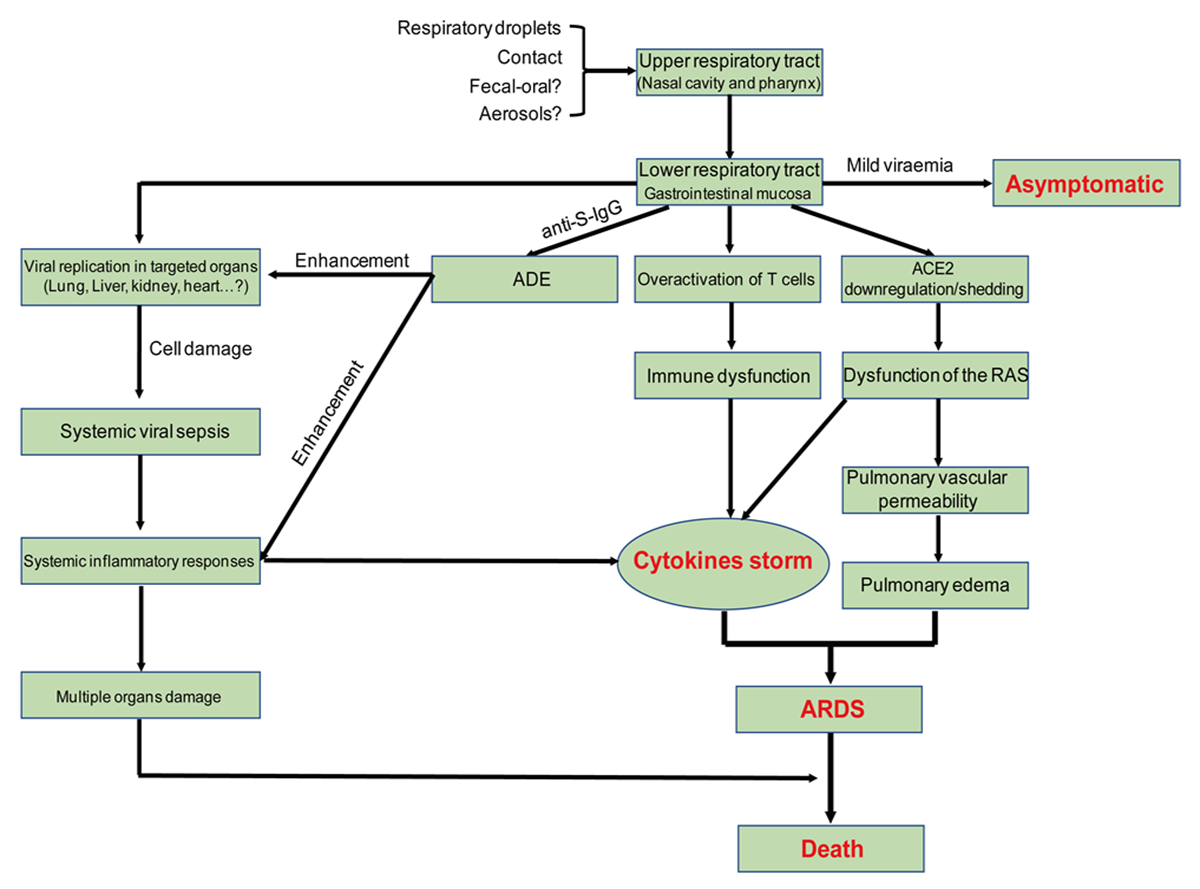
Virus Entry and Spread: SARS-CoV-2 is transmitted predominantly via respiratory droplet, contact and potential in fecal-oral. Primary viral replication is presumed to occur in mucosal epithelium of upper respiratory tract (nasal cavity and pharynx), with further multiplication in lower respiratory tract and gastrointestinal mucosa, giving rise to a mild viremia. Few infections are controlled at this point and remain asymptomatic. Some patients have also exhibited non-respiratory symptoms such as acute liver and heart injury, kidney failure, diarrhea, implying multiple organ involvement. ACE2 is broadly expressed in nasal mucosa, bronchus, lung, heart, esophagus, kidney, stomach, bladder and ileum and these human organs are all vulnerable to SARS-Co 2. Recently, potential pathogenicity of the SARS-CoV-2 to testicular tissues has also been proposed by clinicians, implying fertility concerns in young patients [9].
Pathological Findings
The first report of pathological findings from a severe COVID-19 showed pulmonary bilateral diffuse alveolar damage with cellular fibro myxoid exudates. The right lung showed evident desquamation of pneumocytes and hyaline membrane formation, indicating acute respiratory distress syndrome. The left lung tissue displayed pulmonary edema with hyaline membrane formation, suggestive of early-phase Acute Respiratory Distress Syndrome (ARDS). Interstitial mononuclear inflammatory infiltrates, dominated by lymphocytes, could be observed in both lungs. Multinucleated syncytial cells with atypical enlarged pneumocytes characterized by large nuclei, amphophilic granular cytoplasm and prominent nucleoli were identified in the intra-alveolar spaces, indicating viral cytopathic-like changes. These pulmonary pathological findings extremely resemble those seen in SARS and MERS. Moderate microvascular steatosis and mild lobular and portal activity were observed in liver biopsy specimens, which might be caused by either SARS-CoV-2 infection or drug use. In addition, only a few interstitial mononuclear inflammatory infiltrates were found in the heart tissue, which means that SARS-CoV-2 might not directly impair the heart. Massive mucus secretion in both lungs was found in death cases with COVID-19, which was different from SARS and MERS [10] (Figure 3).
Figure 3: Postulated Pathogenesis of SARS-CoV-2 Infection
Acute Respiratory Distress Syndrome (ARDS)
It is a life-threatening lung condition that prevents enough oxygen from getting to the lungs and into the circulation, accounting for mortality of most respiratory disorders and acute lung injury. In fatal cases of human SARS-CoV, MERS-CoV and SARS-CoV-2 infections, individuals exhibit severe respiratory distress requiring mechanical ventilation and the histopathology findings also support ARDS [11].
Cytokine Storm
Clinical findings showed exuberant inflammatory responses during SARS-CoV-2 infection, further resulting in uncontrolled pulmonary inflammation, likely a leading cause of case fatality. Rapid viral replication and cellular damage, virus-induced ACE2 down regulation and shedding and Antibody Dependent Enhancement (ADE) are responsible for aggressive inflammation caused by SARS-CoV-2, as concluded in a recently published review article. A possible underlying mechanism of Antibody-Dependent Enhancement (ADE) has been proposed recently. ADE, a well-known virology phenomenon, has been confirmed in multiple viral infections. ADE can promote viral cellular uptake of infectious virus–antibody complexes following their interaction with Fc receptors (FcR), FcγR, or other receptors, resulting in enhanced infection of target cells. The interaction of FcγR with the virus-anti-S protein-neutralizing antibodies (anti-S-IgG) complex may facilitate both inflammatory responses and persistent viral replication in the lungs of patients [12].
Immune Dysfunction
Peripheral CD4 and CD8 T cells showed reduction and hyper-activation in a severe patient. High concentrations of pro-inflammatory CD4 T cells and cytotoxic granules CD8 T cells were also determined, suggesting antiviral immune responses and over-activation of T cells. Additionally, several studies have reported that lymphopenia is a common feature of COVID-19, suggestive of a critical factor accounting for severity and mortality.
Diagnostic Parameters of COVID-19
These are different types of coronavirus tests:
Swab Test: In this case, a special swab is used to take a sample from your nose or throat
Nasal Aspirate: In this case, a saline solution will be injected into nose and, then a sample is taken with a light suction
Tracheal Aspirate: In this case, a thin tube with a torch, also known as a bronchoscope, is put into mouth to reach lungs from where a sample is collected
Sputum Test: Sputum is thick mucus that gets accumulated in the lungs and comes out with a cough. During this test, individual required to cough up sputum in a special cup or a swab is used to take a sample from nose
Blood Test: Blood sample is taken from a vein in the arm
Rapid Diagnostic Tests Based on Antigen Detection: A rapid test has also been started for the COVID-19, which involves taking samples from the nose, throat and lungs. Rapid Diagnostic Test (RDT) of a sample of the respiratory tract of a person helps to detect the viral proteins (antigens) related to COVID-19 virus
Rapid Diagnostic Tests Based on Host Antibody Detection: This test detects the presence of antibodies in the blood of COVID-19 infected people. The strength of antibody response depends on several factors like age, medications, infections and severity of disease etc
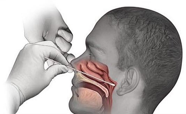
Naso-Pharyngeal Swab Test
A nasal swab is collected from a patient at a hospital or other testing site
The swab is placed into a tube that contains liquid media. Some of the media is transferred to a special, barcoded tube provided by the Broad Institute’s Genomics Platform
The tube is sent to the Board Institute Clinical Research Sequencing Platform, a CLIA-certified clinical lab, where it is unpacked in a clean room and registered in the laboratory’s electronic system for verification and tracking
Within a safety hood in the lab, an automated liquid handling machine transfers the liquid from each barcoded tube into one well of a 96-well plate. This allows dozens of individual samples to be processed at once
The liquid handling machine extracts any viral RNA from the samples
Automating this step speeds up the RNA extraction, allowing for up to 2000 tests to be processed each day
Each plate is inserted into one of four high-throughput quantitative Polymerase Chain Reaction (qPCR) machines
Inside the qPCR machine, a fluorescent dye that sticks to viral complementary DNA is mixed with each sample. The machine measures the fluorescent signal, which indicates the presence of the COVID-19 virus
Results are sent to hospital and physicians through a secure network [13] (Figure 4)
Figure 4: Diagrammatic Representation of Nasopharyngeal Swab Test
Polymerase Chain Reaction (PCR) is a process that amplifies (replicates) a small, well-defined segment of DNA many hundreds of thousands of times, creating enough of it for analysis. Test samples are treated with certain chemicals that allow DNA to be extracted. Reverse transcription converts RNA into DNA
Reverse transcription polymerase chain reaction (RT-PCR) first uses reverse transcription to obtain DNA, followed by PCR to amplify that DNA, creating enough to be analyzed. RT-PCR can thereby detect SARS-CoV-2, which contains only RNA. The RT-PCR process generally requires a few hours
Real-time PCR (qPCR) provides advantages including automation, higher-throughput and more reliable instrumentation. It has become the preferred method
The combined technique has been described as real time RT-PCR or quantitative RT-PCR and is sometimes abbreviated qRT-PCR rRT-PCR or RT qPCR, although sometimes RT-PCR or PCR are used. The Minimum Information for Publication of Quantitative Real-Time PCR Experiments (MIQE) guidelines propose the term RT-Qpcr, but not all authors adhere to this Samples can obtained by various methods, including a nasopharyngeal swab, sputum (coughed up material), throat swabs, deep airway material collected via suction catheter or saliva. Drostenet al. remarked that for 2003 SARS, "from a diagnostic point of view, it is important to note that nasal and throat swabs seem less suitable for diagnosis, since these materials contain considerably less viral RNA than sputum and the virus may escape detection if only these materials are tested”
The likelihood of detecting the virus depends on collection method and how much time has passed since infection. According to Drosten tests performed with throat swabs are reliable only in the first week. Thereafter the virus may abandon the throat and multiply in the lungs. In the second week, sputum or deep airways collection is preferred
Collecting saliva may be as effective as nasal and throat swabs although this is not certain. Sampling saliva may reduce the risk for health care professionals by eliminating close physical interaction. It is also more comfortable for the patient. Quarantined people can collect their own samples. A saliva test's diagnostic value depends on sample site (deep throat, oral cavity, or salivary glands) [14]
Treatment for COVID-19
It has been over six months since the first case of corona virus was detected in China's Wuhan province. More than half a million people across have since lost their lives to the killer disease.
Azithromycin: This must be the most widely prescribed and misused antibiotic in this pandemic. Azithromycin, as with all other antibiotics, does not work in viral infections. Antibiotics are only warranted in patients who have evidence of a secondary bacterial infection as some hospitalized patients will have in the later stages of their disease
Blood Thinners: Hospitalized Covid-19 patients have been observed to have a very high incidence of blood clots. There is current global consensus that all hospitalized Covid-19 patients will benefit from blood thinners injected daily just under their skin (like insulin injections). Though there is sound mechanistic reasoning, randomized controlled trials are awaited
Favipiravir: This is an oral antiviral drug which was fast tracked by the Indian Drug Controller but is not yet approved in the EU or US. Its use should be restricted to mild or moderate infections only. Available data to support its use is sparse but Indian trials have just been completed and the results are awaited
Hydroxy-Chloroquine Sulphate: We now have compelling data from multiple large clinical trials including WHO’s SOLIDARITY and the UK’s RECOVERY trials to categorically say: HCQS does not work
Convalescent Plasma: Our blood is composed of cells and plasma. Plasma from those who have recovered from Covid-19 carries naturally acquired antibodies and when transfused to critically ill patients with Covid-19, may help improve outcomes. This form of therapy is being used across the globe and trials to access its efficacy are under way
Remdesivir: An intravenously administered antiviral medication, it has been shown to be effective in well-designed studies. It seems to shorten recovery time and hospital stay but does not reduce the chance of death. It is currently to be used only in hospitalized patients with severe disease
Steroids: Current evidence shows that dexamethasone can reduce deaths by one-third in patients with severe Covid-19 infection who need oxygen therapy or ventilators
Methyprednisolone: Methyprednisolone is another steroid being used by Indian doctors to handle moderate to severe covid-19 cases
Tocilizumab: This drug is an injection originally used in patients with rheumatoid arthritis. It is being widely used to counter the severe inflammation (cytokine storm) that occurs in some Covid-19 patients. Its use can increase the risk of bacterial infections and it must therefore be used with caution, if at all, in carefully selected patients
Itolizumab: Biocon's Hospital-only, Injectible drug earlier approved for the treatment of Psoriasis. Has got Emergency Use Authorisation for treatment of moderate to severe COVID-19 patients. Approval based on 30 patients’ clinical trial where 20 patients were out on Itolizumab, 10 randomised on standard care
Arbidol: This antiviral was tested along with the drug lopinavir/ritonavir as a treatment for COVID-19. Researchers reported in mid-April that the two drugs didn’t improve the clinical outcomes for people hospitalized with mild to moderate cases of COVID-19
Heparin: A common anti-coagulant drug this is used in treating covid -19 patients. The SARS-CoV2 virus works by using its spike protein to attach to human cells and begin infection. According to recent findings, heparin can bind with the surface spike protein and can potentially bock the infection
Miscellaneous “Cures”: The state machinery has been used to distribute unproven herbal and Ayurvedic potions (Ukalo), homeopathic drops (Arsenicum album) and “treatments” peddled by god-men. Anecdotes and observations do not constitute scientific evidence. In the absence of evidence generated from rigorously vetted clinical trials, the distribution of these substances must be condemned. Pushing unproven and supposedly harmless “treatments” and distributing them to hundreds of thousands is not only disingenuous, but provides people false hope and risks them lowering their guard [15]
Precautions
Until a COVID-19 vaccine is developed, it's crucial to slow the spread of the COVID-19 virus and protect individuals at increased risk of severe illness, including older adults and people of any age with underlying health conditions. To reduce the risk of infection:
Avoid large events and mass gatherings
Avoid close contact (within about 6 feet, or 2 meters) with anyone who is sick or has symptoms
Stay home as much as possible and keep distance between yourself and others
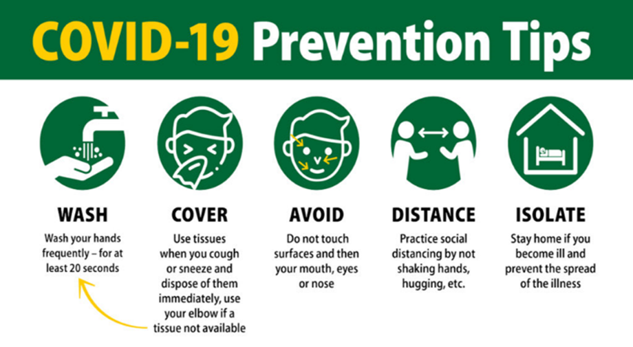
Wash your hands often with soap and water for at least 20 seconds, or use an alcohol-based hand sanitizer that contains at least 60% alcohol
Wear a cloth face covering in public spaces, such as the grocery store, where it's difficult to avoid close contact with others, especially if you're in an area with ongoing community spread. Only use nonmedical cloth masks-surgical masks and N95 respirators should be reserved for health care providers
Cover your mouth and nose with your elbow or a tissue when you cough or sneeze. Throw away the used tissue
Avoid touching your eyes, nose and mouth
Avoid sharing dishes, glasses, bedding and other household items if you're sick
Clean and disinfect high-touch surfaces, such as doorknobs, light switches, electronics and counters, daily
Stay home from work, school and public areas if you're sick [16] (Figure 5)
Figure 5: Key COVID-19 Prevention Measures Including Hand Hygiene, Respiratory Etiquette, Social Distancing and Isolation
Environmental Cleaning and Disinfection
Environmental decontamination
Cleaning of medical equipment
Cleaning soiled bedding, towels and clothes from patients with COVID-19
Cleaning and disinfection of occupied patient rooms
Cleaning and disinfection after patient discharge and transfer
Reduces the spread of microorganisms that cause respiratory infections (colds, flu)
Turn head away from others when coughing/sneezing
Cover the nose and mouth with a tissue
If tissues are used, discard immediately into the trash
Cough/sneeze into sleeve if no tissue is available
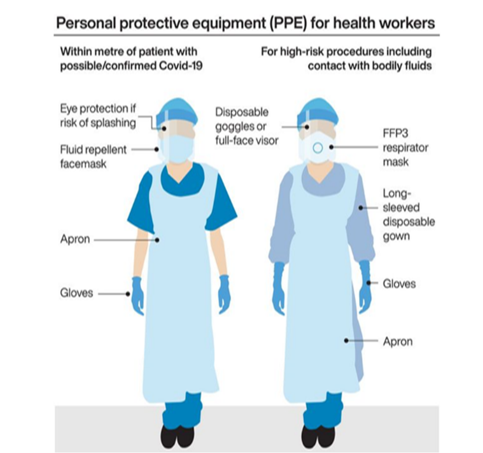
Clean hands with soap, water or alcohol based products. Do not spit here and there [17] (Figure 6)
Figure 6: Personal Protective Equipment for Health Workers
Herd Immunity
Herd immunity occurs when a large portion of a community (the herd) becomes immune to a disease, making the spread of disease from person to person unlikely. As a result, the whole community becomes protected not just those who are immune. Often, a percentage of the population must be capable of getting a disease in order for it to spread. This is called a threshold proportion. If the proportion of the population that is immune to the disease is greater than this threshold, the spread of the disease will decline. This is known as the herd immunity threshold [18].
How is herd immunity achieved?
There are two paths to herd immunity for COVID-19- vaccines and infection.
Vaccines
A vaccine for the virus that causes COVID-19 would be an ideal approach to achieving herd immunity. Vaccines create immunity without causing illness or resulting complications. Herd immunity makes it possible to protect the population from a disease, including those who can't be vaccinated, such as newborns or those who have compromised immune systems. Using the concept of herd immunity, vaccines have successfully controlled deadly contagious diseases such as smallpox, polio, diphtheria, rubella and many others. People who object to vaccines often live in the same neighborhoods or attend the same religious services or schools. If the proportion of vaccinated people in a community falls below the herd immunity threshold, exposure to a contagious disease could result in the disease quickly spreading. Measles has recently resurged in several parts of the world with relatively low vaccination rates, including the United States. Opposition to vaccines can pose a real challenge to herd immunity.
Natural Infection
However, there are some major problems with relying on community infection to create herd immunity to the virus that causes COVID-19. First, it isn't yet clear if infection with the COVID-19 virus makes a person immune to future infection.
Research suggests that after infection with some coronaviruses, reinfection with the same virus though usually mild and only happening in a fraction of people is possible after a period of months or years. Further research is needed to determine the protective effect of antibodies to the virus in those who have been infected. Even if infection with the COVID-19 virus creates long-lasting immunity, a large number of people would have to become infected to reach the herd immunity threshold. Experts estimate that in the U.S, 70% of the population more than 200 million people would have to recover from COVID-19 to halt the epidemic. If many people become sick with COVID-19 at once, the health care system could quickly become overwhelmed. This amount of infection could also lead to serious complications and millions of deaths, especially among older people and those who have chronic conditions [19].
Role of Clinical Pharmacist in Reducing Disease Transmission:
Be a role model and educate others about infection prevention basics
Provide sound guidance to patients and customers
Offer strategies for symptom management for patients with minor viral illnesses
Direct people to reliable resources
Keep up to date on the situation as it evolves
Develop guidance for providing pharmacy services
Create formulary manuals and medicine news to assist clinicians in better
Establish drug registration research
Establish evidence-based drug evaluation and guideline
Remote inpatient order review and dispensing
Tele-pharmaceutical care in fangcang shelter hospital
Develop individual treatment with multidisciplinary team
Telehealth counselling and patient education [20]
Acknowledgment
The authors of the study would like to express their sincere thanks to Dr. S Nelson Kumar, (Principal), M. Venkata Subbaiah, (HOD), P. Gowtham Kumar Reddy & S. Padmakar, (faculty members), department of pharmacy practice of P. Rami reddy Memorial College of Pharmacy, Kadapa, Andhra Pradesh.
Declaration of Conflicting Interests
The author(s) declared no potential conflicts of interest with respect to the research, authorship, and/or publication of this article.
REFERENCES
Dos Santos, W.G. “Natural history of COVID-19 and current knowledge on therapeutic actions.” 2020, published online July 2020.
Chaplin, S. “COVID-19: A brief history and treatments in development.” 2020, published online May 2020.
Shereen, M.A. et al. “COVID-19 infection: Origin, transmission and characteristics of human coronaviruses.” Journal of Advanced Research, vol. 24, 2020, pp. 91–98.
Guo, Y.R. et al. “The origin, transmission and clinical therapies on coronavirus disease 2019 (COVID-19) outbreak an update on the status.” Military Medical Research, vol. 7, no. 1, 2020, pp. 1–10.
Dhama, K. et al. “Coronavirus disease 2019 COVID-19.” Clinical Microbiology Reviews, vol. 33, no. 4, 2020.
Dawood, F.S. et al. “Observations of the global epidemiology of COVID-19 from the pre-pandemic period using web-based surveillance: A cross-sectional analysis.” The Lancet Infectious Diseases, vol. 20, no. 11, 2020, pp. 1255–1262.
Srivastava, N. et al. “Global trends in epidemiology of coronavirus disease 2019 (COVID-19).” Coronavirus Disease 2019 (COVID-19), Springer, Singapore, 2020, pp. 9–21.
Zheng, Z. et al. “Risk factors of critical and mortal covid-19 cases: A systematic literature review and meta-analysis.” Public Health, vol. 81, no. 2, 2020, pp. 16–25.
Jin, Y. et al. “Virology, epidemiology, pathogenesis and control of covid-19.” Viruses, vol. 12, no. 4, 2020, pp. 372.
Yang, M. et al. “Pathological findings in the testes of covid-19 patients: Clinical implications.” European Urology Focus, vol. 6, no. 5, 2020, pp. 1124–1129.
Xu, Z. et al. “Pathological findings of covid-19 associated with acute respiratory distress syndrome.” The Lancet Respiratory Medicine, vol. 8, no. 4, 2020, pp. 420–422.
Mustafa, M.I. et al. “Cytokine storm in covid-19 patients: Its impact on organs and potential treatment.” Journal of Mediators of Inflammation, vol. 18, no. 2, 2020, pp. 7.
Hamilton, S. et al. “COVID-19 Diagnostic Testing at the Broad.” Broad Institute Journal, vol. 8, no. 12, 2019, pp. 35–45.
Abbasi, J. “The promise and peril of antibody testing for covid-19.” JAMA, vol. 323, no. 19, 2020, pp. 1881–1883.
Das, S. et al. “COVID-19 treatment: A lowdown on possible cure and drugs being used.” 2020, published online July 2020.
Cascella, M. et al. “Features, evaluation and treatment of coronavirus (COVID-19).” 2020, published online July 2020.
Tobaiqy, M. “Therapeutic management of patients with covid-19: A systematic review.” Journal of Infection Prevention in Practice, vol. 2, no. 3, 2020, pp. 51–56.
John, T.J. and Samuel, R. “Herd Immunity and herd effect: New insights and definitions.” European Journal of Epidemiology, vol. 16, no. 7, 2000, pp. 601–606.
Randolph, H.E. and Barreiro, L.B. “Herd immunity: Understanding covid-19.” Immunity, vol. 52, no. 5, 2020, pp. 737–741.
Gross, A.E. and MacDougall, C. “Roles of clinical pharmacists during the covid-19 pandemic.” Journal of the American College of Clinical Pharmacy, vol. 3, no. 3, 2020, pp. 564–566.
Recommended Articles
Research Article
An evaluation of Profile of Medico-legal autopsies conducted at Dr. Radhakrishnan Govt. Medical College, Hamirpur, Himachal Pradesh
Download PDF
Research Article
The Efficacy of 5- Fluorouracil Cream and 25% Podophyllin Solution in Treatment of Plantar Warts in Alternative Day Regimen ; An Open Therapeutic Trial.
Download PDF
Research Article
Imaging Evaluation of Ultrasonography Versus Magnetic Resonance,
Imaging in Diagnosis of Carpal Tunnel Syndrome among Female Patients with Positive Nerve Conduction Study
Download PDF
Research Article
Usage of Therapeutic Offloading In Diabetic Foot- A Clinical Audit
Amit Kumar C Jain,
Apoorva HC
Published: 05/05/2021
Download PDF
PO Box 101, Nakuru
Kenya.
Email: office@iarconsortium.org
Editorial Office:
J.L Bhavan, Near Radison Blu Hotel,
Jalukbari, Guwahati-India
Kareemulla, S., Tejaswini, P., Vamaliya, S. Y., Yasmeen, S. & Bharathi, K. C. (2021). Coronavirus Disease (Covid-19) Outbreak: A Narrative Review of Current Knowledge. International Academic Research Journal of Internal Medicine & Public Health, 2(1), 1-8.
MLA
Kareemulla, Shaik, et al. "Coronavirus Disease (Covid-19) Outbreak: A Narrative Review of Current Knowledge." International Academic Research Journal of Internal Medicine & Public Health 2.1 (2021): 1-8.
Chicago
Kareemulla, Shaik, P. Tejaswini, S. Y. Vamaliya, S. Yasmeen and K. C. Bharathi. "Coronavirus Disease (Covid-19) Outbreak: A Narrative Review of Current Knowledge." International Academic Research Journal of Internal Medicine & Public Health 2, no. 1 (2021): 1-8.
Harvard
Kareemulla, S., Tejaswini, P., Vamaliya, S. Y., Yasmeen, S. and Bharathi, K. C. (2021) 'Coronavirus Disease (Covid-19) Outbreak: A Narrative Review of Current Knowledge' International Academic Research Journal of Internal Medicine & Public Health 2(1), pp. 1-8.
Vancouver
Kareemulla S, Tejaswini P, Vamaliya SY, Yasmeen S, Bharathi KC. Coronavirus Disease (Covid-19) Outbreak: A Narrative Review of Current Knowledge. International Academic Research Journal of Internal Medicine & Public Health. 2021 Jan;2(1):1-8.


 Download PDF
 Download XML