One hundred and twelve swab samples of burn and wounds were collected from patients incoming to the Specialized Center for Burns and Plastic Wounds in Mosul City/ Iraq, from September 2022 to the end of January 2023, for different ages ranging 18 months to 55 years and for both sexes. After culturing 112 wound swabs, traditional biochemical methods and the (API 20E) system for investigation, as well as the Vitek-2 compact system were used to diagnose the bacterial isolates. A molecular diagnostic of Citrobacter species was carried out based on the outcomes of the PCR method by the 16SrRNA gene. This involved sequential analysis of the nitrogenous bases to complete the investigative genes of the bacterial isolates. The results indicated that 5 bacterial isolates of Citrobacter spp which included three isolates belonging to the species C. freundii, and one isolate of both C.werkmanii and C. farmeri. Antimicrobial susceptibility test(AST) of all isolates of Citrobacter spp were carried out by using the Kirby-Bauer’s method showed that all isolates were 100% resistant to antibiotics Doxycycline, Tetracycline and Oxytetracycline whereas the study showed that Amikacin, Trimethoprim, Nalidixic acid Norfloxaine, Levofloxacine and Ciprofloxacin best antibacterial effect against of Citrobacter spp.
Keywords
Burn Patients
Citrobacter Spp
Molecular Identification
Antimicrobial Susceptibility Test
INTRODUCTION
Members of the genus Citrobacter belonged to the family Enterobacteriaceae, which are gram-negative bacilli, length ranges between (2-4) micrometers and their width (0.4-0.6) micrometers [1].
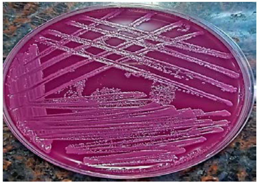
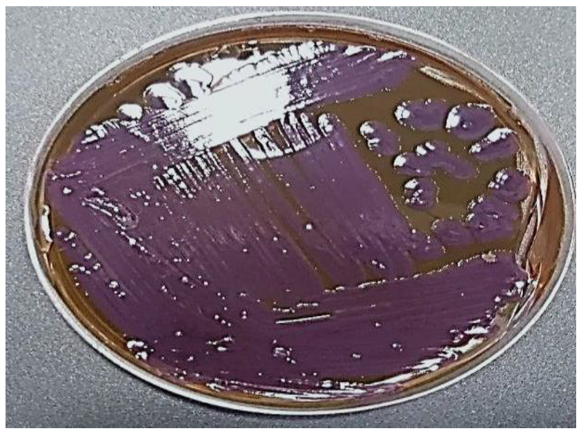
For cultural characteristics, the colonies are smooth, soft, slightly convex, moist, and non-mucous colonies. It is pink color on solid MacConkey medium due to its fermentation of lactose, while on EMB medium a bright purple color [2].
The genus of (Citrobacter) consists of (11 genotypes that can be separated by their biochemical characteristics. Citrobacter bacteria are human opportunistic pathogens that can cause pathological infections such as urinary tract, respiratory tract, central nervous system, skin, and soft tissue infections [3].
The bacteria also can cause osteomyelitis, pyogenic arthritis, bacteremia, endocarditis and intra-abdominal infections, especially in neonates and immunocompromised individuals [4].
The ability of intestinal bacteria to spread from their normal habitat in the digestive tract to other parts of the body, such as the bloodstream, wounds, or urinary tract, determines their pathogenicity to humans and other living organisms [5].The mortality rate due to infection with the Enterobacteriaceae family ranges between 27-44% [6].
One of the most dangerous illnesses that can be fatal is burns. The most recent reports from the World Health Organisation state that burns rank as the fourth most common traumatic injury worldwide, resulting in 180,000 deaths annually. In low- and middle-income countries, burn injuries are on the rise. Numerous physical and psychological issues have a substantial impact on the lives of burn patients [7].
Based on the degree of damage a burn caused, burns were categorised into three degrees or levels: A burn of the first degree only damages the epidermis; a burn of the second degree damages the dermis as well as leaving deep scars. Finally, a third-degree burn to the fatty layer under the skin. The burnt areas are black in colour and cause complete tissue damage [9].
The discovery of antibiotics was one of the most important medical interventions in the history of global health, and they were used to reduce morbidity and mortality caused by bacterial infections [10]. Antibiotics have been used for decades not only for medical purposes, but also as a preventive measure in a variety of fields, including animal husbandry and agriculture [11]. Antibiotics have prevented millions of deaths. However, a serious health and environmental problem, in particular the emergence of multiple antimicrobial resistance, has resulted from inappropriate use of antibiotics. More than 750,000 deaths annually are thought to be caused by antimicrobial resistance [12].
According to what mentioned before about the risk of Citrobacter spp. with human infections, and because of importance the subject of antibiotics resistance generally, the research aimed to know the extent the invasion of Citrobacter species to burn and wounds, as well as to detect the antibiotics profile analysis.
MATERIALS AND METHODS
Sample Collection
Burn and wound samples 112 were obtained from Specialized Center for Burns and Plastic Wounds in Mosul City/ Iraq, between September 2022 and the end of January 2023, ranging in age from 18 months to 55 years, and for both sexes. The bacterial isolates were cultivated on selective media (MacConkey agar and EMB agar) aerobically.
Routine Identification
First of all morphology of suspected colonies belonged to Citrobacter spp. was noticed and registed, then gram stain was done to determin the gram reaction and cell shap. Then some biochemical tests were achieved such as oxidase, catalase, citrate utilization test, Hydrogen-Sulfide-Indole production motility test [13].
Then, API 20E and Vitek-2 compact system utilizing ID-cards (BioMerieux, France) were used to confirm the diagnosis of the species of Gram-negative bacterial isolates of Citrobacter spp.
Molecular Identification
Genomic DNA Extraction: DNA extraction was conducted for 5 suspected Gram-negative bacterial isolates of Citrobacter, identified using the device of Vitek-2 compact system. The kit was used to extract DNA and the extraction method was followed based on the manufacturer’s instructions (Macrogen, Korea). Then purity and concentration were measured by Nanodrop device.
Polymerase Chain Reaction
PCR technique is carried out using the 16SrRNA gene in bacterial isolates in order to accurately diagnose them at the molecular level, which requires:
DNA: (which has been previously extracted) was used in concentration <250ng
Primers: Supplied by the Korean company Macrogen, consisting of the
Following sequences:
27F AGAGTTTGATCMTGGTCAG
1522R AAGGAGGTGATCCARCCGCA [14]
For each primer 10 μM was put in 250 μl of volum
Taq green master mix solution (1X) prepared by the American company (Promega). Table 1 shows the PCR program the followed for implified the 16SrRNA
Table 1: PCR Program for 16srrna Gene Amplification
Number of cycles
Temperature C˚
Time min
Main steps
1
95
3
Initial Denaturation
Step1
30
95
0.30
DNA Denaturation
Step2
55
1
Primer annealing
72
1
Extension Primer
1
72
5
Final Extension
Step3
Electrophoresis
Agarose gel 2% was prepared in a beaker, 2 μl of red safe dye were added with continuous stirring to ensure the distribution of the dye in the gel and poured into the plate placed on its support in the electrical relay device
TAE buffer solution was used to covering the surface of the gel, 6 μl of Ladder were added to the first well, and 8 μl of the reaction product (PCR product) of each sample, and load them into the agarose gel hole starting from the second well
The electric was at 50 volts for 75 minutes a transilluminator at 320 nm was used to observe the bands and photograph them
For the purpose of completing the molecular diagnosis, sequence of the nitrogenous bases of the 16SrRNA gene was performed, the PCR products were sent to the United States of America with the forward primer, then the results were analyzed using the BLAST program located at the National Center for biotechnology information (NCBI) through a sequence comparison. Nitrogenous bases with international strains registered in it and finally determining the bacterial species diagnosis
Antibiotic Susceptibility Test
The disc diffusion method was used to detect antibiotic sensitivity of the isolates Citrobacter spp using the Kirby-Bauer method. The results were compared with the standard diameter of inhibition zones for each antibiotic [15]. 12 antibiotics prepared by the Turkish company Bioanalyse were used, as shown in Table 2.
Table 2: Limits of Sensitivity and Resistance of Citrobacter Spp to the Antibiotics
R
I
S
Antibiotic concentration (µg/disc)
Antibiotic symbol
Antibiotics
≤14
16-20
≥19
10 μg
TE
Tetracycline
≤12
14-16
≥16
10 μg
D0
Doxycycline
≤14
15-18
≥19
30 µg
T
Oxytetracycline
≤15
16-20
≥21
10 µg
CIP
Ciprofloxacin
≤13
14-16
≥17
5 µg
LVX
Levofloxacin
≤1
13-16
≥17
10 µg
NOR
Norfloxacin
≤15
16-18
≥19
10 µg
SXT
Trimethoprim- sulfamethoxazole
≤14
15-16
≥17
10 µg
AK
Amikacin
≤ 12
13-14
≥ 15
10 µg
GM
Gentamicin
≤ 17
18-20
≥21
30 µg
CAZ
Ceftazidime
≤ 24
25-26
≥ 27
10 µg
CRO
Ceftriaxone
≤ 13
14-18
≥ 19
30 µg
NA
Nalidixic acid
A number of pure bacterial colonies were transferred to a test tube containing a saline solution to obtain a bacterial suspension with a concentration of 1.5 x 10⁸ colony-forming units/ml, compared to a McFarland tube No. 0.5, 100 microliters of the suspension were spread on solid Mueller-Hinton agar medium. The discs were placed using sterile forceps on the medium (6 discs in each Dish), the dishes were incubated at 37°C for 24 hours. After completing the incubation period, the results were read by ruller and compared with the standard diameter of inhibition zones for each antibiotic shown in Table 2 [15]. Sensitivity or resistance of this bacteria to the antibiotics was determined.
Isolation and Identification of Citrobacter Spp
After the sample collection and cultivation, the results showed that 18 samples 16.1% had no bacterial growth, while 94 samples 83.9% had growth, 49 of which were non-lactose fermenter isolates, excluded during the study. However, 42 bacterial isolates 44.7% appeared as Pseudomonas aeruginosa based on their distinctive odor and its pigment.
45 bacterial isolates that remained fermented the sugar lactose. 12 bacterial isolates belonging to the genus Klebsiella spp due to the apparent mucous on the plate these were also directly excluded.
For the distinguishing of Citrobacter and differentiate it from 33 remaining bacterial isolates, the biochemical tests and the (API 20E) system, as well as diagnosis with the Vitek device, revealed that 28 isolates were E. coli and 5 isolates of Citrobacter spp which included three isolates belonging to the species C. freundii, and one isolate of both C. werkmanii and C. farmer. The molecular diagnosis using the 16srRNA gene by PCR technique and sequencing revealed that all species were C. freundii.
Citrobacter colonies appeared fermented the sugar lactose on the MacConkey medium, as indicated by their pink appearance. The colonies were also small in size, slightly convex, moist, mucous or not mucous [16], as illustrated in
Figure 1. Furthermore, in order to differentiate Citrobacter from E.coli, the lactose fermenter isolates were subcultured on EMB medium for 24hr. at 37Co. Violet colonies were the positive result colonies of Citrobacter, with large, smooth, shiny colonies [17], as shown in Figure 2.
Microscopic examination of cells stained with Gram stain showed that the cells were negative for this stain, bacilli in shape,
Figure 1: Citrobacter Colonies on Macconkey Agar Medium
Figure 2: Citrobacter Colonies on Eosin-Methylene Blue Agar
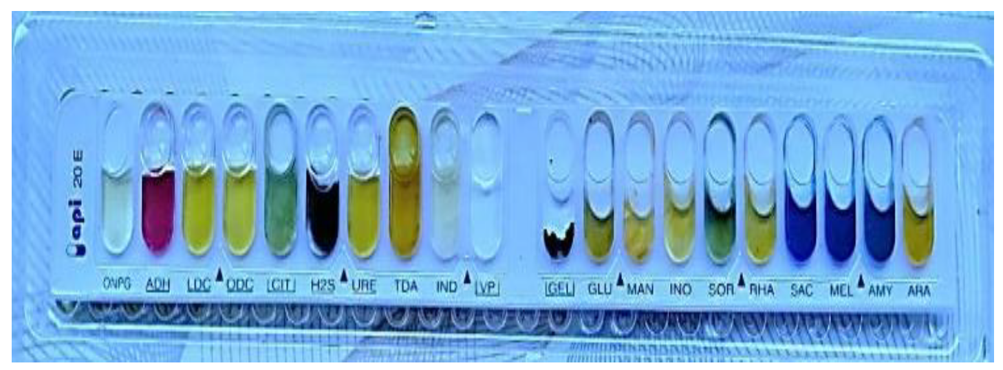
The API-20E strip results as shown in Figure 4 for Citrobacter revealed that the five bacterial isolates were positive for the glucose and mannitol fermentation tests, negative for the sucrose test, and positive for the arabinose sugar test.
The isolates, on the other hand, were negative for the Ortho-nitrophenylgalactopyranoside (ONPG) test, positive for citrate consumption, and negative for hydrogen sulphide gas production and negative to urease and indole enzyme production to the test.
The report introduced by [18] mentioned the same characterization of isolate of C. fruendii which isolated from one hundred samples of choleocystitis cases from the hospital of Mosul city/Iraq in 2019.
Our findings were consistent with those of other researchers [19] when they isolated Citrobacter spp. from Iraqi patients in the city of Samarra. All isolates were identified by the API 20E system. The obtained results for C. freundii are shown in Figure 3.
Figure 3: ApI-20E Test Result for Citrobacter Freundii
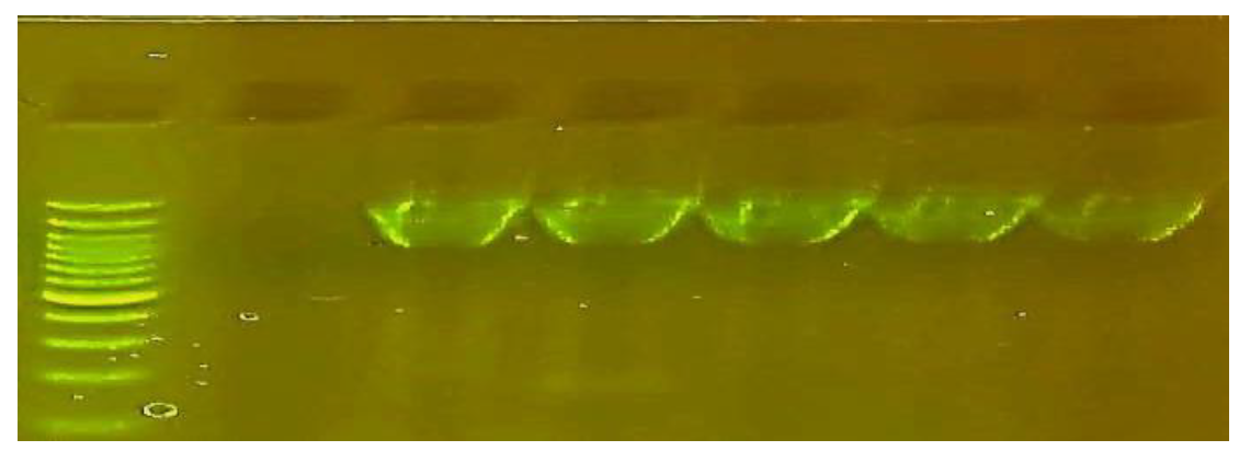
Figure 4: 2% Agarose Gel Electrophoresis of the Ribosomal Gene 16srrna Amplification Products at a Voltage of 50 Volts for 75 Min
Identification by Using the Vitek 2Compact System
Vitek-2 compact system very useful in investigating bacterial isolates and determining their sensitivity and resistance to many antibiotics, particularly in health institutions. In our study, five isolates of Citrobacter were identified by using this device: three isolates of C. freundii 97% and one isolate each of C.werkmanii 99% and C. farmeri 99%.
Molecular Identification
In order to diagnosis Citrobacter spp, precisely, molecular technique was used. After extracting the DNA of suspected Citrobacter isolates and determining the concentrations between (182-326) ng/µl using the Nanodrop device, the purity was ranging from (1.6-1.9), following that, a PCR programme using primers of 16SrRNA was carried out. the DNA products were electrophoresed on an agarose gel, and the results showed the appearance of fluorescent bands with molecular size 1500 bp as shown in Figure 4 indicates the presence of the 16SrRNA gene. Identification of the ribosomal gene 16SrRNA is the most accurate method for identifying bacterial species because it has a fixed sequence and a conserved gene that does not change over time [20].
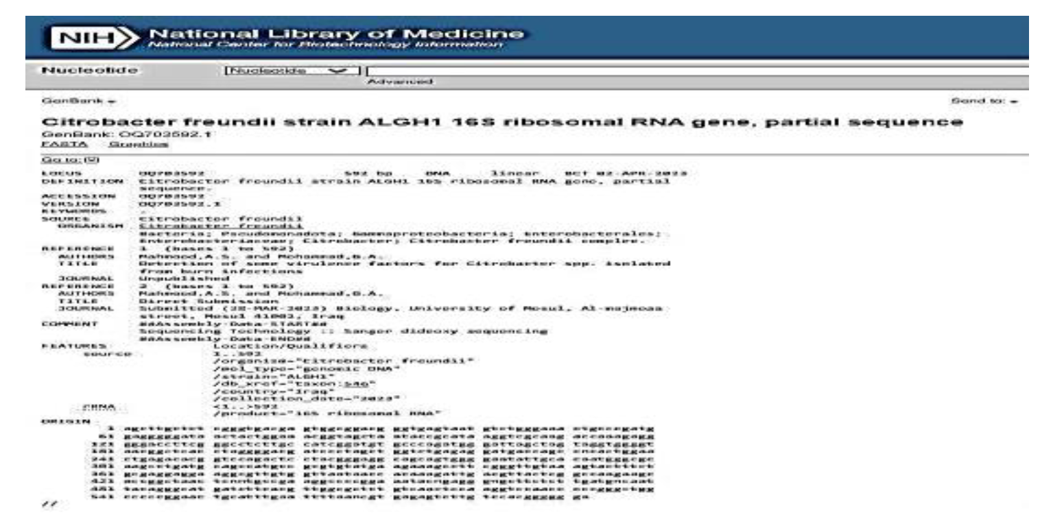
In order to complete the molecular diagnosis, a sequential analysis of the PCR product for the gene 16SrRNA was performed, and these products were sent to the United States of America. After obtaining the sequences, the alignment process was applied at the global site NCBI and the results showed that the three isolates belong to the genus C.freundii and one isolate belong to Citrobacter sp. The sequences of the nitrogenous bases of our isolates were compared to those of the isolates registered at (NCBI). The four isolates were found to be genetically identical to the isolates recorded globally in NCBI by 98-99%. Our isolates were given a unique accession number, as shown below:
C.freundii strain ALGH1under accession number OQ703592.1
Citrobacter sp strain ALGH2 under accession number OQ703593.1
C.freundii strain ALGH3 under accession number OQ703594.1
C.freundii strain ALGH3 under accession number OQ703595.1
The results showed that Vitek-2 system diagnosis was approximately identical to molecular diagnosis using the PCR technique for the Citrobacter species. Our results agreed with the results of researchers [21] and [18] who used the 16SrRNA gene in their study in the rapid and direct detection of Citrobacter bacteria.
The use of 16SrRNA gene sequences allows for differentiation and distinction between organisms at the level of genus, species, and strain, but more than one species can share the sequence of a particular gene. As a result, when diagnosing a newly discovered species, it is preferable to rely on the entire piece, which has a molecular weight of 1,500 base pairs) [22].
Figure 5: Registration of Citrobacter Freundii in Gene Bank under Accession Number OQ703592.1
Susceptibility of C. Freundii Strains to Antibiotics
Fortunately, our result inTtable (3) demonstrate bacterial species were sensitive to most antibiotics, except the resistance against three antibiotics (Doxycyclin, Tetracycline, Oxytetracycline) belonging to the Tetracycline family, while there was an intermediate sensitivity to Gentamycin, Ceftazidime, and ceftriaxone, The threatening human health and the ecosystem, the spread of antibiotic resistance genes among bacterial species is a major factor in the elimination of tetracycline from their natural habitat
The results of susceptibility test for antibiotics showed that strains of C. Freundii showed 100% resistance to antibiotics (Tetracycline, Doxycycline and Oxytetracycline) whereas 100% sensitivity to (Ciprofloxacin, Levofloxacine, Norfloxaine, Trimethoprim-sulfamethoxazole, Amikacin, Gentamycin and Nalidixic acid). The strains of C. freundii also showed moderate sensitivity and resistance to antibiotics (Ceftazidime and Ceftriaxone).
Our result of the resistant to Doxycyclin agree with [17] who said that one bacterial isolate from Citrobacter intermedius was MDR which resisted Doxycyclin, Piperacillin and Cefotaxime, while nine strains were XDR Tetracycline is a broad-spectrum antibiotic that inhibits bacterial protein synthesis. It inhibits bacterial growth by inhibiting translation. It prevents aminoacyl-tRNA from interacting or association with bacterial ribosomes. This antibiotic done this work by binding to the 16s unit of the 30s ribosomal unit and prevents the tRNA molecule charged from binding to an amino acid to the A site of the ribosome [23].
According to Roberts and Schwarz [24], there are three main ways in which bacteria develop resistance to tetracycline: enzymatic inactivation of the compound, excretion of the compound outside the bacterial cell through efflux pumps, and genes present on chromosomes or plasmids that encode for the production of cytoplasmic proteins that protect ribosomes from tetracycline's action and provide ribosomal protection. One of the most important reasons that leads to the establishment of bacterial resistance is the indiscriminate use of antibiotics because bacteria adapt themselves to changes in their surrounding environment, as well as the incorrect use of antibiotics by the patient himself or failure to complete the specified time period to complete taking the antibiotic, which acts as stimulates genetic mutations in bacteria, represented by resistance-related mutations [25].
Table 3: The Sensitivity for the Isolates of the C.Freundii toward Antibiotics
C.freundii 3
C.freundii 2
C.freundii 1
Antibiotic symbol
Antibiotic
R
R
R
TE (10µg)
Tetracycline
R
R
R
DO (10µg)
Doxycycline
R
R
R
T (30 µg)
OXYtetracycline
S
S
S
CIP (10µg)
Ciprofloxacin
S
S
S
LEV (5µg)
Levofloxacine
S
S
S
NOR (10µg)
Norfloxaine
S
S
S
TMP (10µg)
Trimethoprim-sulfamethoxazole
S
S
S
AK (10µg)
Amikacin
S
S
S
CN (10µg)
Gentamycin
I
I
R
CAZ (30µg)
Ceftazidime
S
I
R
CRO (10µg)
Ceftriaxone
S
S
S
NA (10µg)
Nalidixic acid
REFERENCE
Oliveira, J. and Reygaert, W.C. “Gram negative bacteria.” StatPearls, StatPearls Publishing, 2022.
Carroll, K.C. et al.Review of medical microbiology. 27th ed., McGraw-Hill, 2016.
Riedel, S. et al.Jawetz, Melnick and Adelberg’s medical microbiology. 28th ed., McGraw-Hill Education, 2019.
Sarah, S.L. et al.Principles and practice of pediatric infectious diseases. Elsevier, 2022.
Pakbin, B. et al. “Virulence factors of enteric pathogenic Escherichia coli: A review.” International Journal of Molecular Sciences, vol. 22, no. 18, 2021, pp. 9922.
Falagas, M.E. et al. “Deaths attributable to carbapenem-resistant Enterobacteriaceae infections.” Emerging Infectious Diseases, vol. 20, no. 7, 2014, pp. 1170–1175.
Parvizi, A. et al. “A systematic review of life satisfaction and related factors among burns patients.” International Wound Journal, 2023.
Warby, R. and Maani, C.V. “Burn classification.” StatPearls, StatPearls Publishing, 2022.
Hollyer, I. and Ison, M.G. “The challenge of urinary tract infections in renal transplant recipients.” Transplant Infectious Disease, vol. 20, no. 2, 2018, e12828.
Gajdacs, M. and Albericio, F. “Antibiotic resistance: From the bench to patients.” Antibiotics, vol. 8, no. 3, 2019, pp. 129.
Patangia, D.V. et al. “Vertical transfer of antibiotics and antibiotic-resistant strains across the mother/baby axis.” Trends in Microbiology, vol. 30, no. 1, 2022, pp. 47–56.
Granato, P.A. et al.Laboratory manual and workbook in microbiology: Application to patient care. McGraw-Hill Education, 2019.
Khaleel, A.M. et al. “The efficiency of molecular methods compared to traditional methods in identifying bacteria from blood and cerebrospinal fluid samples.” Malaysian Journal of Microbiology, vol. 19, no. 2, 2023.
Clinical and Laboratory Standards Institute. Performance standards for antimicrobial susceptibility testing. CLSI, 2022.
Shinu, P. “Antimicrobial resistance, phenotypic characteristics and biofilm production in Citrobacter freundii isolates obtained from urinary tract infections.” Journal of Pharmacology and Pharmacotherapeutics, vol. 13, no. 4, 2022, pp. 375–381.
Hashim, M.H. and AlKhafaji, M.H. “Isolation and identification of Citrobacter freundii from chicken meat samples using cultural and molecular techniques.” Iraqi Journal of Science, 2018, pp. 1216–1224.
Selah, T.M. and Mohammad, G.A. “Detecting antibiotics profile analysis of Citrobacter species isolated from cholecystitis.” Proceedings of the 4th Al-Noor International Conference for Science and Technology, 2022, pp. 50–57.
Mahmood, A.E. and Atyah, A.A. “Determination of some virulence factors of Citrobacter freundii isolated from Iraqi patients.” Iraqi Journal of Science, 2021, pp. 3358–3365.
Church, D.L. et al. “Performance and application of 16S rRNA gene cycle sequencing for routine identification of bacteria in the clinical microbiology laboratory.” Clinical Microbiology Reviews, vol. 33, no. 4, 2020.
Rondini, S. et al. “Characterization of Citrobacter sp. line 328 as a source of Vi for a Vi-CRM197 glycoconjugate vaccine against Salmonella Typhi.” Journal of Infection in Developing Countries, vol. 6, no. 11, 2012, pp. 763–773.
Woo, P.C.Y. et al. “Identification by 16S rRNA gene of Enterobacteriaceae species from bone marrow transplant recipients.” Journal of Clinical Pathology: Molecular Pathology, vol. 53, 2000, pp. 211–215.
Chen, X. et al. “A comprehensive review on biodegradation of tetracyclines: Current research progress and prospect.” Science of the Total Environment, vol. 814, 2022, pp. 152852.
Roberts, M.C. and Schwarz, S. “Tetracycline and chloramphenicol resistance mechanisms.” Antimicrobial Drug Resistance: Mechanisms of Drug Resistance, vol. 1, 2017, pp. 231–243.
Kakoullis, L. et al. “Mechanisms of antibiotic resistance in important gram-positive and gram-negative pathogens and novel antibiotic solutions.” Antibiotics, vol. 10, no. 4, 2021, p. 415.
Recommended Articles
Review Article
Hyperuricemia and Cardiovascular Diseases: A Comprehensive Review
Neha Verma,
Ritika Kashyap
Published: 25/07/2024
Download PDF
Research Article
Role of Maternal Serum Lactate Dehydrogenase in Determining the Severity of Preeclampsia: A Case-Control Study
Alhan Ali Salih,
...
Sabah Hasan Mohammed
Published: 04/10/2024
Download PDF
Research Article
Euthanasia is a Medical Act Which Remove Any Pain
Siniša Franjić
Published: 30/11/2021
Download PDF
Research Article
C-Reactive Protein, Vitamin D and Calcium as Biomarkers for the Diagnosis in some Osteoarthriris Patients who Attending Al-Refai Teaching Hospital
Ahlam Mohsen Kudaier
Published: 30/06/2024
Download PDF
PO Box 101, Nakuru
Kenya.
Email: office@iarconsortium.org
Editorial Office:
J.L Bhavan, Near Radison Blu Hotel,
Jalukbari, Guwahati-India
Mahmoud, A. S. & A. Mohammad, G. (2024). Revealing antibiotics analysis against Citrobacter frundii isolated from burn and wound cases. IAR Journal of Clinical & Medical Biochemistry, 4(1), 1-6.
MLA
Mahmoud, Ali S. and Ghada A. Mohammad. "Revealing antibiotics analysis against Citrobacter frundii isolated from burn and wound cases." IAR Journal of Clinical & Medical Biochemistry 4.1 (2024): 1-6.
Chicago
Mahmoud, Ali S. and Ghada A. Mohammad. "Revealing antibiotics analysis against Citrobacter frundii isolated from burn and wound cases." IAR Journal of Clinical & Medical Biochemistry 4, no. 1 (2024): 1-6.
Harvard
Mahmoud, A. S. and A. Mohammad, G. (2024) 'Revealing antibiotics analysis against Citrobacter frundii isolated from burn and wound cases' IAR Journal of Clinical & Medical Biochemistry 4(1), pp. 1-6.
Vancouver
Mahmoud AS, A. Mohammad G. Revealing antibiotics analysis against Citrobacter frundii isolated from burn and wound cases. IAR Journal of Clinical & Medical Biochemistry. 2024 Jan;4(1):1-6.


 Download PDF
 Download XML