Contents
Download PDF
pdf Download XML
188 Views
72 Downloads
Share this article
Research Article | Volume 6 Issue 1 (January-June, 2026) | Pages 1 - 6
The Relationship Between Maternal Levels of Perfluoroalkyl Substances (PFAS) During Early Pregnancy and the Development of Preeclampsia
 ,
1
M.B.Ch.B. Iraqi Board for Medical Specialization in Obstetrics and Gynecology, Ministry of Higher Education and Scientific Research, Iraq
2
M.B.Ch.B. F.I.C.O.G, College of Medicine, Tikrit University, Tikrit, Iraq
Under a Creative Commons license
Open Access
Received
Feb. 3, 2026
Revised
March 9, 2026
Accepted
April 19, 2026
Published
May 1, 2026
Abstract

Background: Hypertensive disorder of pregnancy is one of the most common complications in pregnancy forming a triad together with hemorrhage and infection. It affects about 10% of pregnancies and contributes for a significant maternal and perinatal mortality Aim: The study aimed to investigate potential associations between polyfluoroalkyl substances (PFASs) serum levels and preeclampsia, considering disease severity. Patients and methods: This case-control study, conducted at Salah Al-Din General Hospital in Tikrit city from 15th of January to 1st of December, 2023, included 120 pregnant women who attended the hospital in to 2 visits: the first study visit (median gestational age of 24 weeks) and the second visit (median gestational age of 35 weeks), divided into two groups: 60 with diagnosed preeclampsia or eclampsia (Preeclamptic group) and 60 healthy, normotensive pregnant women without dipstick proteinuria (Control group). Blood samples were collected from each pregnant women for determination of polyfluoroalkyl substances (PFASs) using standard laboratories procedure. Results: The study investigates the association between Polyfluoroalkyl Substances (PFAS) concentrations and preeclampsia, comparing polyfluoroalkyl substances levels in two groups at different gestational time points. The study showed a significant differences in mean polyfluoroalkyl substances concentrations between women with preeclampsia and a control group at both the 24th and 35th weeks, with statistically significant p-values of 0.001. Specifically, at the 1st visit (week 24), women with preeclampsia exhibit a lower mean polyfluoroalkyl substances concentration of 8.68 ng/ml compared to the control group's mean of 13.56 ng/ml. Similarly, at the 2nd visit (week 35), the mean polyfluoroalkyl substances concentration for women with preeclampsia is 6.88 ng/ml, lower than the control group's mean of 10.78 ng/ml. The 95% confidence intervals for the mean differences further underscore the robustness of these findings, providing additional statistical support for the observed differences in mean PFAS concentrations between the two groups at different time points. Additionally, the stusy demonstrated that the mean polyfluoroalkyl substances concentrations in relation to the severity of preeclampsia at the 24th and 35th weeks, reinforcing the significant associations between polyfluoroalkyl substances levels and disease severity. Conclusions: The study findings strongly suggest a potential association between lower Polyfluoroalkyl Substances (PFAS) levels and an increased risk of developing preeclampsia, particularly in severe cases. This underscores the significance of PFAS monitoring as a potential predictive marker for specific subtypes of preeclampsia.

Keywords
INTRODUCTION

Hypertensive disorders of pregnancy (HDP) represent a major global health concern and remain one of the leading causes of maternal and perinatal morbidity and mortality worldwide. Among these disorders, preeclampsia is particularly significant due to its complex pathophysiology and potential for severe complications affecting both the mother and fetus. Clinically, preeclampsia is defined by the new onset of hypertension after 20 weeks ofgestation accompanied by proteinuria or evidence of maternal organ dysfunction, including renal, hepatic, neurological, or hematological abnormalities [1-3]. Despite advances in obstetric care, the exact etiology of preeclampsia remains incompletely understood; however, it is widely accepted that abnormal placentation plays a central role. Inadequate trophoblastic invasion of the spiral arteries leads to reduced uteroplacental perfusion, placental ischemia, and the subsequent release of antiangiogenic factors, inflammatory mediators, and oxidative stress markers into the maternal circulation. These changes contribute to widespread endothelial dysfunction, vasoconstriction, and multiorgan involvement, which characterize the clinical manifestations of the disease [4-6]. In recent years, growing attention has been directed toward environmental factors that may contribute to the development of preeclampsia, particularly exposure to endocrine-disrupting chemicals. Among these, per- and polyfluoroalkyl substances (PFAS) have emerged as a group of concern due to their persistence, bioaccumulation, and potential adverse health effects. PFAS are synthetic fluorinated compounds widely used in industrial processes and consumer products, including non-stick cookware, water-resistant textiles, and firefighting foams. Their strong carbon–fluorine bonds render them highly resistant to environmental degradation, leading to their classification as “forever chemicals” [7,8]. Consequently, PFAS have been detected in water, soil, wildlife, and human biological samples worldwide, including serum, placenta, and breast milk. Epidemiological studies have demonstrated associations between PFAS exposure and various adverse health outcomes, including dyslipidemia, immune dysfunction, endocrine disruption, and reproductive toxicity [9,10]. Importantly, increasing evidence suggests a potential link between maternal PFAS exposure and hypertensive disorders of pregnancy, including preeclampsia. Large population-based studies, such as those conducted in highly exposed communities, have reported higher odds of preeclampsia among women with elevated serum concentrations of PFAS compounds such as perfluorooctanoic acid (PFOA) and perfluorooctanesulfonic acid (PFOS) [11,12]. However, findings from studies conducted in populations with lower exposure levels have been inconsistent, indicating that the relationship may be influenced by exposure intensity, timing, and individual susceptibility. Several biological mechanisms have been proposed to explain the association between PFAS exposure and preeclampsia. PFAS may interfere with normal placental development by disrupting immune regulation and impairing trophoblast invasion, which is essential for proper remodeling of uterine spiral arteries. Additionally, PFAS have been shown to activate peroxisome proliferator-activated receptors (PPARs), leading to alterations in lipid metabolism, increased oxidative stress, and vascular inflammation, all of which are implicated in the pathogenesis of preeclampsia. Furthermore, PFAS exposure may affect the balance of angiogenic and antiangiogenic factors, such as soluble fms-like tyrosine kinase-1 (sFlt-1) and placental growth factor (PlGF), thereby contributing to endothelial dysfunction and hypertension during pregnancy. Given the significant burden of preeclampsia and the widespread exposure to PFAS in modern environments, investigating the potential relationship between these environmental contaminants and hypertensive disorders of pregnancy is of critical importance. Understanding this association may provide valuable insights into the underlying mechanisms of disease development, identify modifiable risk factors, and contribute to improved prevention and management strategies for affected women.

PATIENTS AND METHODS

The case-control study was carried out in Tikrit city (Salah Al-Din General Hospital) from 15th of January to 1st of December 2023.


The number of pregnant women under study was 120, all carrying a live fetus and attending two visits: the first study visit (median gestational age of 24 weeks) and the second visit (median gestational age of 35 weeks). The sample was divided into the following groups:


  • Preeclamptic Group: comprised 60 pregnant women who met the criteria for preeclampsia or eclampsia

  • Control Group: consisted of 60 pregnant women in good health, normotensive, and without dipstick proteinuria


Inclusion Criteria

Women are eligible for the study if they are:


  • At least 18 years of age and 

  • Sought prenatal care before 10 weeks of gestation.


Exclusion Criteria

Exclusion criteria include:


  • Pregnant women with chronic hypertension, diabetes mellitus, chronic kidney disease, liver disease, cardiovascular disease, thyroid or other endocrine disorders, and malignancy

  • Pregnancies with major maternal or fetal abnormalities, including hydatidiform mole, placenta accrete, and abrupt placenta

  • Pregnant women with a history of smoking or consumption of a drug that might affect their blood pressure


Ethical Approval

Approval from the Council of the College of Medicine / Tikrit University was obtained for the study proposal. Approval permission was presented to the director of Salah Al-Din Health Directorate / Salah Al-Din General Hospital. 


Maternal serum samples were collected at the first study visit (median= 24 wk gestation). After enrollment, participants attend one more study visit (median = 35 wk gestation), where serum samples are also collected. All specimens are stored in deep freeze until analysis. 


Gestational age was estimated according to the American College of Obstetrics and Gynecologists (ACOG) using the last menstrual period with verification by ultrasound measures


Preeclampsia was diagnosed according to ACOG guidelines at the time of recruitment: new or worsening hypertension (≥140 mmHg systolic or ≥90  mm Hg diastolic blood pressure) and proteinuria (>300mg /24h or protein/creatinine ratio of >0.20) after 20 week of gestation (8). All cases of preeclampsia were deidentified and reviewed by a panel of Maternal–Fetal Medicine–certified physicians.. Gestational age at disease onset was recorded. Cases were further categorized as early- or late-onset, depending on whether disease onset occurred prior to 34 wk gestation. Severe preeclampsia is defined as having one or more of the following criteria: blood pressure of at least 160/110 mmHg measured on two occasions each 6 hours apart, proteinuria of at least 5 g per 24 hours, or at least 3+ on dipstick testing, oliguria of lesser than 500 ml per 24 hours, cerebral or visual disturbances, pulmonary edema or cyanosis, epigastric or right upper quadrant pain, impaired liver function, thrombocytopenia, fetal growth restriction (8).


Routine clinical information (e.g., maternal weight or blood pressure) was also collected at study visits. Body mass index (BMI) was calculated using self-reported maternal prepregnancy weight and height measured at the first study visit. 


Blood pressure was taken by auscultator method in sitting position after making patient comfortable. Patients were considered hypertensive if diastolic BP was greater than or equal to 90 mm Hg on two occasions 4 hours apart or single reading of >110 mm Hg. They were further divided into mild and severe preeclampsia. 


Cases with systolic BP between 140 and 160 mm Hg and diastolic BP between 90 and 110 mm Hg were considered as mild and those with systolic BP greater than 160 mm Hg and diastolic greater than 110 mm Hg were considered as severe preeclampsia. 


Maternal serum concentrations perfluorinated alkyl substances (PFAS) were measured in samples collected at both visit of the studied women. Concentrations of PFAS were measured using a high-performance liquid chromatography (HPLC)-isotope dilution tandem mass spectrometry (LC-MS-MS). Specifically, samples were concentrated by on-line SPE interfaced with a Thermo Scientific Transcend system using a Cyclone-P extraction column. This method was validated and found to perform within acceptance criteria established within the CDC method. 


Statistical Analysis 

Computerized statistically analysis was performed using SPSS version 26 statistic program. Comparison was carried out using Chi-square (X2) and T-Test for determination of probability value (Pvalue). Receiver operating characteristic (ROC) analysis was conducted to evaluate the predictive capacity of serum PFAS. The P value> 0.05 was considered statistically significant, while for those which its P value was greater than 0.05 considered non-significant statistically.

RESULTS

There was no significant difference in basic characteristics between two groups (at weeks 24). Specifically, age, gestational age, parity, and BMI, while there was a significant difference between studied cases and the control group regarding systolic and diastolic blood pressure (p<0.05) Table 1.


Table 1: Characteristics of Women with Preeclampsia and the Control Groups

Parameters

Women with preeclampsia (n:60)

Control group (n:60)

P-value

Age (years)

28.91±4.65

28.99±5.18

0.67

Parity

2.4±1.3

2.6 ± 3.1

0.17

Gestational age, weeks

24.8± 3.1

24.1 ± 3.1

0.61

Body mass index, kg/m2

26.14 ± 4.1

26.76 ± 4.7

0.13

Mean 24 h SBP, mm Hg

139.6±13.1

109.8±6.86

0.018

Mean 24 h DBP, mm Hg

94.3±8.45

71.3±5.6

0.023

Maximal SBP, mm Hg

168.1±19.4

115.6±9.35

0.012

Maximal DBP, mm Hg

120.1±9.25

76.4±11.6

0.015

SBP, systolic blood pressure; DBP, diastolic blood pressure


In Table 2, the association between Polyfluoroalkyl Substances (PFAS) concentrations and preeclampsia is investigated through the comparison of PFAS levels in two studied groups at different time points. The data reveals a significant difference in PFAS concentrations between women with preeclampsia and the control group at both the 1st visit (week 24) and the 2nd visit (week 35). At the 1st visit, women with preeclampsia exhibited a lower mean PFAS concentration (8.68 ng/ml) compared to the control group (13.56 ng/ml) with a statistically significant p-value of 0.001. Similarly, at the 2nd visit, women with preeclampsia had a lower mean PFAS concentration (6.88 ng/ml) compared to the control group (10.78 ng/ml) with a significant p-value of 0.001. The 95% confidence intervals for the mean differences further emphasize the robustness of these findings.


Table 2: Association between Polyfluoroalkyl Substances (PFAS) Concentrations and Preeclampsia

PFAS (ng/ml)Studied groupsNo.Mean95% CI for Differencep-value

1st visit 

(week 24)

Women with preeclampsia

60

8.68±2.67

(-5.93, -3.82)

0.001

Control group

60

13.56±3.16

2nd visit

 (week 35)

Women with preeclampsia

60

6.88±2.23

(-9.263, -7.112)

0.001

Control group

60

10.78±2.16


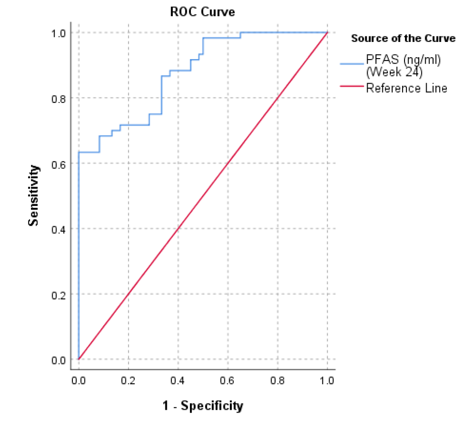
In Figure 1, the significance of the cut-off value of 11.79 ng/ml for serum PFAS becomes apparent in the context of predicting preeclampsia during the 24th week of gestation. When the serum PFAS level exceeds this particular threshold, it indicates the presence of preeclampsia. The sensitivity of the test at this cut-off is reported to be 87%, denoting that the test accurately identifies 87% of cases where preeclampsia is genuinely present. However, the specificity is noted as 35%, indicating that the test is less precise in correctly identifying cases where preeclampsia is absent.


Figure 1: Cut off value Serum PFAS in Prediction of Preeclampsia using ROC Curve


The cut-off value 11.79 ng/ml for serum PFAS in the prediction of preeclampsia, sensitivity of 87% and specificity 35%.


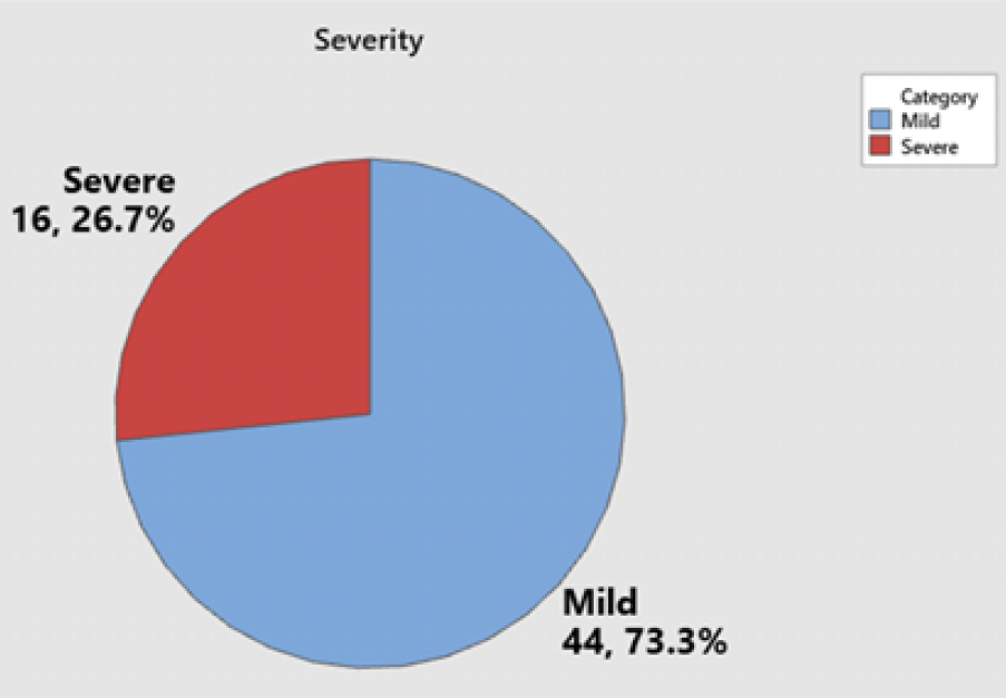
The presented findings indicate that in the study, 16 cases in the 1st visit, constituting 26.7%, were identified as severe preeclampsia, while 44 cases, accounting for 73.3%, were classified as mild preeclampsia, Figure 2.


Figure 2: Distribution of Preeclamptic Women according to the Severity of Preeclampsia


The data presented suggests a progression in the severity of preeclampsia over the course of the follow-up period, with an increase in the proportion of severe cases from the 1st visit (24 weeks of gestation) to the 2nd visit (35 weeks of gestation). At the 1st visit, 26.67% of cases were classified as severe, while this percentage increased to 41.67% at the 2nd visit. Conversely, the percentage of mild cases decreased from 73.33% at the 1st visit to 58.33% at the 2nd visit. This shift in severity distribution suggests a potential worsening of the condition as the pregnancy progresses, Table 3.


Table 3: Progression in the severity of preeclampsia over the course of the follow-up period

Preclamesia in the 1st visit (24 week of gestation)in the 2nd visit (35 week of gestation)

No. 

 %

No. 

 %

Severe

16

26.67

25

41.67

Mild

44

73.33

35

58.33

Total

60

100

60

100

P-value: 0.021


Table 4 reveals a noteworthy association between Polyfluoroalkyl Substances (PFAS) concentrations and the severity of preeclampsia at the 24th week of gestation. Strikingly, the mean PFAS concentration in pregnant women with severe preeclampsia (7.37 ng/ml) is significantly lower than in those with mild preeclampsia (12.27 ng/ml), as evidenced by the low p-value of 0.001.


Table 4: Mean of PFAS Concentrations in Preeclamptic Women in Relation to the Severity of Preeclampsia in 24th Wweek of Gestation

Preeclampetic pregnant women

No.

PFAS concentrations in 24th week of gestation

Severe preeclampsia

16

7.37±1.71

Mild preeclampsia

44

12.27±0.97

P-value: 0.001


Table 5 presents compelling evidence of the association between Polyfluoroalkyl Substances (PFAS) concentrations and the severity of preeclampsia at the 35th week of gestation. The mean PFAS concentration in women with severe preeclampsia (5.94 ng/ml) is significantly lower than in those with mild preeclampsia (9.88 ng/ml), as indicated by the p-value of 0.001. This inverse relationship suggests that lower PFAS concentrations are associated with a more severe form of preeclampsia at this later stage of gestation.


Table 5: Mean of PFAS Concentrations in Preeclamptic Women in Relation to the Severity of Preeclampsia in 35th Week of Gestation

Preeclampetic pregnant women

No.

PFAS concentrations in 35th week of gestation

Severe preeclampsia

25

5.94±1.38

Mild preeclampsia

35

9.88±0.78

P-value: 0.001

DISCUSSION

The present study found significant differences in maternal serum per- and polyfluoroalkyl substances (PFAS) concentrations between women with preeclampsia and normotensive pregnant controls. Women with preeclampsia showed lower PFAS concentrations across the first, second, and third visits, suggesting an inverse association between PFAS level and preeclampsia occurrence. Although PFAS are commonly considered environmental toxicants with potential adverse metabolic, endocrine, and vascular effects, the lower concentrations observed in preeclamptic women may reflect complex physiological changes during pregnancy, including altered renal clearance, plasma volume expansion, placental transfer, and disease-related metabolic redistribution rather than a simple direct protective or harmful effect. This interpretation is important because preeclampsia is not a single uniform disease but a heterogeneous syndrome with different maternal, placental, vascular, and metabolic pathways, as emphasized by Rana et al. [1], Jung et al. [3], and Roberts et al. [13].


The association between PFAS and preeclampsia should be interpreted in relation to the established pathophysiology of the disease. Preeclampsia is mainly characterized by abnormal placentation, endothelial dysfunction, oxidative stress, inflammation, and imbalance between angiogenic and antiangiogenic factors. Chappell et al. [6] and Nirupama et al. [7] described preeclampsia as a multisystem disorder in which placental dysfunction leads to maternal vascular injury. Therefore, the observed relationship between PFAS levels and preeclampsia may reflect PFAS interaction with vascular, metabolic, and placental pathways rather than a direct causal relationship. The finding of lower PFAS concentrations in preeclamptic women appears biologically complex. PFAS are persistent chemicals that can accumulate in human tissues and influence metabolic pathways. Beale et al. [14] reported that PFAS exposure is associated with common biochemical response pathways, while Cakmak et al. [15] found associations between blood PFAS concentrations and clinical biochemical markers of organ function and metabolism. These findings support the possibility that PFAS may interact with maternal metabolic status during pregnancy. However, the inverse association in the present study may also be explained by physiological changes in preeclampsia, particularly altered renal function and reduced glomerular filtration, which are recognized features of the disease. Rana et al. [4] and Gyselaers [16] highlighted that preeclampsia involves renal endothelial dysfunction and systemic vascular changes, which may influence circulating biomarker concentrations. The ROC analysis showed that a serum PFAS cut-off value of 11.79 ng/mL at the 24th week of gestation had predictive value for preeclampsia, with high sensitivity. This suggests that PFAS measurement may have potential value as a supportive biomarker in preeclampsia prediction. However, this result should not be interpreted as a standalone diagnostic tool. MacDonald et al. [17] and Chaemsaithong et al. [18] emphasized that prediction of preeclampsia is most accurate when multiple clinical, biochemical, and placental biomarkers are combined. Therefore, PFAS may be useful as part of a broader predictive model rather than as an isolated marker. The association between PFAS and early-onset preeclampsia in the present study is clinically important. Early-onset preeclampsia is generally considered more severe and more strongly related to placental dysfunction than late-onset disease. Roberts et al. [13] noted that preeclampsia subtypes differ in their pathophysiology and clinical implications. Similarly, Chatzakis et al. [19] highlighted the importance of disease timing in determining maternal and fetal outcomes. The lower PFAS concentrations observed in early-onset cases may therefore indicate that PFAS-related changes are linked to placental or maternal vascular dysfunction occurring earlier in pregnancy. The results are also consistent with the concept that preeclampsia is a syndrome with a cascade of pathophysiological events rather than a single disease entity. Gyselaers [16] described preeclampsia as a progressive disorder involving maternal cardiovascular maladaptation, endothelial activation, and placental stress. From this perspective, PFAS concentrations may reflect underlying disease processes, including altered lipid metabolism, vascular inflammation, and organ dysfunction. This is supported by Yao et al. [20] and Labine et al. [21], who demonstrated that PFAS exposure can influence biochemical and metabolic pathways.


Some findings in the present study may appear different from the traditional assumption that higher PFAS exposure increases the risk of adverse pregnancy outcomes. This disagreement may be explained by differences in exposure level, population characteristics, timing of sample collection, type of PFAS measured, and disease subtype. Romero et al. [22] emphasized that preeclampsia research requires careful phenotyping because different subtypes may have distinct mechanisms. Therefore, inconsistent findings across studies may result from combining early- and late-onset preeclampsia or mild and severe forms into one outcome group.


The possible role of maternal metabolic status should also be considered. Obesity, hypertension, and metabolic dysfunction are established risk factors for preeclampsia. Broughton-Pipkin and Loughna [23] discussed the strong relationship between obesity, hypertension, and preeclampsia. PFAS have also been linked to metabolic changes and organ function markers, as reported by Cakmak et al. [15] and Huang et al. [24]. Therefore, the association between PFAS and preeclampsia may be modified by maternal BMI, lipid profile, renal function, and inflammatory status.


The present study also suggests that PFAS levels may differ according to disease severity. Lower PFAS concentrations in mild cases compared with severe cases across pregnancy stages may indicate that PFAS distribution or clearance varies with disease progression. Severe preeclampsia is associated with greater endothelial dysfunction, renal impairment, hepatic involvement, and hematological abnormalities. Vidaeff et al. [12] argued that preeclampsia requires a more biological definition because clinical criteria alone may not fully capture disease heterogeneity. Therefore, PFAS may represent one part of a larger biomarker profile reflecting biological severity. Overall, the findings of this study support the potential role of PFAS as a biomarker associated with preeclampsia risk, onset, and severity. However, the inverse association observed should be interpreted cautiously. PFAS concentrations may be influenced by pregnancy physiology, placental transfer, renal function, maternal metabolism, and timing of sample collection. Further longitudinal studies are needed to clarify whether low PFAS levels are a cause, consequence, or marker of altered maternal–placental physiology in preeclampsia. In conclusion, this study adds to the growing evidence that environmental chemicals may be involved in pregnancy-related hypertensive disorders. The observed lower PFAS concentrations among preeclamptic women, especially in early-onset cases, suggest a possible relationship between PFAS dynamics and preeclampsia development. Nevertheless, because preeclampsia is a complex and heterogeneous syndrome, PFAS should be studied alongside established clinical risk factors and biomarkers such as blood pressure, proteinuria, angiogenic factors, renal function, and placental indicators. Future studies with larger sample sizes, repeated PFAS measurements, and stratification by disease subtype are recommended to validate these findings.

REFERENCE
  1. Rana, S. et al. “Preeclampsia: pathophysiology, challenges, and perspectives.” Circulation Research, vol. 124, no. 7, 2019, pp. 1094–1112.

  2. Christiansen, C.H. et al. “Multivitamin use and risk of preeclampsia: a systematic review and meta-analysis.” Acta Obstetricia et Gynecologica Scandinavica, vol. 101, no. 1, 2022, pp. 233–239.

  3. Jung, E. et al. “The etiology of preeclampsia.” American Journal of Obstetrics and Gynecology, vol. 226, no. 2, 2022, pp. S844–S866.

  4. Rana, S. et al. “Preeclampsia: pathophysiology, challenges, and perspectives.” Circulation Research, vol. 124, no. 7, 2019, pp. 1094–1112.

  5. Rose, C. et al. “Hypertension in pregnancy.” Medical Roundtable Cardiovascular Edition, 2020, pp. 358–366.

  6. Chappell, L.C. et al. “Pre-eclampsia.” The Lancet, vol. 398, no. 10297, 2021, pp. 341–354.

  7. Nirupama, R. et al. “Preeclampsia: pathophysiology and management.” Journal of Gynecology Obstetrics and Human Reproduction, vol. 50, no. 2, 2021, p. 101975.

  8. Roberts, J.M. et al. “Task Force on Hypertension in Pregnancy.” Obstetrics & Gynecology, vol. 122, no. 5, 2013, pp. 1122–1131.

  9. Vial, F. et al. “Preeclampsia. Eclampsia.” EMC Anesthésie Réanimation, vol. 46, no. 3, 2020, pp. 1–9.

  10. Karrar, S.A. and Hong, P.L. “Preeclampsia.” In: StatPearls. StatPearls Publishing, 2023.

  11. Erez, O. et al. “Preeclampsia and eclampsia: the conceptual evolution of a syndrome.” American Journal of Obstetrics and Gynecology, vol. 226, no. 2, 2022, pp. S786–S803.

  12. Vidaeff, A.C. et al. “Preeclampsia: the need for a biological definition and diagnosis.” American Journal of Perinatology, vol. 38, no. 9, 2020, pp. 976–982.

  13. Roberts, J.M. et al. “Subtypes of preeclampsia: recognition and determining clinical usefulness.” Hypertension, vol. 77, no. 5, 2021, pp. 1430–1441.

  14. Beale, D.J. et al. “A review of omics-based PFAS exposure studies reveals common biochemical response pathways.” Science of the Total Environment, vol. 845, 2022, p. 157255.

  15. Cakmak, S. et al. “The association between blood PFAS concentrations and clinical biochemical measures of organ function and metabolism in participants of the Canadian Health Measures Survey.” Science of the Total Environment, vol. 827, 2022, p. 153900.

  16. Gyselaers, W. “Preeclampsia is a syndrome with a cascade of pathophysiologic events.” Journal of Clinical Medicine, vol. 9, no. 7, 2020, p. 2245.

  17. MacDonald, T.M. et al. “Clinical tools and biomarkers to predict preeclampsia.” EBioMedicine, vol. 75, 2022, p. 103780.

  18. Chaemsaithong, P. et al. “First trimester preeclampsia screening and prediction.” American Journal of Obstetrics and Gynecology, vol. 226, no. 2, 2022, pp. S1071–S1097.

  19. Chatzakis, C. et al. “Early delivery or expectant management for late preterm preeclampsia: a meta-analysis of randomized controlled trials.” Acta Obstetricia et Gynecologica Scandinavica, vol. 100, no. 8, 2021, pp. 1392–1400.

  20. Yao, J. et al. “Novel perfluoroalkyl ether carboxylic acids and sulfonic acids: occurrence and association with serum biochemical parameters in residents living near a fluorochemical plant in China.” Environmental Science & Technology, vol. 54, no. 21, 2020, pp. 13389–13398.

  21. Labine, L.M. et al. “Comparison of sub-lethal metabolic perturbations of select legacy and novel perfluorinated alkyl substances in Daphnia magna.” Environmental Research, vol. 212, 2022, p. 113582.

  22. Romero, R. “Introduction to the guest editors of the American Journal of Obstetrics & Gynecology supplement on preeclampsia 2022.” American Journal of Obstetrics and Gynecology, vol. 226, no. 2, 2022, pp. S781–S785.

  23. Broughton-Pipkin, F. and Loughna, P. “Obesity, hypertension, and preeclampsia.” In: Obesity and Obstetrics. Elsevier, 2020, pp. 117–124.

  24. Huang, J.K. et al. “Decreased levels of perfluoroalkyl substances in patients receiving hemodialysis treatment.” Science of the Total Environment, vol. 896, 2023, p. 165184.

Recommended Articles
Research Article
Evaluation of Mortality at One Year Follow-Up of Patients Having Early Tumors of Oropharynx Undergoing Trans-Oral Robotic Surgery in a Tertiary Care Hospital
Published: 27/01/2025
Download PDF
Research Article
Molecular Study to Detect Some Virulence Factors in Methicillin – Resistance Staphlococcus Aureus Isolated from Eye Infections in Wasit Province
Published: 15/12/2023
Download PDF
Research Article
Occurrence of Multi-Drug Resistance of E. coli Isolated from Urinary Tract Infection in Najaf City
...
Published: 05/10/2025
Download PDF
Research Article
Estimation Of Hba1c in Non-Diabetic Patients with Myocardial Infarction
Published: 19/07/2024
Download PDF
Chat on WhatsApp
Flowbite Logo
PO Box 101, Nakuru
Kenya.
Email: office@iarconsortium.org

Editorial Office:
J.L Bhavan, Near Radison Blu Hotel,
Jalukbari, Guwahati-India
Useful Links
Order Hard Copy
Privacy policy
Terms and Conditions
Refund Policy
Shipping Policy
Others
About Us
Team Members
Contact Us
Online Payments
Join as Editor
Join as Reviewer
Subscribe to our Newsletter
+91 60029-93949
Follow us
MOST SEARCHED KEYWORDS
Copyright © iARCON International LLP . All Rights Reserved.