Contents
Download PDF
pdf Download XML
217 Views
44 Downloads
Share this article
Research Article | Volume 6 Issue 1 (January-June, 2026) | Pages 1 - 4
Blood Parasites of Budgerigar (Melopsittacus undulatus) in Maysan Governorate, Iraq
1
Ministry of Education, Open Educational College, Iraq
Under a Creative Commons license
Open Access
Received
Nov. 3, 2025
Revised
Dec. 9, 2025
Accepted
Jan. 2, 2026
Published
Jan. 28, 2026
Abstract

Melopsittacus undulatus, a budgerigar of the family Psittacidae, is widely distributed in most parts of the world. The present research aimed to characterize the blood parasites associated with this species by analyzing fifty-three blood samples collected from budgerigars of both sexes in Maysan Governorate during the period from October to July 2024. The study focused on determining the prevalence, infection pattern, and distribution between sexes, as well as characterizing the detected parasitic species. The data showed that the overall prevalence of haemosporidian parasites was 30.18%. The relative distribution of infection with Plasmodium, Haemoproteus, and Leucocytozoon species was 68.75%, 18.75%, and 25%, respectively. In addition, the study revealed a significant difference in infection rates between the sexes, with a higher rate recorded in females than in males. In terms of infection patterns, the results showed that single infections constituted the majority (68.75%), while co-infections were less common (43.75%). These results indicate that budgerigars in Maysan Governorate represent a potential host for blood parasites, with varying rates recorded depending on the parasite species and sex. This study represents the first documentation of the presence of hemosporidian parasites in budgerigars (M. undulatus) in the region, contributing to enriching the epidemiological database on blood parasites of ornamental birds in Iraq and paving the way for more comprehensive future studies on the transmission dynamics of these parasites and their impact on the health of birds and other potential host species.

Keywords
INTRODUCTION

Plasmodium, Haemoproteus, and Leucocytozon, belonging to the phylum Apicomplexa, specifically the order Haemosporida, are among the most important causes of blood infections in birds. More than 200 known species have been described within this group, with thousands of other undescribed species likely to exist. Despite displaying a high degree of genetic diversity, most of these parasites display remarkable similarity in morphological characteristics, which limits the ability to rely solely on morphological characteristics to distinguish between different species [1-3]. These parasites can lead to significant increases in mortality rates among some wild bird species [4].


Hemoproteus parasites are unable to reproduce in the bird's peripheral blood, making their transmission to new birds through direct blood inoculation unlikely, unlike Plasmodium parasites, which commonly rely on this method. Instead, the development of their sexual stages is restricted to the louse fly, making this insect the crucial intermediary for transmission and ensuring the continuity of the parasite's life cycle between hosts [5]. Of the more than 100 described species of Plasmodium, Plasmodium relectum is the most common infecting birds, each characterized by varying degrees of host specificity and a specific geographic range. These parasites represent a group of blood pathogens, which include proteiosis, avian malaria, and avian leukemia, and exhibit a remarkable ability to influence the physiological and biological functions of their avian hosts. The effects of these parasites range from minor hematological disturbances to severe effects on survival and reproduction [6-8].


Birds infected with haemosporid parasites exhibit marked deterioration in their physiological and behavioral characteristics. Evidence indicates a marked reduction in lifespan and its ability to survive, as well as its reduced ability to mate and produce eggs, with increased susceptibility to secondary microbial infections. Studies also indicate that malaria parasites contribute to the decline in physical health of birds, reflecting the overall negative impact of these parasites on host fitness [9-11]. In many cases, bird deaths are attributed to the destructive effects of parasites on internal tissues, outside the red blood cells, where death may occur suddenly and without previous clinical signs [12].


Budgerigars are among the most common [traded] and widespread birds in various parts of the world, belonging to the parrot family. Their popularity as pets lies in a combination of their aesthetic and behavioral characteristics, including their bright colors, quiet voices, and ability to interact with humans and mimic words [13,14]. Although most parrot species are at risk of extinction, budgerigars have stable populations and are not considered a threatened species. These birds typically live between ten and fifteen years and have a relatively short breeding period [15,16].


Although the budgerigar M. undulatus, Shaw is native to Australia, its range has now spread to many regions around the world. This bird is known for its a large increase in bird numbers can cause overcrowding in cages, negatively impacting their overall health and increasing the likelihood of disease, especially parasitic diseases., if environmental and nutritional conditions are not carefully observed [17]. 


It comes right after common pets like cats and dogs, budgies (M. undulatus) have captured the hearts of pet lovers global thanks to their small build, playful spirit, and low cost. These intelligent birds are not only beautiful in color; they also possess an amazing ability to learn sounds even after reaching adulthood, making them a wonderful partner for human interaction and a unique model for scientific studies on auditory and vocal learning and development. Each budgie possesses a personality that is both curious and playful, making them an unforgettable addition to any home [18-20].


Hemoprotozoa injury are insect-borne diseases in parrots, with Plasmodium and Hemoproteus parasites playing a major role in their spread. Molecular studies have revealed a great diversity of Leucocytozoan species, which are the main cause of death in these birds [21]. The parasite Plasmodium relictum causes malaria in birds, deaths occur in parrots and cause widespread pathogenic effects on many bird species worldwide [22]. Microscopy continues to be the primary tool for detecting parasites in blood and tissue samples [23]. The current study, conducted in Maysan Governorate, aims to examine blood parasites in M. undulatus, determine infection rates of the most common species, and evaluate differences in infection rates between females and males.

MATERIALS AND METHODS

Between February 2024 and March 2025, a total of 53 parrots (M. undulatus) of genders together were collected from the shops in Maysan Governorate. Following sample collection, thin blood smears were prepared and allowed to air dry for approximately 20–35 seconds. These smears were then fastened by pure methanol within 10 to 30 minutes on the day of the examination. After drying, the slides were dyed with (1:10) giemsa staining solution and placed inside paper strips for preservation. The slides were examined under a microscope at 100x oil magnification for two to eight hours to identify blood parasites, including Plasmodium, Haemoproteus, and Leucocytozoon species [24,25]. Microscopic images were obtained by two different digital cameras mounted on a photic microscope: AmScope MU1003 (10 M.P.) and Tomaltic/Hayear (12 M.P.). The parasite diagnosis was made using [26-29].

RESULTS

The outcomes showed that the overall incidence of blood parasites among budgerigars in Maysan Governorate was 30.18% (Table 1). 


Table 1: Evaluation of the Incidence of Haemosporidian in M. Undulatus in Maysan Governorate

Number of bird samples examined

birds affected by infection

Incidence proportion

53

16

30.18


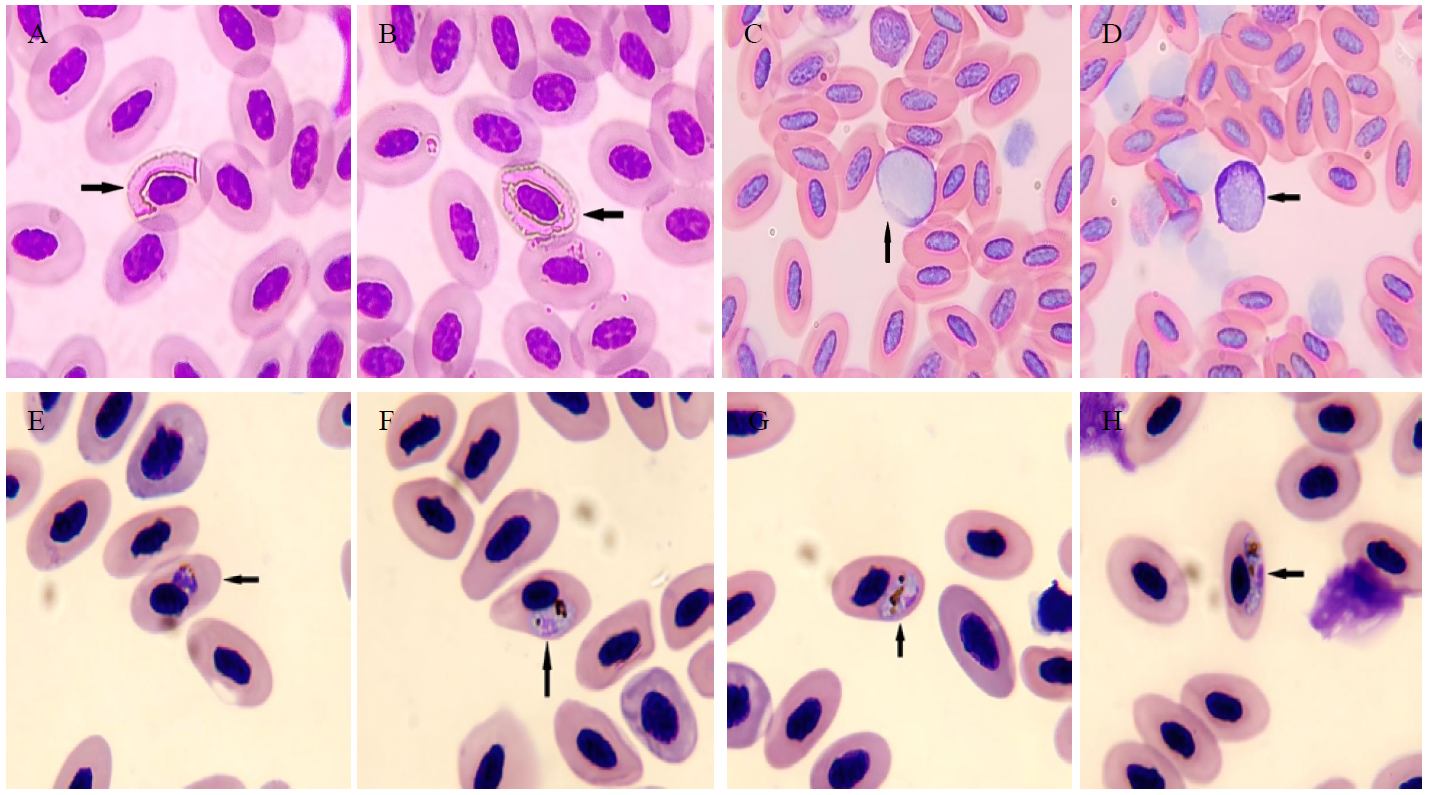
It was also found that these birds their bodies are exposed to three different genera of parasites: Haemoproteus spp., Plasmodium spp., and Leucocytozoon spp. 18.75% for the first type, 68.75% for the second type, and 25% for the third type (Table 2, Figure 1). 


Figure 1: Haemoproteus spp., A, B: Gametocytes, Leucocytozoon spp., C, D: Gametocytes, Plasmodium spp., E: Schizont, F-H: Gametocytes


Table 2: The Level of Infection of Budgies Infected with Blood Parasites

Types of Blood Parasites

Birds affected by infection

Incidence proportion

Haemoproteus species

3

18.75

Plasmodium species

11

68.75

Leucocytozoon species

4

25


It was noted that the infection rate was more pronounced in females than in males. On the other hand, single infections constituted the largest percentage, amounting to 68.75%, while double infections did not exceed 43.75% (Table 3). 


Table 3: Haemosporidian Prevalence Rates in Parrots According to Infection Type

Infection class

Birds affected by infection

Incidence proportion

Single infection

11

68.75%

Double infection

7

43.75%


Overall, a higher infection rate was recorded in females, amounting to 68.75%, compared to 45.45% in males (Table 4).


Table 4: Prevalence Rates of Blood Parasites among Male and Female Budgerigars

Sexual identity

Birds affected by infection

Incidence proportion

Females

11

68.75%

Males

5

45.45%

DISCUSSION

Many wild and domestic birds are infectived with various species of haemosporidian, which constitute a source of transmission to other species. Despite the importance of these birds in transmitting infection, there are few studies on these parasites in Iraq. When comparing the infection rate in our current study, which indicated that budgerigars (M. undulatus) were infectived with three genera of haemosporidian, with the rates in other studies, there is only one study by Khalaf [29] on this species of bird in Erbil, with a higher infection rate of 35.48% and two types of blood parasites. In New Zealand, Baron et al. [30] conducted their study on M. undulatus, and the infection rate in the same species was 30% with Plasmodium spp., while in the current study, the infection rate was 73.33%.


The infection rate in the current study is lower than in several studies, including Dimitrov et al. [31], who reported a 58% infection rate across several bird species. Ali et al. [32] reported a 60% infection rate in partridges. Berthová et al. [33] reported a 33.9% infection rate across several bird species in Slovakia. However, the rate was lower than in several studies, including Marzal et al. [34], who reported that 18.15% of parrots were infected with blood parasites. Rodríguez and Mata [35] reported that 15.9% of birds were infected with blood parasites in Colombia. Mohammad and Al-Moussawi [36] reported a 10.5% infection rate in passerines


Regarding the Leucocytozoon parasite, the infection rate was higher than that recorded by Lotta et al. [28] at 1.2% in South America, and Mirzaei et al. [37] at 11.64% in Iranian birds. There are several factors that control differences in infection rates in birds, including the season, the bird's sex, and the bird's geographic location, as well as the birds' ages and susceptibility to infection, in addition to feeding areas and the presence of vectors [38].

CONCLUSION

The current study found that ornamental birds (Melopsittacus undulatus) were infected with several types of haemosporidian, it falls under three species: Plasmodium, Haemoproteus, and Leucytozoon, with an infection rate of 30.18%. Females were more affected than males, and the incidence of double infection was lower than single infection.


Declaration of conflict of interest

The authors certify that this study is free from any conflict of interest in the context of its publication.

REFERENCE
  1. Bensch, S. et al. “MalAvi: A public database of malaria parasites and related haemosporidians in avian hosts based on mitochondrial cytochrome b lineages.” Molecular Ecology Resources, vol. 9, no. 5, 2009, pp. 1353–1358. https://doi.org/10.1111/j.1755-0998.2009.02692.x.

  2. Martinsen, E.S. et al. “A three-genome phylogeny of malaria parasites (Plasmodium and closely related genera): evolution of life-history traits and host switches.” Molecular Phylogenetics and Evolution, vol. 47, no. 1, 2008, pp. 261–273. https://doi.org/10.1016/j.ympev.2007.11.012.

  3. Ferreira, F.C. et al. “Diptera vectors of avian haemosporidians: with emphasis on tropical regions.” Avian Malaria and Related Parasites in the Tropics, 2020. https://doi.org/10.1007/978-3-030-51633-8_6.

  4. Mandour, A.M. et al. “Atoxoplasma (Lankesterella) columbae sp. nov. in the domestic pigeon Columba livia in Assiut Province.” Assiut Veterinary Medical Journal, vol. 17, no. 34, 1986, pp. 95–97. https://linksshortcut.com/wqyXC.

  5. Cosgrove, C. “Avian malaria parasites and other haemosporidia.” Systematic Biology, vol. 54, no. 5, 2005, pp. 860–863. https://www.jstor.org/stable/20061291.

  6. Santiago-Alarcon, D. et al. “Bloodmeal analysis reveals avian Plasmodium infections and broad host preferences of Culicoides (Diptera: Ceratopogonidae) vectors.” PLOS ONE, vol. 7, no. 2, 2012, pp. 1–5. https://doi.org/10.1371/journal.pone.0031098.

  7. Perkins, S.L. “Malaria's many mates: past, present and future of the systematics of the order Haemosporida.” Journal of Parasitology, vol. 100, no. 1, 2014, pp. 11–25. https://doi.org/10.1645/13-362.1.

  8. Nourani, L. et al. “Dynamics of prevalence and distribution pattern of avian species and its vectors in diverse zoogeographical areas: a review.” Infection, Genetics and Evolution, vol. 81, 2020, pp. 1–21. https://doi.org/10.1016/j.meegid.2020.104244.

  9. Dranzoa, C. et al. “The ecto-, gastro-intestinal and haemo-parasites of live pigeons (Columba livia) in Kampala, Uganda.” Avian Pathology, vol. 28, no. 2, 1999, pp. 119–124. https://doi.org/10.1080/03079459994830.

  10. Ilgūnas, M. et al. “Mortality and pathology in birds due to Plasmodium (Giovannolaia) homocircumflexum infection with emphasis on the exoerythrocytic development of avian malaria parasites.” Malaria Journal, vol. 15, 2016, pp. 1–11. https://doi.org/10.1186/s12936-016-1310-x.

  11. Coon, C.A. et al. “Malaria infection negatively affects feather growth rate in the house sparrow Passer domesticus.” Journal of Avian Biology, vol. 47, no. 6, 2016, pp. 779–787. https://doi.org/10.1111/jav.00942.

  12. Vanstreels, R.E.T. et al. “Outbreak of avian malaria associated to multiple species of Plasmodium in magellanic penguins undergoing rehabilitation in southern Brazil.” PLOS ONE, vol. 9, no. 4, 2014, pp. 1–11. https://doi.org/10.1371/journal.pone.0094994.

  13. Roldán-Clarà, B. et al. “Literature review of the use of birds as pets in Latin-America with a detailed perspective on Mexico.” Ethnobiology and Conservation, vol. 3, no. 5, 2014, pp. 1–18. https://doi.org/10.15451/ec2014-10-3.5-1-18.

  14. Baghkheirati, A.A. et al. “Papillary cystadenocarcinoma in a budgerigar (Melopsittacus undulatus).” Iranian Journal of Veterinary Medicine, vol. 17, no. 4, 2023, pp. 409–414. https://doi.org/10.32598/ijvm.17.4.1005214.

  15. Kalmar, I.D. et al. “Guidelines and ethical considerations for housing and management of psittacine birds used in research.” Institute for Laboratory Animal Research Journal, vol. 51, no. 4, 2010, pp. 409–423. https://doi.org/10.1093/ilar.51.4.409.

  16. Kubiak, M. “Budgerigars and cockatiels.” Handbook of Exotic Pet Medicine, 2021, pp. 141–164. https://doi.org/10.1002/9781119389934.

  17. Wyndham, E. “Diurnal cycle, behaviour and social organization of the budgerigar Melopsittacus undulatus.” Emu, vol. 80, no. 1, 1980, pp. 25–33. https://doi.org/10.1071/MU9800025.

  18. Striedter, G.F. et al. “For whom the male calls: an effect of audience on contact call rate and repertoire in budgerigars, Melopsittacus undulatus.” Animal Behaviour, vol. 65, no. 5, 2003, pp. 875–882. https://doi.org/10.1006/anbe.2003.2135.

  19. Hara, E. et al. “Neural FoxP2 and FoxP1 expression in the budgerigar, an avian species with adult vocal learning.” Behavioural Brain Research, vol. 283, 2015, pp. 22–29. https://doi.org/10.1016/j.bbr.2015.01.017.

  20. Ajay, D.T.P. and Rahana, M.K.V.K. “A study on ecology and behavior of budgerigar (Melopsittacus undulates) under captivity at Edavanna Malappuram district, Kerala.” Aureole, vol. 12, 2020, pp. 95–119. https://linksshortcut.com/MSpcK.

  21. Olias, P. et al. “Avian malaria deaths in parrots, Europe.” Emerging Infectious Diseases, vol. 17, no. 5, 2011, pp. 950–952. https://doi.org/10.3201/eid1705.101618.

  22. Marzal, A. et al. “Invasive avian malaria as an emerging parasitic disease in native birds of Peru.” Biological Invasions, vol. 17, 2015, pp. 39–45. https://doi.org/10.1007/s10530-014-0718-x.

  23. Rosenblatt, J.E. et al. “Laboratory diagnosis of infections due to blood and tissue parasites.” Clinical Infectious Diseases, vol. 49, no. 7, 2009, pp. 1103–1108. https://doi.org/10.1086/605574.

  24. Burry-Caines, J.R. and Bennett, G.F. “The Haemoproteidae (Apicomplexa: Haemosporina) of the avian families Fringillidae and Emberizidae sl.” Canadian Journal of Zoology, vol. 70, no. 6, 1992, pp. 1149–1160. https://doi.org/10.1139/z92-16.

  25. Abdulzahra, A.I. and Abdullah, B.H. “Survey of some blood parasites in three species of wild birds in Babylon Province, Iraq.” Journal of Basrah Researches (Sciences), vol. 48, no. 1, 2022, pp. 49–59. https://doi.org/10.56714/bjrs.48.1.5.

  26. Valkiūnas, G. et al. “Characterization of Plasmodium relictum, a cosmopolitan agent of avian malaria.” Malaria Journal, vol. 17, 2018, pp. 1–21. https://doi.org/10.1186/s12936-018-2325-2.

  27. Valkiūnas, G. and Iezhova, T.A. “Keys to the avian malaria parasites.” Malaria Journal, vol. 17, 2018, pp. 1–24. https://doi.org/10.1186/s12936-018-2359-5.

  28. Lotta, I.A. et al. “Disentangling Leucocytozoon parasite diversity in the neotropics: descriptions of two new species and shortcomings of molecular diagnostics for leucocytozoids.” International Journal for Parasitology: Parasites and Wildlife, vol. 9, 2019, pp. 159–173. https://doi.org/10.1016/j.ijppaw.2019.05.002.

  29. Khalaf, W.K. “Detection of blood parasites in different pet birds in Erbil City.” Al-Anbar Journal of Veterinary Sciences, vol. 15, no. 2, 2022, pp. 28–36. https://doi.org/10.37940/AJVS.2022.15.2.4.

  30. Baron, H.R. et al. “Disease screening of three breeding populations of adult exhibition budgerigars (Melopsittacus undulatus) in New Zealand reveals a high prevalence of a novel polyomavirus and avian malaria infection.” Avian Diseases, vol. 58, no. 1, 2014, pp. 111–117. https://doi.org/10.1637/10604-063013-REG.1.

  31. Dimitrov, D. et al. “Genetic diversity of avian blood parasites in SE Europe: cytochrome b lineages of the genera Plasmodium and Haemoproteus (Haemosporida) from Bulgaria.” Acta Parasitologica, vol. 55, no. 3, 2010, pp. 201–209. https://doi.org/10.2478/s11686-010-0029-z.

  32. Ali, K.N. et al. “Prevalence of haemosporidian parasites of Chukar Partridge birds in Sulaimani Province/Kurdistan Region of Iraq.” Al-Anbar Journal of Veterinary Sciences, vol. 12, no. 2, 2019, pp. 81–88. https://2u.pw/VNebzJHk.

  33. Berthová, L. et al. “The first report of nine species of haemosporidian parasites (Haemosporida: Haemoproteus, Plasmodium and Leucocytozoon) in wild birds from Slovakia.” Biologia, vol. 67, no. 5, 2012, pp. 931–933. https://doi.org/10.2478/s11756-012-0094-x.

  34. Marzal, A. et al. “Prevalence and diversity of avian malaria parasites in illegally traded white-winged parakeets in Peruvian Amazonas.” Animal Conservation, vol. 23, no. 3, 2024, pp. 364–373. https://doi.org/10.1111/acv.12913.

  35. Rodríguez, O.A. and Matta, N.E. “Blood parasites in some birds from eastern plains of Colombia.” Memórias do Instituto Oswaldo Cruz, vol. 96, no. 8, 2001, pp. 1173–1176. https://doi.org/10.1590/S0074-02762001000800026.

  36. Mohammad, M.K. and Al-Moussawi, A.A. “Blood parasites of some passeriform birds in Baghdad area, central Iraq.” Bulletin of the Iraq Natural History Museum, vol. 12, no. 1, 2012, pp. 29–36. https://2u.pw/bthbN3Gd.

  37. Mirzaei, F. et al. “Blood parasites in domestic birds in Central Iran.” Veterinary Sciences, vol. 7, no. 3, 2020, pp. 1–7. https://doi.org/10.3390/vetsci7030126.

  38. Al-Barwari, S. and Saeed, I. “The parasitic communities of the rock pigeon Columba livia from Iraq: component and importance.” Türkiye Parazitolojii Dergisi, vol. 36, no. 4, 2012, pp. 232–239. https://2u.pw/NWIXL4bI.
Recommended Articles
Research Article
The Impact of Medicinal Plant Extracts on Entamoeba histolytica Parasite in Rats: A Study on Hematological and Biochemical Parameters
...
Published: 28/02/2026
Download PDF
Research Article
Determination of the Factors that Define the Tropism of Astroviruses in Established Cell Lines
...
Published: 15/12/2025
Download PDF
Research Article
The use of dual spectrum reflectivity (NDSI, SI) in diagnosing the distribution of some types of salts in the founding organelle in the sura and wasit contacts.
Published: 30/10/2023
Download PDF
Research Article
Genetics study of Leptin Gene Polymorphism for Adolescent obese of Thi-Qar Province
Published: 25/08/2023
Download PDF
Chat on WhatsApp
Flowbite Logo
PO Box 101, Nakuru
Kenya.
Email: office@iarconsortium.org

Editorial Office:
J.L Bhavan, Near Radison Blu Hotel,
Jalukbari, Guwahati-India
Useful Links
Order Hard Copy
Privacy policy
Terms and Conditions
Refund Policy
Shipping Policy
Others
About Us
Team Members
Contact Us
Online Payments
Join as Editor
Join as Reviewer
Subscribe to our Newsletter
+91 60029-93949
Follow us
MOST SEARCHED KEYWORDS
Copyright © iARCON International LLP . All Rights Reserved.