Contents
Download PDF
pdf Download XML
149 Views
25 Downloads
Share this article
Research Article | Volume 7 Issue 1 (January-June, 2026) | Pages 1 - 6
Evaluation of Fetal Thymic Thoracic Ratio in pregnancies complicated with Preeclampsia
 ,
1
Department of Obstetrics and Gynecology, Al-Elwiya Maternity Teaching Hospital, Iraq
2
M.B.Ch.B, C.A.B.O.G, Consultant in Obstetrics and Gynecology, Al-Yarmouk Teaching Hospital, Baghdad, Iraq
Under a Creative Commons license
Open Access
Received
April 3, 2026
Revised
May 9, 2026
Accepted
June 10, 2026
Published
June 20, 2026
Abstract

Objective: Preeclampsia is a serious hypertensive disorder of pregnancy and a leading cause of maternal and perinatal morbidity and mortality. Its exact etiology remains unclear, though immunological factors are implicated. Emerging evidence suggests the fetal thymic thoracic ratio (TTR) may serve as a novel marker for predicting preeclampsia. Aim of the Study: To investigate the relationship between fetal TTR and preeclampsia, and to assess its association with disease severity. Patients and Methods: This case control study was conducted at the Department of Obstetrics and Gynecology, Al Elwiya Maternity Teaching Hospital, between January 2024 and January 2025. A total of 200 third trimester pregnant women were enrolled: 100 with preeclampsia and 100 healthy controls. Preeclamptic cases were subdivided into mild and severe groups. Maternal demographic, clinical, and ultrasound data were collected. Fetal TTR was measured and analyzed for its diagnostic utility in predicting severe preeclampsia. Results: Fetal TTR was significantly lower in preeclamptic pregnancies compared with controls, with the greatest reduction in severe cases. Mean fetal TTR was 0.362±0.024 in severe preeclampsia, 0.394±0.014 in mild cases, and 0.425±0.03 in controls (p<0.001). Receiver operating characteristic (ROC) analysis confirmed strong predictive value, with an area under the curve of 0.921. A cutoff of 0.396 yielded 90% sensitivity and 71.3% specificity. Conclusion: Fetal TTR decreases significantly in preeclampsia, particularly in severe disease. Its clinical integration may enhance early detection and management, offering a valuable tool for assessing disease severity.

Keywords
INTRODUCTION

Hypertensive disorders of pregnancy [HDP] are a major cause of maternal and fetal morbidity and mortality, with consequences extending beyond pregnancy and the postpartum period [1]. Chronic hypertension, gestational hypertension, preeclampsia, eclampsia and chronic hypertension with superimposed preeclampsia are all forms of hypertensive disorders of pregnancy (HDP). [2] 


Preeclampsia (PE) is defined as new-onset hypertension (≥140/90 mmHg) after 20 weeks of gestation, resolving after delivery, and accompanied by proteinuria and/or maternal organ dysfunction such as renal, hepatic, neurological, or hematological complications, and/or fetal growth restriction [3]. Untreated preeclampsia can lead to pulmonary edema, eclampsia, brain injury or death. Impaired perfusion of the uterus and placenta contributes to fetal growth restriction, placental abruption, preterm delivery, or stillbirth [4]. 


Globally, PE affects 2–8% of pregnancies and accounts for significant maternal mortality, particularly in Africa, Asia, and Latin America [5,6]. In Iraq, prevalence is reported at 4.79%, with hypertensive disorders ranking as the third leading cause of maternal death. Mortality related to HDP was 8–14% in recent years [7,8]. The incidence has risen in the U.S. since the 1990s, linked to chronic hypertension, diabetes, and obesity. WHO estimates the incidence is seven times higher in developing countries [9].


PE is a complex, multifactorial disorder. The two-stage model proposes that shallow invasion of trophoblasts leads to inadequate placentation [4]. When the placenta does not receive enough blood flow, it leads to oxidative stress and the release of inflammatory cytokines, angiotensin autoantibodies and anti-angiogenic factors, causing endothelial dysfunction [10]. 


In normal pregnancy, cytotrophoblasts invade maternal spiral arteries, transforming them into low resistance vessels that ensure adequate placental perfusion. This process is mediated by angiogenic factors, notably VEGF (Vascular Endothelial Growth Factor) and PlGF (Placental Growth Factor), which belong to the VEGF family and are essential for vascular remodeling and new vessel formation [11]. Their activity secures sufficient oxygen and nutrient delivery to support fetal growth.     


In pregnancies complicated by PE (Preeclampsia), trophoblastic invasion is shallow and incomplete, resulting in inadequate arterial remodeling, placental ischemia, and impaired oxygen transfer [12]. This disturbance initiates oxidative stress and stimulates the release of anti angiogenic mediators.


A key factor is sFlt 1 (Soluble FMS like Tyrosine Kinase 1), a circulating anti angiogenic protein that functions as a decoy receptor. By binding VEGF and PlGF, sFlt 1 prevents their interaction with endothelial receptors, thereby suppressing angiogenesis and abolishing their protective vascular effects [13].


Among these protective effects is the stimulation of NO (Nitric Oxide) synthesis. NO is a potent vasodilator that relaxes vascular smooth muscle, neutralizes reactive oxygen species, and reduces resistance. Elevated sFlt 1 diminishes VEGF and PlGF activity, leading to reduced NO production, heightened vasoconstrictor sensitivity, and maternal hypertension [14,15].


Collectively, the interplay of impaired cytotrophoblast invasion, disrupted VEGF/PlGF signaling, sFlt 1 elevation, and nitric oxide deficiency underpins the complex pathophysiology of PE. These vascular, placental, and immunological mechanisms converge to produce the endothelial dysfunction, hypertension, and fetal compromise characteristic of the disorder.


Beyond vascular and maternal factors, immunological mechanisms are increasingly recognized in PE. A long held belief is that inadequate trophoblast invasion and shallow placentation are influenced not only by maternal comorbidities but also by immune dysregulation, including decidual natural killer cell activity and regulatory T cell imbalance [4,10]. These changes contribute to abnormal placental perfusion, oxidative stress, and the release of inflammatory cytokines and anti angiogenic factors such as sFlt 1, which perpetuate endothelial dysfunction and hypertension [13–15].


The fetal thymus involved in the development of the adaptive immune system, and it is vital for both the maturation of T cells and the development of immune tolerance. The fetal thymus is extremely sensitive to stress in utero, as well as to disease processes in the mother. In pregnancies complicated by PE, the growth of the fetal thymus may be impaired. Data from sonographic evaluations indicate that fetal thymus size is reduced in fetuses of mothers with preeclampsia. This finding indicates that fetal immune maturation is impaired and that intrauterine programming may be altered [11,13]. These data demonstrate the relationship between maternal vascular pathologies and fetal immune development.


The Fetal Thymus Thoracic Ratio (TTR) has been developed as a reproducible and standardized sonographic index for quantifying fetal thymus size. The TTR is obtained by comparing the anteroposterior (AP) diameter of the thymus to the thoracic (TH) diameter at the standard three-vessel and tracheal view. The TTR serves as a reliable means of determining the size of the thymus, because it takes into account the size of the fetal thorax, reducing the variability of absolute measurements of thymic size and enhancing the ability to use the results for clinical diagnosis [12].


Studies show that a decrease in TTR corresponds with the overall severity of the disease and corresponding adverse outcomes in the patient with PE. From previous reports, TTR values of fetuses from mothers suffering from severe PE, were significantly less than those of fetuses from mothers suffering mild disease or normal controls; hence, it was thought that thymic involution might represent a marker for intrauterine compromise [13,16]. In addition, the decreased TTR value reflects the combined impact of placental ischemia, maternal endothelial dysfunction, and chronic inflammation on impaired fetal immune organ development.


Therefore, measuring fetal thymic thoracic ratios will provide the clinician with a unique perspective on the immunologic effects of preeclampsia. By adding together all vascular, maternal, and fetal immune factors, TTR measurement has the potential to improve the accuracy of predicting the severity of disease, assist practitioners in their clinical management, and increase the likelihood of positive maternal to fetal outcomes [17-20].


Traditional diagnostic markers such as blood pressure and proteinuria are known to have limited sensitivity and specificity [21]. Therefore, there is a need for new sonographic markers to increase prediction and risk stratification. The purpose of the present study is to determine the fetal TTR in pregnancies complicated by preeclampsia and to assess how this relates to the severity of the disease. Therefore, incorporation of TTR measurement into clinical practice should lead to more timely identification of preeclampsia, better directing medical management, and improved fetal-maternal outcomes.

MATERIALS AND METHODS

Study Design

 A prospective case control study was conducted in the Obstetrics and Gynecology Department in Al-Elwiya Maternity Teaching Hospital between first of January 2024 to January 2025 after gaining approval from Scientific Council of Obstetrics& Gynecology / Iraqi Board for Medical Specialization for the study protocol. All the enrolled women gave their verbal consent after explaining the purpose of the study to them, and they answered a questionnaire prepared to gather their data.


Study Population

A total of 200 pregnant women in the third trimester were enrolled. Participants were divided into two groups:


•      Study Group (n = 100): Women diagnosed with preeclampsia according to the criteria of the American College of Obstetricians and Gynecologists (ACOG). This group was further subclassified into mild and severe preeclampsia based on blood pressure             thresholds, proteinuria, and presence of maternal organ dysfunction

•      Control Group (n = 100): Healthy pregnant women matched for gestational age, with no history of hypertension, diabetes, renal disease, or autoimmune disorders.


Inclusion Criteria


  • Singleton pregnancy in the third trimester (≥28 weeks)

  • Women with clinically confirmed preeclampsia (study group)

  • Healthy normotensive women (control group)


Exclusion Criteria


  • Multiple gestations

  • Known fetal anomalies or intrauterine infections

  • Maternal chronic systemic diseases (e.g., chronic hypertension, diabetes mellitus, renal disease, autoimmune disorders)

  • Poor visualization of the fetal thymus on ultrasound.


Data Collection

Maternal demographic and clinical data were recorded, including age, parity, body mass index (BMI), blood pressure, and laboratory investigations. Clinical classification of preeclampsia was performed according to ACOG guidelines.


Ultrasound Assessment

All participants underwent standardized sonographic examination using a high‑resolution ultrasound machine equipped with a 3.5–5 MHz transducer.


  • The fetal thymus was visualized in the upper mediastinum at the level of the three‑vessel and trachea (3VT) view

  • The anteroposterior diameter of the thymus and the anteroposterior diameter of the thorax were measured

  • The thymic thoracic ratio (TTR) was calculated as

  • TTR=AP diameter of thymus/ AP diameter of thorax

  • Measurements were performed by an experienced sonographer blinded to maternal clinical status. Each measurement was repeated twice, and the mean value was used for analysis


Statistical Analysis

Data were analyzed using SPSS (Statistical Package for Social Sciences), version 25. Continuous variables were expressed as mean±standard deviation (SD), and categorical variables as frequencies and percentages.


  • Independent t‑test was used to compare mean TTR between groups

  • ANOVA was applied for comparison across mild, severe, and control groups

  • Receiver Operating Characteristic (ROC) curve analysis was performed to assess the diagnostic utility of TTR in predicting severe preeclampsia, with calculation of sensitivity, specificity, and area under the curve (AUC)

  • A p‑value <0.05 was considered statistically significant


Ethical Considerations and Official Approvals

Written informed consent was obtained from all participants prior to data collection with maintaining strict confidentiality throughout the study. The study protocol was reviewed and approved by the Ethics Committee of the Iraqi Board of Medical Specialization and the Department of Obstetrics and Gynecology at Al-Elwiya Maternity Teaching Hospital. Approval was granted under reference number 18, dated 3/1/2024.

RESULTS

The study included 200 pregnant women categorized into three groups: severe preeclampsia, mild preeclampsia, and healthy controls. Maternal age was similar across groups, with mean values of 30.78±8.22 years in the severe group, 32.4±8.55 years in the mild group, and 30.89±9.09 years in the control group (P = 0.319), indicating no statistically significant difference. Body mass index (BMI) also showed no significant variation, with mean values of 23.1±1.53 kg/m², 22.36±1.23 kg/m², and 23.76±1.44 kg/m² in the severe, mild, and control groups respectively (P > 0.3 for all comparisons). Gestational age at the time of study was comparable across groups, averaging 33.1±1.25 weeks (severe), 32.6±1.78 weeks (mild), and 33.28±1.53 weeks (control), with no significant differences observed (P > 0.6).


Table 1: Maternal and Fetal Characteristics

Variable

Severe (Mean±SD)

Mild (Mean±SD)

Control (Mean±SD)

P1 (Severe vs. Mild)

P2 (Severe vs. Control)

P3 (Mild vs. Control)

Age (years)

30.78±8.22

32.4±8.55

30.89±9.09

0.319

0.906

0.392

Parity

1.3±1.63

2.34±1.76

2.46±1.86

0.003

0.724

0.755

Gestational age (weeks)

33.1±1.25

32.6±1.78

33.28±1.53

0.658

0.787

0.632

BMI (kg/m²)

23.1±1.53

22.36±1.23

23.76±1.44

0.732

0.645

0.344


However, parity differed significantly. Women with severe preeclampsia were more frequently nulliparous, reflected in a lower mean parity (1.3±1.63) compared to mild cases (2.34±1.76) and controls (2.46±1.86). The difference between severe and control groups was statistically significant (P = 0.003), while comparisons between severe vs. mild and mild vs. control were not.


Fetal thymic thoracic ratio (TTR) showed a consistent and significant decline with increasing severity of preeclampsia. Mean TTR values were 0.362±0.024 in the severe group, 0.394±0.014 in the mild group, and 0.425±0.030 in controls. All pairwise comparisons were statistically significant (P < 0.001). Minimum and maximum TTR values also followed this trend, with the lowest values observed in the severe group and the highest in controls.


Table 2: Distribution of Thymic Thoracic Ratio

ParameterSevereMildControlP1P2P3
Mean0.3620.3940.425<0.001<0.001<0.001
Standard Dev.0.0240.0140.030
Minimum0.3220.3660.375
Maximum0.3990.4170.473


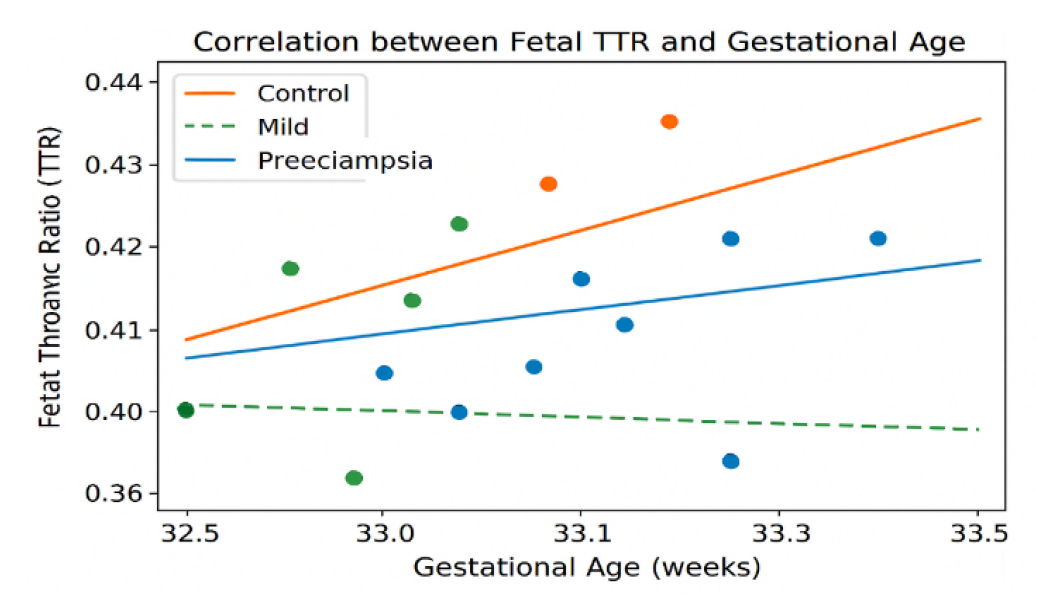
A scatter plot analysis revealed a strong positive correlation between fetal TTR and gestational age (R = 0.736, P < 0.001). However, for any given gestational age, TTR values were consistently higher in the control group compared to preeclamptic pregnancies. The regression lines confirmed that TTR increased more steeply in controls, with the orange line (controls) lying above the blue line (preeclampsia), as shown in Figure 1.


Figure 1: Correlation Between Fetal TTR and Gestational Age


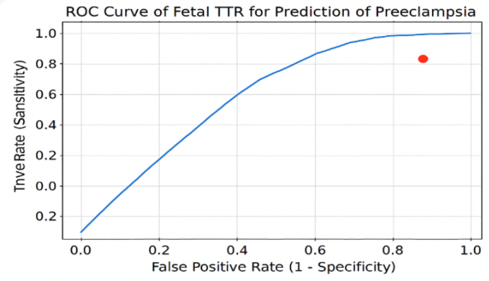
To evaluate the diagnostic performance of fetal TTR in predicting preeclampsia, a receiver operating characteristic (ROC) curve was constructed. The area under the curve (AUC) was 0.921, indicating excellent discriminative ability. The optimal cutoff point was 0.396, yielding a sensitivity of 90% and specificity of 71.3%. The positive predictive value was 51.1%, while the negative predictive value reached 95.5%, with an overall accuracy of 76%.


Table 3: ROC Curve Analysis of TTR for Preeclampsia Prediction

Parameter

Value

Area under the curve (AUC)

0.921

Cutoff point

0.396

Sensitivity (%)

90

Specificity (%)

71.3

Positive predictive value

51.1

Negative predictive value

95.5

Accuracy (%)

76


Figure 2: ROC Curve of Fetal TTR for Prediction of Preeclampsia

DISCUSSION

The baseline characteristics of mothers and fetuses in this study were similar across study groups, without statistically significant differences in either maternal age or BMI, suggesting that pregnancies were carefully matched to limit external variables associated with them. The only baseline difference between parity (multiparous vs nulliparous) was found to be significant, with women diagnosed with severe pre-eclampsia having a higher tendency to be nulliparous, which agrees with previous studies showing that nulliparity is a risk factor for developing pre-eclampsia [22]. The restriction of study entry criteria for women to only those with normal BMI will also assist in eliminating the confounding effect of obesity on fetal thymus size.


This study's primary objective was to explore whether pre-eclampsia impacts fetal thymus size, hypothesizing that pre-eclampsia results in the atrophy of the thymus due to the physiological stressors of the intrauterine environment. Overall, our study resulted in an increase in the number of patients with decreased mean fetal TTR in pregnancies complicated by pre-eclampsia. Women classified as mildly pre-eclamptic had decreased mean TTR compared with controls, while those classified as having pre-eclampsia had even lower mean TTRs. These findings were substantiated with statistical evidence supporting the association between both severity of disease and thymic atrophy.


Our results corroborate previous studies. Gok et al. found reduced fetal thymic ratios in pregnancies complicated by preeclampsia compared with control (healthy) pregnancies [23]. Mohamed et al. reported decreased thymic diameter and perimeter in pregnancies complicated by preeclampsia, indicating impaired thymic development [24]. In support of these findings, Ozlem Ece et al. demonstrated a lower fetal thymus volume at 11–14 weeks' gestation in pregnancies that later developed into preeclampsia [25]. Additionally, Eviston et al. showed decreased thymic ratios at 17–21 weeks' gestation (before clinical onset) in pregnancies affected by preeclampsia [26]. These studies collectively support the theory that abnormal placentation and maternal stress response lead to thymic involution.


The biological process of hypoxic and metabolic stress likely stimulates the hypothalamic-pituitary-adrenal (HPA) axis, leading to increased cortisol secretion, which acts on receptors in cortical thymocytes, triggering apoptosis and, ultimately, thymic involution. This pathway is thought to mediate the effects of exogenously administered steroids in pregnancies. The condition of severe preeclampsia likely compounds these processes by inducing increased levels of maternal inflammation and placental dysfunction, causing an even greater degree of fetal stress and more extensive thymic involution. Thus, fetal thymic size is a reflection of both maternal disease severity and fetal immune competence.


Beyond pathophysiological insights, our study provides evidence that fetal TTR could be a useful test to diagnose preeclampsia through ROC analysis finding an AUC of 0.921, demonstrating very good discriminative ability. At the optimal cutoff of 0.396 the fetal TTR test has demonstrated sensitivity of 90% and a specificity of 71.3%, as well as a negative predictive value of 95.5%. These results support the use of fetal TTR as a biomarker for predicting preeclampsia. Compared with the cutoff of 0.409 reported by Gök et al., which achieved sensitivity of 94.2% and specificity of 90.8% [23], our slightly lower threshold may reflect the focus on severe preeclampsia rather than preeclampsia in general. Regardless of this difference, both studies highlight TTR's potential as a promising non-invasive biomarker.


Limitations

Although conducted by a single center limit the generalizability of the findings, it will also limit the establishment of temporal or causal relationships. It is important to note that the case-control design only allows comparisons between preeclamptic versus healthy pregnancies but cannot establish the timing or causation of the relationships between the groups being compared.


BMI was controlled for in this study to reduce confounding; however, neither maternal comorbidities nor biochemical markers were assessed to determine their effects on fetal thymic development. Additionally, while the ultrasound assessment of the fetal thymic thoracic ratio was validated in terms of its reproducibility and reliability, operator dependence may introduce bias to the measurement process.

CONCLUSION

The fetal thymic thoracic ratio in preeclamptic pregnancies was significantly reduced compared to healthy pregnancies and the greatest amount of reduction was associated with severe cases of preeclampsia. The correlation between TTR and severity of disease supports the hypothesis that abnormal placentation and stress-induced apoptosis play a role in fetal thymic involution through preeclampsia. Results from the ROC curve showed excellent diagnostic validity for the TTR as a viable, non-invasive marker for identifying pregnancies at risk for developing preeclampsia.


Recommendations

Future studies will need larger multicenter cohorts to validate these findings and establish normative (cut-off) values for clinical usage, as well as longitudinal/prospective studies to further establish fetal thymus size throughout pregnancy and its prognostic value before the onset of clinical disease. Moreover, including additional biochemical and immunological measures with TTR may help improve the diagnostic accuracy of fetal thymic size. Clinically, fetal thymus size monitoring should be incorporated into clinical practice to potentially assist in risk stratification and early intervention for at-risk pregnancies.

REFERENCE
  1. Bisson, C. et al. “Preeclampsia pathophysiology and adverse outcomes during pregnancy and postpartum.” Frontiers in Medicine, vol. 10, 2023, article 1144170. 

  2. Hurrell, A. et al. “The assessment of blood pressure in pregnant women: Pitfalls and novel approaches.” American Journal of Obstetrics and Gynecology, vol. 226, no. 2, 2022. 

  3. American College of Obstetricians and Gynecologists. “Gestational hypertension and preeclampsia: ACOG Practice Bulletin, Number 222.” Obstetrics and Gynecology, vol. 135, no. 6, 2020, pp. e237–e260. 

  4. Chaemsaithong, P. et al. “First trimester preeclampsia screening and prediction.” American College of Obstetricians and Gynecologists, 2020. 

  5. Armaly, Z. et al. “Preeclampsia: Novel mechanisms and potential therapeutic approaches.” Frontiers in Physiology, vol. 9, 2018, pp. 973–977. 

  6. Vera-Ponce, V.J. et al. “Global prevalence of preeclampsia, eclampsia, and HELLP syndrome: A systematic review and meta-analysis.” Frontiers in Reproductive Health, vol. 7, 2025, article 1706009. 

  7. Majeed, B.A. et al. “Iraqi women with preeclampsia: Maternal and neonatal outcomes.” Open Access Macedonian Journal of Medical Sciences, vol. 8, 2020, pp. 866–870. 

  8. Ministry of Health. Annual statistical report. Ministry of Health, 2022, pp. 93. 

  9. Jawad, K. Incidence and determinants of hypertensive disorders of pregnancy in the US: Hospitalization discharge rate for preeclampsia, eclampsia, and gestational hypertension, 2016–2018. 2023. 

  10. Phipps, E.A. et al. “Pre-eclampsia: Pathogenesis, novel diagnostics and therapies.” Nature Reviews Nephrology, vol. 15, 2019, pp. 275–289. 

  11. Edmonds, K.D. et al. “Hypertensive disorders.” Dewhurst’s Textbook of Obstetrics and Gynaecology. 9th ed., Blackwell Publishing, 2018, pp. 73–82. 

  12. Cunningham, F.G. et al. “Preeclampsia syndrome.” Williams Obstetrics. 26th ed., McGraw Hill, 2022. 

  13. Wewers, T.M. et al. “Circulating soluble Fms-like tyrosine kinase in renal diseases other than preeclampsia.” Journal of the American Society of Nephrology, vol. 32, no. 8, 2021, pp. 1853. 

  14. Jena, M.K. et al. “Pathogenesis of preeclampsia and therapeutic approaches targeting the placenta.” Biomolecules, vol. 10, no. 6, 2020, pp. 953. 

  15. Jim, B., and S.A. Karumanchi, editors. “Preeclampsia: Pathogenesis, prevention, and long-term complications.” Seminars in Nephrology. Elsevier, 2017. 

  16. Breetveld, N. et al. “Cardiovascular disease risk is only elevated in hypertensive, formerly preeclamptic women.” BJOG: An International Journal of Obstetrics and Gynaecology, vol. 122, no. 8, 2015, pp. 1092–1100. 

  17. Overton, E. et al. “Preeclampsia diagnosis and management.” Best Practice and Research Clinical Anaesthesiology, vol. 36, 2022, pp. 107–121. 

  18. National Institute for Health and Care Excellence. Hypertension in pregnancy: Diagnosis and management. NICE, 2019. 

  19. McGinnis, R. et al. “Variants in the fetal genome near FLT1 are associated with risk of preeclampsia.” Nature Genetics, vol. 49, no. 8, 2017, pp. 1255–1260. 

  20. Fox, R. et al. “Preeclampsia: Risk factors, diagnosis, management, and the cardiovascular impact on the offspring.” Journal of Clinical Medicine, vol. 8, no. 10, 2019, pp. 1625. 

  21. National Guideline Alliance. Hypertension in pregnancy: Diagnosis and management (NG133). National Guideline Alliance, 2019. 

  22. Lin, Y.C. et al. “A comprehensive and bias free machine learning approach for risk prediction of preeclampsia with severe features in a nulliparous study cohort.” BMC Pregnancy and Childbirth, vol. 24, 2024, pp. 853. 

  23. Gök, K. and S. Ozden. “Ultrasonographic evaluation of the fetal thymic-thoracic ratio in preeclampsia.” Journal of Maternal-Fetal and Neonatal Medicine, vol. 36, no. 1, 2023, article 2183739. 

  24. Mohamed, H.T. et al. “Doppler changes in pre-eclampsia.” Medical Journal of Cairo University, vol. 90, no. 3, 2022, pp. 271–278. 

  25. Basaran, O.E., and E.S. Guvendag Guven. “First trimester fetal thymus volume may predict preeclampsia.” Pregnancy Hypertension, vol. 26, 2021, pp. 116–120. 

  26. Eviston, D.P. et al. “Impaired fetal thymic growth precedes clinical preeclampsia: A case-control study.” Journal of Reproductive Immunology, vol. 94, no. 2, 2012, pp. 183–189.

Recommended Articles
Research Article
Oral Manifestations of Myasthenia Gravis, Rheumatoid Arthritis, Pernicious Anemia, Graft Vs Host Disease, Linear IgA Disease and Chronic Bullous Disease of Childhood
Download PDF
Research Article
Tlics Score & Denis Scale and Their Association with Various Variables among Patients Undergoing Operative Treatment for the Thoraco-Lumbar Fracture at Tertiary Care Hospital
...
Published: 10/04/2022
Download PDF
Research Article
Socio-Demographic Characteristics, Classification, Management, Complication and Implant Status of Patients Undergoing Operative Treatment for the Thoraco-Lumbar Fracture at Tertiary Care Hospital.
...
Published: 10/04/2022
Download PDF
Research Article
Association of Maternal Age with Risk of Neonatal Sepsis: A Mono-Centric Retrospective Study in Lebanon
...
Published: 29/12/2020
Download PDF
Chat on WhatsApp
Flowbite Logo
PO Box 101, Nakuru
Kenya.
Email: office@iarconsortium.org

Editorial Office:
J.L Bhavan, Near Radison Blu Hotel,
Jalukbari, Guwahati-India
Useful Links
Order Hard Copy
Privacy policy
Terms and Conditions
Refund Policy
Shipping Policy
Others
About Us
Team Members
Contact Us
Online Payments
Join as Editor
Join as Reviewer
Subscribe to our Newsletter
+91 60029-93949
Follow us
MOST SEARCHED KEYWORDS
Copyright © iARCON International LLP . All Rights Reserved.