Background: Hodgkin disease is characterized by progressive enlargement of lymph nodes. It is considered unicentric in origin and has predictable pattern of spread by extension to contiguous nodes. Materials and Methods: Over a 6-year period, from January 1st 2003 to December 31st 2009 a retrospective study was done for 86 children with Hodgkin’s lymphoma who were admitted to the pediatric oncology unit in Children Welfare Teaching Hospital, The follow up period extended to February 10th 2011. Results: The patients were referred to from 11 governorates, 42 patients (48.8%) were from Baghdad. Highest rate of referral occurred during 2009 compared to previous years of the study. There were 63 (73.3%) males and 23(47.8%) females; male to female ratio was 2.7:1. Fifty eight patients (67.4%) presented with age group between 6-10 years, with a median age of 8 years and mean age of 7.8 years, ranged 2 years to 12.4 years. Cervical lymph node enlargement was found in 64 (74.4%) patients, followed by supraclavicular lymph node enlargement in 8 (9.3%) patients. B symptoms (weight loss, fever and night sweating), were found in 37/80 patients (43%) The mean duration of illness was 7.5 months. Stage III was seen in 42 (48.8%) patients, followed by stage II in 33 (39.5%) patients, stage I in 10 (11.6%) patients and nil for stage IV.The commonest histopathological finding was mixed cellularity in 71(82.6%) patients, then lymphocyte predominant in 12 (14%) patients, nodular sclerosis in 2 (2.3%) patients and lymphocyte depleted in 1 (1.2%) patients. Seven patients were excluded from treatment outcome (4 treated with Non-Hodgkin’s Lymphoma protocol, 1 died before treatment, 1 refused treatment by the family and 1 treated in another institute. From 79 patients who received chemotherapy; fifty six (70.9%) patients finished treatment and remained free of disease till time of last follow up, 1 (1.3%) patient died during treatment, 12 (15.2%) patients relapsed after finishing treatment, 4 (5%) patients abandoned treatment and 6 (7.6%) patients got progressive disease during treatment. At median follow up period of 36 months, ranged from 1 month – 82 months; Event free survival is 70.9%. Sevently-two (89.9%) patients were alive till time of last follow up, 2 (2.5%) died and 5 (6.3%) lost follow up. Conclusion: Males were more commonly affected than females and age group between 6-10 years was the most common. Stage III and mixed cellularity histopathological subtype were most common. Event free and overall survival is improving, yet it is below the standard reported in other countries.
Keywords
Hodgkin’s
Lymphoma
Protocol
INTRODUCTION
Hodgkin's lymphoma, previously known as Hodgkin's disease, is a type of lymphoma, which is a cancer originating from white blood cells called lymphocytes. It was named after Thomas Hodgkin, who first described abnormalities in the lymph system in 1832 [1,2].
Hodgkin's lymphoma is characterized by the orderly spread of disease from one lymph node group to another and by the development of systemic symptoms with advanced disease. When Hodgkin's cells are examined microscopically, multinucleated Reed-Sternberg cells (RS cells) are the characteristic histopathologic finding. Hodgkin's lymphoma may be treated with radiation therapy, chemotherapy or hematopoietic stem cell transplantation, the choice of treatment depending on the age and sex of the patient and the stage, bulk and histological subtype of the disease. The disease occurrence shows two peaks: The first in young adulthood (age 15-35) and the second in those over 55 years old [3]. The 10-year overall survival rate is more than 90% for any stages, early stage may help more (stage I or II) Hodgkin's lymphoma. Since many patients are young, they often live 40 years or more after treatment. However, few studies follow patients as long as 25 years and those studies are of older treatments with more life-threatening adverse effects, so it is impossible to predict long-term outcomes of newer, less harmful treatments.
Radiation treatments and some chemotherapy drugs, pose a risk of causing potentially fatal secondary cancers, heart disease and lung disease 40 or more years later. Modern treatments greatly minimize the chances of these late effects [4]. Patients with a history of infectious mononucleosis due to Epstein-Barr virus may have an increased risk of HL.
Review of Literatures
Etiology and Epidemiology:
Etiology is unknown
Comprises 8.8% of all childhood cancers under the age of 20
Incidence increases to 32 per million for adolescents 15–19 years
Bimodal age-incidence curve with one peak at 15–35 years of age and the other above 50 years of age (incidence is highest among 15–19 year olds)
Association with Epstein–Barr Virus (EBV)
Incidence increased among consanguineous family members and among siblings of patients with HL [5]
The childhood form of Hodgkin lymphoma tends to increase with increasing family size and decreasing socioeconomic status. In contrast, the young adult form of Hodgkin lymphoma is associated with a higher socioeconomic status in industrialized countries [6].
Familial Hodgkin Lymphoma
Clustering of cases of Hodgkin lymphoma within families or races may suggest a genetic predisposition to the disease or a common exposure to an etiologic agent studies of affected families have suggested an increased association of Hodgkin lymphoma with specific Human Leukocyte Antigen (HLA) [7]. Hodgkin lymphoma is diagnosed more commonly in persons, whose immune system is abnormal, a finding that may reflect the slight increase in familial incidence [8].
The etiologic factors underlying the immune deficiency include genetic (e.g., ataxia-telangiectasia), infectious {e.g., Human Immunodeficiency (HIV) and iatrogenic agents [8].
Epstein–Barr Virus and Hodgkin Lymphoma
In the young adult form, delayed exposure to an infectious agent has been proposed as a risk factor for the development of Hodgkin lymphoma because its epidemiologic features are similar to that seen with paralytic poliomyelitis. Epstein-Barr Virus (EBV) has been implicated in the causation of Hodgkin lymphoma by both epidemiologic and serologic studies [9].
In cases associated with EBV, the virus is localized to the Reed-Sternberg cell, EBV latent gene products are expressed, ad the EBV infection is clonal. Hodgkin lymphoma that is EBV-positive at initial diagnosis is usually also positive for EBV at relapse with persistence of the same EBV strain, [10].
Pathology
Hodgkin lymphoma is characterized by a small number of clonal tumor cells {HRS cells, Lymphocytic and Histiocytic (L&H) cells and their variants}, surrounded by a polymorphous inflammatory cell population that constitutes the bulk of the tumoral tissue. These studies have shown that the HRS cells contain clonal Immunoglobulin (Ig) rearrangements and therefore are neoplasms of lymphoid origin [11]. In exceedingly rare cases, the HRS cells have been found to contain T-cell receptor gene rearrangement suggesting that some of these tumors may be of T-lymphoid lineage [12]. These cases are somewhat controversial [13] and remain to be further investigated. Thus, Hodgkin lymphoma is a unique type of B-cell neoplasm and hence the currently used World Health Organization (WHO) classification [14] has abandoned the term" Hodgkin disease" thought to reflect the lack of knowledge regarding the true origin of the HRS cells.
Macroscopic Features
The spread of HL occurs mostly by contiguity from one chain of lymph nodes to another.
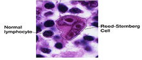
Involvement of the left supraclavicular nodes often follows abdominal para-aortic node involvement with spread up the thoracic duct, whereas involvement of the right supraclavicular nodes tends to be associated with mediastinal adenopathy. Para-aortic node involvement commonly occurs in association with involvement of the spleen, which in turn is commonly followed by liver or bone marrow involvement, or both. The R-S cell is the hallmark of classical HL and is characterized by a binucleated or a multinucleated giant cell that is often characterized by a bilobed nucleus, with two large nucleoli, giving the classically described “owl’s eye” appearance. The R-S cells are embedded within a benign- appearing reactive infiltrate of lymphocytes, macrophages, granulocytes and eosinophils [5] (Figure 1).
Figure 1: Pattern of Lymph Node Spread and Histopathological Features of Hodgkin Lymphoma
Hodgkin lymphoma is characterized by a minority of malignant cells that account for 0.1 to 10% of the total cell population of the tumor [13]. The majority of the tumor is composed of an infiltrate of inflammatory cells (e.g. histiocytes, plasma cells, lymphocyte, eosinophils, neutrophils) and fibrosis, which develop as a result of cytokine release in its most typical forms, Hodgkin lymphoma must therefore be differentiated from several subtype of NHL with similar morphologic characteristics(i.e. T-cell rich large B-cell lymphoma and anaplastic large cell lymphoma) and from benign lymphoid hyperplasia with similar cellular composition (e.g. infectious mononucleosis, progressive transformation of germinal centers [15].
Immunophenotypic Features
Tumor cells in classic HL usually do not express B-cell antigens such as CD20 and CD79a, but virtually all express CD30 and most express CD15 [5].
Histological Classification
Hodgkin Lymphomas (HL) consist of two disease groups, as was proposed in the Revised European-American Classification of Lymphoid Neoplasms (REAL classification) and incorporated into the 2008 4th edition of the WHO Classification of Tumors of Haematopoietic and Lymphoid Tissues [5]. The current WHO histologic classification of Hodgkin lymphoma is largely based on the Rye modification of the Lukes and Bulter classification schema [16].
The WHO classification [15] recognizes two major subtypes of Hodgkin lymphoma cHL and nodular LPHL, based on their biologic and clinical features the cHL contains HRS cells with the classical morphology and immunophenotype.
Histologic Classification of Hodgkin Lymphoma According to the Who Classification:
Nodular lymphocyte predominant Hodgkin lymphoma
Classical Hodgkin lymphoma:
Nodular sclerosis subtype
Mixed cellularity subtype
Lymphocyte-rich subtype
Lymphocyte-depleted subtype
In the nodular LP HL, the tumor has at least partially nodular architecture. The neoplastic nodules contain classic L&H cells and only rare classic HRS CELLS [16] Nodular LP HL affects 10-15% of patients, is more common among male and younger patients and usually presents as clinically localized disease affecting approximately 40% of younger patients and 70% of adolescents, Nodular Sclerosis (NS) cHL is the most common subtype [17]. In the MC subtype of cHL, HRS cells and their variants are often numerous (5 to 15 per high-power filed), this type is observed in approximately 30% of patients, is more common in children aged 10 years or younger and frequently presents as advanced disease with extranodal involvement. LD Hodgkin lymphoma is rare in children, but it is common in HIV-infected patients [18]. The Lymphocyte Rich (LR) variant of cHL is characterized by classical HRS cells and a cellular background that consists mostly of small B lymphocytes [19].
This subtype comprises approximately 5% of all Hodgkin lymphoma and closely overlaps with the nodular LP subtype in presenting clinical features and prognosis.
Clinical Presentation
Lymphadenopathy (90% of All Cases):
Painless swelling of one or more groups of superficial lymph nodes; rarely painful
Cervical nodes involved in 60–80% of cases; associated with mediastinal involvement in 60% of cases
Axillary, inguinal, mediastinal and retroperitoneal nodes also frequently involved
Involved nodes are discrete, elastic, painless and usually “rubbery”; tenderness is rare
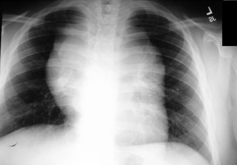
The bulks of palpable lymph nodes are defined by the single largest dimension (in centimeters) of the single largest lymph node or conglomerate node mass in each region of involvement. A node or nodal mass 10 cm is defined as bulky. Abdominal nodal bulk is generally determined using CT (Figure 2)
Figure 2: Clinical Features and Distribution of Lymphadenopathy in Hodgkin Lymphoma
Mediastinal Adenopathy (60% of All Cases)
Approximately 20% of patients have bulky mediastinal disease defined as a large mediastinal mass (10 cm in maximum dimension) or which, on a posterior–anterior chest radiograph has a maximum width equal to or greater than one-third of the internal transverse diameter of the thorax at the level of the T5–6 interspace (mass to thoracic ratio >0.33).
Adolescents and young adults present with a mediastinal mass in 75% of cases, as opposed to children less than 10 years of age where mediastinal disease is present in only 35% of cases.
This is thought to be due to these younger patients having either mixed cellularity or lymphocyte predominant histology, where peripheral adenopathy is more common.
Splenomegaly
The spleen is commonly enlarged on physical examination or by 99mTc-sulfur colloid scan. However, its size is not indicative of splenic involvement with Hodgkin lymphoma.
In 13% of cases, the spleen is the only site of subdiaphragmatic disease. Spleen is involved in 26% of spleens resected in staging laparotomy.
Pulmonary Disease (17% of All Cases)
The mediastinal and hilar nodes are the pathway to the lungs for the spread of HL. Pulmonary HL does not occur without mediastinal or hilar disease. About two-thirds of patients with HD have intrathoracic involvement. There is a striking predilection of the nodular sclerosis type for mediastinal involvement.
Neurologic Manifestations
Neurologic dysfunction is usually a late manifestation. The disease can reach the nervous system in the following ways:
Spread from paravertebral lymph nodes along nerve roots, blood vessels, or perineural lymphatics into the spinal canal by way of the intervertebral foramina and similarly into the intracranial region
Hematogenous dissemination
Bone Disease (2% of All Cases)
Osseous HL typically presents with bone pain and the majority of patients have concurrent non osseous lesions detected at staging. The radiographic features of osseous Hodgkin lymphoma vary but indicate an aggressive malignant process.
Bone Marrow Involvement (5% of All Cases)
Symptomatic patients, particularly those with anemia, leukopenia, or thrombocytopenia, may have bone marrow involvement. Multiple biopsies are indicated, because HL tends to involve the marrow in a focal fashion.
Liver Disease (2% of All Cases) Mild Hepatomegaly Nonspecific abnormalities on liver scan and abnormal liver function tests do not correlate with actual histologic involvement of the liver. Liver biopsy is the only method for accurate diagnosis of liver involvement.
To Establish Liver Involvement, the Following Findings Should be Present:
Evidence of discrete lesions that features one of the histologic subtypes of HL
Conclusive evidence of a Reed–Sternberg cell or one of its variants
Renal Manifestations
HL may directly involve the kidney in 13% of cases. Renal involvement may be unilateral or bilateral and may be present as diffuse involvement, discrete nodules, or microscopic disease. Renal involvement with HL may result from ureteral obstruction and patients with HL may also have renal dysfunction related to renal vein thrombosis, hypercalcemia and hyperuricemia.
Myelosuppression in HL may be caused by hypersplenism or bone marrow infiltration
A positive Direct Antiglobulin Test (DAT) may or may not be associated with overt hemolysis. Patients with a positive DAT test frequently have advanced-stage disease and systemic symptoms
Immune thrombocytopenia may accompany HL in 1–2% of cases. Thrombocytopenia may develop before, concurrently with, or after the diagnosis of HL
Autoimmune neutropenia has also been described in patients with HL. Neutropenia may occur before the diagnosis
Eosinophilia is frequently associated with HL. This occurs in 15% of patients. The presence of eosinophilia is not related to stage or histology. IL-5 production by Reed- Sternberg cells, may explain the etiology of eosinophilia accompanying HL [5]
The erythrocyte sedimentation rate and serum copper and ferritin levels may be elevated, reflecting activation of the reticuloendothelial system [20]
Constitutional B Symptoms
Approximately 20% of patients have associated B symptoms. B symptoms are defined as:
Unexplained weight loss or more than 10% of body weight in the 6 months preceding the diagnosis
Unexplained fever with temperatures .38_C for more than 3 days
Drenching night sweats [5]
Pruritus is another systemic symptom commonly observed in patients with Hodgkin lymphoma, which lacks the prognostic significance associated with fever, sweats and weight loss
Pruritus generally occurs more frequently in patients with advanced-stage disease, may accompany other systemic symptoms, may be more common in women and is usually generalized.
Proposed explanations of Hodgkin- associated prurites include cholestatic liver disease and peripheral sensory neuropathy pruritus typically resolves when the Hodgkin lymphoma is treated [21].
Prognostic Factors
Several pretreatment factors have been associated with an adverse outcome in one or more studies and these include:
Advanced stage of disease (Stage IIB, IIIB, or IV disease)
The presence of B symptoms
The presence of bulk disease
Extranodal extension
Male sex
Elevated erythrocyte sedimentation rate
White blood cell count 11,500/mm3 or higher
Hemoglobin less than 11.0 g/dL
The combination of B symptoms and bulky disease are inferior prognostic indicators. Histology has been noted to be an important prognostic indicator as well. Patients with Nodular Lymphocyte Predominance HL (NLPHL) appear to have an overall better prognosis than those with classical HL (cHL)
Serum markers are currently being studied as prognostic indicators; those associated with poor prognosis include: tumor necrosis factor, soluble CD30, beta 2 macroglobulin, transferrin and serum IL-10 level. Favorable outcomes have been noted with high levels of caspase 3 in Reed–Sternberg cells
Other prognostic indicators include initial response to chemotherapy. Early response assessment is based on volume reduction of disease, functional imaging status or both. Significant reduction in disease volume and PET negativity after two cycles of chemotherapy is associated with a favorable outcome in children and adolescents and failure to achieve this has been associated with an unfavorable outcome in adults
This therapeutic response may permit titration of therapy based on early response and this paradigm is being studied in cooperative group and consortium trials [5].
Differential Diagnosis
Hodgkin lymphoma must be differentiated from other inflammatory causes of lymphadenopathy, particularly those with an indolent course (e.g., atypical mycobacterium infections and toxoplasmosis).
NHL may have similar presenting signs & symptoms, but the growth rate of the affected lymph nodes is often more rapid than in hodgkin lymphoma. The cervical lymphadenopathy of Hodgkin lymphoma must also be differentiated from metastatic adenopathy of other primary tumors (e.g., nasopharyngeal carcinoma and soft tissue sarcoma).
A more difficult problem is that of a mediastinal mass that must be differentiated from normal thymus in an otherwise asymptomatic patient, the thymus is maximal in size in children aged approximately 10 years and may be differentiated from tumor on thoracic Computed Tomography (CT) scans by its texture [22].
Staging
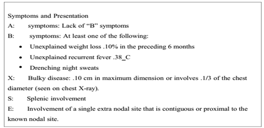
The Ann Arbor Classification was originally adopted in 1971 and was up dated in 1989; it has also been called the Cotswald Classification [5]. Hodgkin lymphoma appears to spread along contiguous lymph nodes until late in the course of disease [23] the currently used Ann Arbor staging system, adopted in 1971, is based on this observation the sub stage classification A, B and E amend each stage based on defined clinical features. Sub stage A indicates "asymptomatic" disease. B symptoms include fever exceeding 38c for 3 consecutive days, drenching night sweats and an unexplained loss of at least 10% of body weight over 6 months, sub stage E denotes minimal extralymphatic disease (Figure 3).5
Figure 3: A Modified Ann Arbor Classification of Hodgkin lymphoma (Continued) Symptoms and Presentation
Disease limited to the spleen or splenic hilar or porta hepatic nodes has been said to have a more favorable prognosis relative to other stage III A disease and is designated III A1 (Table 1) [24].
Table 1: Modified Ann Arbor Classification of Hodgkin lymphoma [5]
Stage
Definition
I
Involvement of single lymph node region (I) or localized involvement of a single extralymphatic organ or site (1E)
II
Involvement of two or more lymph node regions on the same side of the diaphragm (II) or localized contiguous involvement or a single extralymphatic organ or site and its regional lymph node(s)with involvement of one or more lymph node regions on the same side of the diaphragm (IIE)
III
Involvement of lymph node regions on both sides of the diaphragm (III), which may also be accompanied by localized contiguous involvement of an extralymphatic organ or site (IIIE), by involvement of the spleen (IIIS), or both (IIIS1E)
Diagnostic Workup
Recommended for children with equivocal infra diaphragmatic imaging if expertise is available at center
Recommended for all children except those with stages IA/IIA
Recommended for children with Bone pain and elevated alkaline phosphates
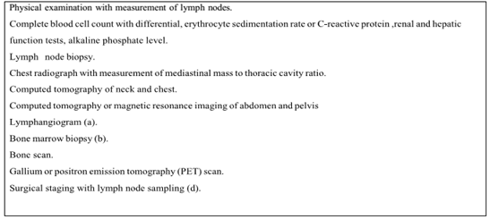
Recommended in selected cases only (Figure 4)
Figure 4: Diagnostic Evaluation for Children with Hodgkin Lymphoma
The chest radiograph provides preliminary information about mediastinal involvement and intrathoracic structures patients with "bulky" mediastinal lymphadenopathy measurement greater than or equal to 33% of the maximum intrathoracic cavity may benefit from a combined-modality treatment approach [25].
Positron Emission Tomography (PET) has been used with increasing frequency over the last decade and will likely replace gallium as the preferred functional imaging modality for lymphoma staging [26].
In PET scanning, uptake of the radioactive glucose analogue 18-Fluoro-2-Deoxyglucose (FDG) correlates with proliferative activity in tumors undergoing anaerobic glycolysis [27].
Bone marrow involvement at the time of initial presentation of Hodgkin lymphoma is uncommon and rarely occurs as an isolated site of extranodal disease.
A bone marrow aspirate alone is not adequate to assess the marrow for disease. A bone marrow biopsy should be performed in any patient with clinical stage III to IV or B symptoms or in any patient at the time of disease recurrence.
A technetium -99 bone scan with corresponding plain radiograph of abnormal areas aids in the assessment of skeletal metastases and should be reserved for the child who has bone pain, a serum alkaline phosphate concentration elevated beyond that expected for age, or extranodal disease identified by other staging studies [28].
Treatment
In general, the use of combined chemotherapy and Low-Dose Involved Field Radiation Therapy (LD-IFRT) is considered standard of care.
This allows for maximal therapy with reduction of individual drug-related or radiation-related toxicities. The volume of radiation and the intensity/duration of chemotherapy are determined by prognostic factors at presentation and include:
The presence of B symptoms
Initial stage of disease
Presence of bulky disease [5]
Combination of Chemotherapy
Current effective multi drugs regimens for Hodgkins and other pediatric malignancies combine non-cross-resistant agents with following properties:
Each agent should be individually active against the tumor
The agents should differ in the mechanism of antineoplastic activitiy, there by targeting different cellular or biochemical events and preventing development of resistance
Toxicities of the agents should not overlap, so that each drug can by an administered at full single-agent dose [29] (Table 2).
Table 2: Lists some of the Regimens used in Hodgkin Lymphoma [29]
Hodgkin lymphoma is a radiosensitive disease. Treatment for HL in children and adolescents employs radiation therapy in the context of multimodality therapy. Most newly diagnosed children and adolescents are treated with risk-adapted chemotherapy alone or in combination with LD-IFRT. In general, doses of 15–25 Gy are used with modification based on patient’s age, the presence of bulk disease, normal tissue concerns and potential acute and long-term effects [5].
Prognosis
Treatment of Hodgkin's disease has been improving over the past few decades. Recent trials that have made use of new types of chemotherapy have indicated higher survival rates than have previously been seen.
In one recent European trial, the 5-year survival rate for those patients with a favorable prognosis was 98%, while that for patients with worse outlooks was at least 85% [30].
A Progression/Relapse of the Disease
Progression if it occurs until three months after the end of therapy (last day of chemotherapy application (including Prednisone/ prednisolone) or last day of radiotherapy respectively)
Early relapse if it occurs between three and twelve months after the end of therapy
Late relapse if it occurs later than twelve months after the end of therapy [31]
Treatment for Relapsed and Progressive Disease
Treatment failure in children and adolescents with HL can be divided into:
Relapsed disease, or
Primary progressive disease
Relapsed patients can still be cured even if relapse occurs after initial treatment. Relapse usually occurs within the first 4 years following treatment. The presence of B symptoms and extra nodal disease at the time of relapse are poor prognostic features. A study conducted by the Society for Pediatric Oncology.
Hematology (GPOH) showed the Following Results:
Patients with an early relapse had a 10-year Event-Free Survival (EFS) of 55% and Overall Survival (OS) of 78%
Patients who had a late relapse had a 10-year EFS and OS of 86 and 90%, respectively [5]
Most relapse in patients with Hodgkin lymphoma occur within the first 3 years, although some patients may relapse as long as 10 years after initial diagnosis.
As many as 50% to 80%; of patients who relapse after radiation therapy alone can be salvaged with chemotherapy or combined-modality therapy [32].
Standard multi agent chemotherapy and radiation therapy may salvage 40% to 50% of children with 1 year or longer initial remissions, but subsequent treatment sequelae, including second malignancies, may reduce ultimate survival [33].
Patients who develop refractory disease during or within 1 year of completing therapy respond poorly to conventional salvage therapy, as does patient with multiple relapses.
For these high-risk patients, consolidation with myeloablative therapy followed by Hematopoietic Stem Cell Transplantation (HSCT) provides the best opportunity for a durable remission.
Several studies support the feasibility and efficacy of this approach, which produce overall survival rates ranging from 30 to 60% in children and adolescents with relapsed Hodgkin lymphoma [34].
Transplant–associated mortality occurs in approximately 10% of patients and most commonly results from infection, cardiopulmonary, or neoplastic complications [33]. Patients have been reported to be at risk for relapse as late as 5 years after HSCT, emphasizing the need for heightened surveillance to ensure continued remission status and monitor for late treatment sequelae.
Autologous HSCT has been preferred for patients with relapsed Hodgkin lymphoma because of the historically high transplant-related mortality associated with allogeneic transplantation [35].
Other novel therapeutic approaches for relapsed Hodgkin lymphoma are currently under investigation. immunotoxin therapy targeting antigens expressed by HRS cell include CD25(the IL-2 receptor) and CD30 (the Ki-1 antigen) clinical trials to date have included only small numbers of patients [36].
Long-Term Complications
Survivors of HL are at risk for numerous late complications of chemotherapy and radiation therapy. Late effects include secondary malignancies, organ dysfunction and psychosocial sequelae.
Secondary Malignancies
The incidence of secondary malignancies in survivors is 7–18 times higher than the general population. Many of these patients had received high-dose alkylating agents and radiation.
Cardiac Toxicity
HL survivors exposed to doxorubicin or thoracic radiation therapy are at an increased risk for long-term toxicity.
Pulmonary Dysfunction
Pulmonary fibrosis is a late complication of HL following radiation therapy. Bleomycin associated pulmonary fibrosis with decreased diffusion capacity.
Thyroid Dysfunction
Primary hypothyroidism is noted following radiation therapy to the neck for HL; though hyperthyroidism, goiter, or nodules have also been noted.
Gonadal Dysfunction and Infertility 6- Growth Dysfunction
Long-term effects on bone and soft-tissue growth among survivors of childhood HL are related to the dose and volume of radiation received, as well as the age of the child when the radiation was received.
Psychosocial Problems
Six percent of HL survivors have symptoms of depression and 15% have somatic distress higher than that for other malignancies or when compared to sibling cohorts. Risk factors associated with increased risk for somatic distress include female gender, lower household income, less than a high school education and lack of current employment [5].
Aim of the Study
To study the clinico-pathological features of Hodgkin Disease in Iraqi children
To compare the results of our study with other studies done inside and outside Iraq
To assess the response to treatment, outcome and survival
MATERIALS AND METHODS
Patients and Methods
Over 6-year period, from January 1st 2003 to December 31st 2009 a retrospective study was done for 86 children with Hodgkin’s lymphoma who were admitted to the pediatric oncology unit in Children Welfare Teaching Hospital.
The information's regarding (age, sex, residence, clinical presentation, diagnostic procedure, staging investigation, treatment modality and its complications, outcome) were taken from hospital registry (Inpatient and outpatient) in addition to own patient’s records.
Pretreatment studies included complete blood count, serum electrolyte, hepatic and renal profile, bone marrow aspirate and biopsy, incision or excision biopsy , Fine needle aspirate, radiography (chest x-ray, abdominal ultrasound, CT scan and/or MRI to the primary site).
Because of lack of the facilities, several investigations were not done like bone scan, serum LDH, immunohistochemical studies, cytogenetic and molecular analysis.
In 8 cases; the paraffin blocks were sent to Pathology department/Sapienza Rome University and re-evaluation was done.
RESULTS
Presenting Features
A total 86 children with Hodgkin disease were seen at CWTH from January 1st 2003 to December 31st 2009. Patients' data were analysed.
The patient were referred to CWTH from 11 governorates, nearly half of them were from Baghdad 42 (48.8%) patients followed by Diala 9 (10.5%) patients, Wasit 8 (9.3%) patients, as shown in Table 3.
Table 3: Referral Area of Patients to CWTH
Governorate
Frequency
Percent
Baghdad
42
48.8
Diyala
9
10.5
Waset
8
9.3
Dewania
6
7.0
Salahalden
6
7.0
Anbar
4
4.7
Karbla
4
4.7
Babylon
3
3.5
Mesan
2
2.3
Najaf
1
1.2
Sulemania
1
1.2
Total
86
100.0
There were an increment in referral of the patients with Hodgkin disease to our hospital in 2003 as compared to 2009 (1.2 Vs 25.6%), as shown in Table 4.
Table 4: Years of Diagnosis
Year
Frequency
Percent
2003
1
1.2
2004
10
11.6
2005
13
15.1
2006
11
12.8
2007
16
18.6
2008
13
15.1
2009
22
25.6
Total
86
100.0
There were 63 (73.3%) males and 23 (47.8%) females; male to female ratio was 2.7:1 as shown in Table 5.
Table 5: Sex Distribution
Sex
Frequency
Percent
Male
63
73.3
Female
23
26.7
Total
86
100.0
Male: Female ratio 2.7:1
Fifty eight (67.4%) patients presented with age group between 6-10 years, 15 (17.4%) patients presented with age group between 11-14 years and 13 (15.1%) patients presented below 5 years, with median age was 8 years; the mean age was 7.8 years, ranged from 2 years to 12.4 years, as shown in Table 6.
Table 6: Age Distribution
Age (year)
Frequency
Percent
<5
13
15.1
6-10
58
67.4
11-14
15
17.4
Total
86
100.0
The median age was 8 years; The mean age was 7.8 years
Neck swelling was the most common sign observed in 42 (48.4%) patients. Neck swelling and fever were present in 16 (18.6%) patients; axillary swelling was present in 3 (3.5%) patients, as shown in Table 7.
Table 7: Presenting Signs
Sign
Frequency
Percent
Neck swelling
42
48.8
Neck swelling & fever
16
18.6
Fever & weight loss
7
8.1
Axillary swelling
3
3.5
Others (dyspnea, abdominal pain, chest 7 8.1
wall mass, generalized oedema, skin rash)
(2,1,1,1,2)
Total
86
100
Regarding lymphadenopathy, cervical lymph node enlargement was found in 64 (74.4%) patients, followed by supraclavicular lymph node enlargement which was found in 8 (9.3%) patients, generalized lymph adenopathy was found in 6 (7%) patients, axillary lymphadenopathy in 4 (4.7%) patients, inguinal lymphadenopathy in 4 (4.7%) patients, as shown in Table 8.
Table 8: Site of Involvement
Frequency
Percent
Cervical LAP 64
74.4
Supraclavicular 8
9.3
Generalized LAP 6
7.0
Axillary LAP 4
4.7
Inguinal LAP 4
4.7
Total 86
100.0
B symptoms (weight loss, fever and night sweating), were found 37/80 patients (43%). The mean duration of illness was 7.57 months.
Diagnosis
Chest X-ray reports were seen in the records of all cases; it was normal in 49 (57.0%) patients, mediastinal mass in 26 (30.2%) patients and hilar lymphadenopathy in 11 (12.8%) patients as shown in Table 9.
Table 9: Radiological Findings
Frequency
Percent
CXR
Normal
49
57.0
Mediastinal mass
26
30.2
Hilar LAP
11
12.8
Abdominal U/S
Normal
44
51.2
Splenic involvement
23
26.7
HSM
12
14.0
Mesentric or paraoartic LAP
3
3.5
Pleural effusion
2
2.3
Pancreatic involvement
1
1.2
Not Recorded
1
1.2
CT chest
Hilar LAP
6
7.0
Normal
4
4.7
Not done
76
88.4
CT abdomen
Normal
2
2.3
Splenic involvement
2
2.3
Not done
82
95.3
Ultrasound reports were found in 85 cases; it was normal in 44 (51.2%) patients, splenic involvement was found in 23 (26.7%) patients, hepatosplenomegaly was found in 12 (14%) patients, mesenteric or para-aortic lymphadenopathy was found in 3 (3.5%) patients, pleural effusion was found in 2 (2.3%) patients, pancreatic involvement was found in 1 (1.2%) patient and one (1.2%) patient has no record of abdominal ultrasound.
CT chest records were found in 10/86 cases; it was normal in 4 (4.7%) patients, hilar lymphadenopathy was found in 6 (7%) patients and 76 (88.4%) patients had not done CT chest.
CT abdomen records were found in 4 cases; it was normal in 2 (2.3%) patients and splenic involvement was shown in 2 (2.3%) patients as shown in Table 9.
Staging
Stage III was the commonest; recognized in 42 (48.8%) patients, followed by stage II in 33 (39.5%) patients and stage I in 10 (11.6%) patients, while no patients presented with stage IV as shown in Table 10.
Table 10: Staging
Stage
Frequency
Percent
I
10
11.6
II
34
39.5
III
42
48.8
IV
0
0.0
Total
86
100.0
Histopathology
All pathological specimens were evaluated in Iraq; 8 cases re-evaluated in Rome (in 4 cases the diagnosed was changed to Hodgkin’s lymphoma after being diagnosed and treated as non-Hodgkin’s lymphoma).
Mixed cellularity was found to be the commonest histopathological group in 71 (82.6%) patients, followed by lymphocyte predominant in 12 (14%) patients, nodular sclerosis in 2 (2.3%) patients and lymphocyte depleted was found in 1 (1.2%) patient as shown in Table 11.
Table 11: Histopathology
Histopathology
Frequency
Percent
Mixed Cellularity
71
82.6
Lymphocyte Predominant
12
14.0
Nodular Sclerosis
2
2.3
Lymphocyte Depleted
1
1.2
Total
86
100.0
Treatment Outcome
Seven patients were excluded from the outcome analysis (4 patients were treated as non-Hodgkin’s Lymphoma, 1 died before treatment, 1 refused treatment by the family and 1 treated in another institute).
Till February 10th 2011; 71 (89.9%) patients alive, 2 (2.5%) died and 6 (7.6%) lost follow up during therapy.
From 79 patients who received chemotherapy; fifty six (70.9%) patients finished treatment and remained free of disease till time of last follow up, 1 (1.3%) patient died during treatment, 12 (15.2%) patients relapsed after finishing treatment (all of them received rescue treatment with more intensive protocols; 11 patients remained alive free of disease and one patient got second relapse and abandoned further treatment), 4 (5%) patients abandoned treatment and never came back and 6 (7.6%) patients got progressive disease during treatment (one died, one abandoned further treatment and 4 received rescue treatment and remained free of disease till time of last follow up), as shown in Table 12.
Table 12: Treatment Outcome
Outcome
Frequency
Percent
Alive free of disease
56
70.9
Relapse
12
15.2
progressive disease
6
7.6
Lost
4
5
Died
1
1.3
Total
79
100.0
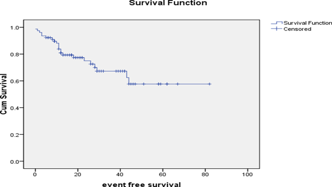
At median follow up period of 36months, ranged from 1 month -82 months; Event free survival is 70.9%, as shown in Figure 5.
Figure 5:Event Free Survival is 70.9%
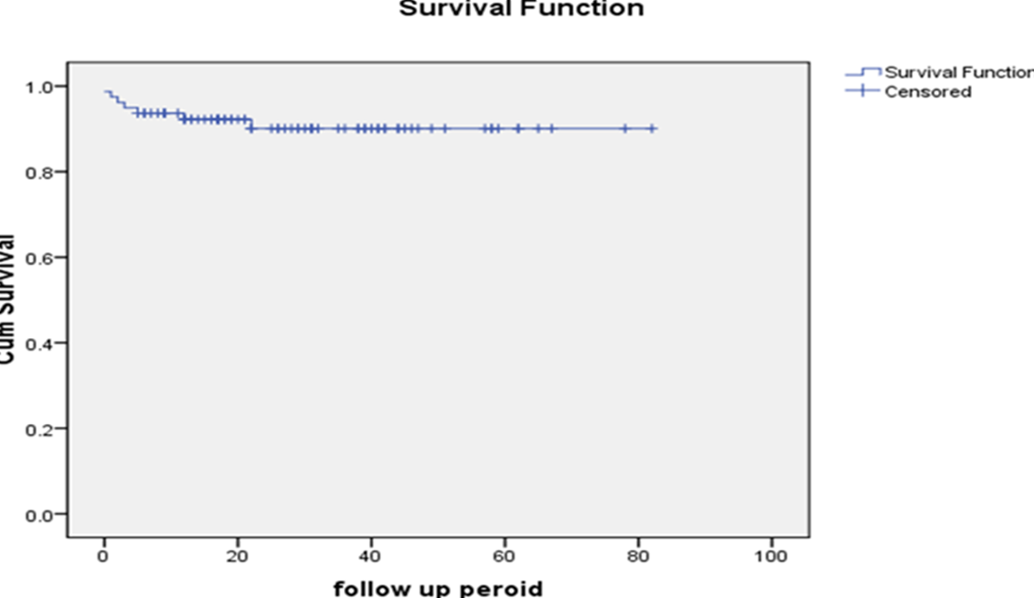
Overall survival was 89.9%, as shown in Figure 6.
Figure 6: Overall Survival for 36 Months is 89.8%
DISCUSSION
This study described outcomes of patients with Hodgkin disease in our institution. Patients had male to female ratio of 2.7:1, comparable to3:1that mentioned in literature [5], Olapuram Jose B study in USA 2004 of 2.6:1 [37] and two previous studies done in the same hospital (1991-1995) of 4:1 and (1997-2003) of 3.1:1[38,39] but lower than 9.6:1 in Arya et al. [40] study from India 2006.
Median age at presentation was 8 years, comparable to previous studies in the same hospital of (8 and 7.5 years) respectively [38,39] and Arya et al. [40], study of 8 years but lower than 14 years in Olapuram Jose B study in USA 2004 although 3% of their patients were above 15 years of age [37], the lower median age in our study might be due to overcrowding and subsequent infection.
Cervical lymphadenopathy was the commonest presentation in our study in 64 (74.4%) patients, similar to literature and previous Iraqi studies [5]; [38,39].
Table 13 shows comparison between 3-study periods in the same hospital; there was a trend towards increasing number of referrals to CWTH over the last 2 decades and in the number of early stages of the disease rather than advanced stages, this might reflect better awareness of the disease by the families, health paramedics and physicians which led to early seeking of medical advice and relatively earlier diagnosis.
Table 13: Overall Survival
Outcome
Frequency
Percent
Alive
71
89.9
Died
2
2.5
Lost
6
7.6
Total
79
100.0
Comparing with other studies; still our study series showing more advanced stages than other studies in developed countries [37,41], but less than studies India and South Africa [40,42] as shown in Table 14. This might be due to increased family sizes, poor socioeconomic status, less awareness of the disease and poor diagnostic facilities in resource limited countries.
Table 14: Comparison between 3 Studies done to Patients with Hodgkin’s Lymphoma’s in the Children Welfare Teaching Hospital
Variables
CWTH
CWTH
CWTH
1991-1995
1997-2003
2004-2009
Total
30
58
86
Average new patient/year
6
8.28
14.3
Median age
8
7.5
8
Male: Female ratio
3.1:1
4:1
2.7:1
Commonest histopathology
MC
MC
MC
Stage I
0%
0%
11.6%
Stage II
26.6%
41.4%
38.4%
Stage III
60%
51.7%
46.5%
Stage IV
13.3%
6.9%
0%
In the current study, mixed cellularity was the commonest histological subtype (82.5%), differ from Hesseling PB study in South Africa 1997 [42], in which nodular sclerosis was the commonest histological subtype (59%) and from Hung study in Taiwan [43] in which the commonest histological subtype was nodular sclerosis (57%). This might be explained by higher incidence of EBV infection in the developing countries, which contribute to the pathogenesis of younger age in Hodgkin’s lymphoma and M.C. subtype (Table 15).
Table 15: Comparison between Current Study and other Studies Analyzing Patients with Hodgkin’s Lymphoma
CWTH
Sandoval
Olapuram
Hesseling
Arya
Variables
2004-2009
USA
USA
South Africa
India
1962-2000
1981-1992
1973-1996 1991-2001
Total
86
51
32
39 148
Patient/year
14.3
1.3
2.6
1.6 14.8
Median age
8
10.5
14
10.8 8
M:F ratio
2.7:1
6.3:1
2.5:1
2.9:1 9.8:1
Commonest
MC
NS
NS MC
Histopathology
(82.5%)
(78%)
(59%) (86%)
No.(%)
Stage I
Stage II
11.6%
38.4%
71%
15%
21.8%
37.5%
5%
36.5%
41%
Stage III
Stage IV
46.5%
0%
8%
6%
28.1%
12.5%
28%
63.5%
26%
In the current study, splenic involvement was found in 23 (26.7%) cases, which was different from that mentioned in Arya et al. study in India [44], in which splenic involvement was found in (15.6%).
In the current study; B symptoms were found in (43%), which was different from that mentioned in Arya et al. study in India [40], in which B symptoms were found in (54.5%).
Event free survival was 70.9% better than (48.3%) in the previous study in CWTH (1997-2003).
Overall survival in the current study was 89.9%, better than 74.1% in the previous CWTH (1997-2003) but lower than 98% in European studies and that mentioned in Fadoo et al. study in Pakistan 2010 [45], in which overall survival was 94%.
There is no follow up system in our setting to detect those who lost to follow up during therapy or post therapy taking in consideration that Hodgkin’s Lymphoma needs follow up beyond five years after finishing therapy.
CONCLUSION
Male affected more than female
Presentation in younger age groups
Advanced stages are less than the previous two studies in the same hospital
Mixed celluarity is the most common histopathologiacl type
High rate of patients presented with B symptoms
Overall survival better than previous studies in CWTH but still below the standard in developed countries
Recommendation
Education of general practitioners for early referral of patients
Improve diagnostic facilities for accurate diagnosis.
Improving medical health services by establishment of trained full time team of physicians and nurses, provision of nurses with specialized training in oncology
Comprehensive treatment with all modalities, chemotherapy and radiotherapy in well-equipped specialized center
REFERENCE
Hellman, S. “A Brief Consideration of Thomas Hodgkin and His Times.” In: Hodgkin’s Disease, S. Hellman (Ed.), Lippincott Williams & Wilkins, 1999, pp. 3–7.
Hodgkin, T. “On Some Morbid Appearances of the Absorbent Glands and Spleen.” Medico-Chirurgical Society Transactions, vol. 17, 1832, pp. 68–114.
Surveillance, Epidemiology and End Results Program. SEER Cancer Statistics Review. National Cancer Institute, www.seer.cancer.gov.
Armitage, J.O. “Early-Stage Hodgkin’s lymphoma.” New England Journal of Medicine, vol. 363, no. 7, 2010, pp. 653–662.
Lanzkowsky, P. “Hodgkin lymphoma and non-Hodgkin lymphoma.” Manual of Pediatric Hematology and Oncology, Elsevier, 2011, pp. 599–646.
Westergaard, T. et al. “Birth Order, Sibship size and risk of Hodgkin’s disease in children and young adults.” International Journal of Cancer, vol. 72, no. 6, 1997, pp. 977–981.
Robertson, S.J. et al. “Familial Hodgkin’s Disease: A Clinical and Laboratory Investigation.” Cancer, vol. 59, no. 7, 1987, pp. 1314–1319.
Rigges, S. and Hagemeister, F.B. “Immunodeficiency State: A Predisposition to Lymphoma.” In: Hodgkin’s Disease and Non-Hodgkin’s Lymphomas in Adults and Children, L.M. Fuller (Ed.), Raven Press, 1988, pp. 451.
Weiss, L.M. et al. “Detection of Epstein–Barr Viral Genomes in Reed–Sternberg Cells of Hodgkin’s Disease.” New England Journal of Medicine, vol. 320, no. 8, 1989, pp. 502–506.
Brousset, P. et al. “Persistence of the Same Viral Strain in Early and Late Relapses of Epstein–Barr Virus-Associated Hodgkin’s Disease.” Blood, vol. 84, 1994, pp. 2447–2451.
Küppers, R. and Rajewsky, K. “The Origin of Hodgkin and Reed/Sternberg Cells in Hodgkin’s Disease.” Annual Review of Immunology, vol. 16, 1998, pp. 471–493.
Seitz, V. et al. “Analysis of BCL-6 Mutations in Classic Hodgkin Disease of the B- and T-Cell Type.” Blood, vol. 97, no. 8, 2001, pp. 2401–2405.
Daus, H. et al. “Hodgkin and Reed–Sternberg Cells Do Not Carry T-Cell Receptor Gamma Gene Rearrangements.” Blood, vol. 85, 1995, pp. 1590–1595.
Jaffe, E.S. “World Health Organization Classification of Tumors.” Pathology and Genetics of Tumours of Haematopoietic and Lymphoid Tissues, WHO, 2001, pp. 185–187.
Nguyen, P.L. et al. “Progressive Transformation of Germinal Centers and Nodular Lymphocyte Predominance Hodgkin’s Disease.” American Journal of Surgical Pathology, vol. 23, no. 1, 1999, pp. 27–33.
Lukes, R.J. and Butler, J.J. “The Pathology and Nomenclature of Hodgkin’s Disease.” Cancer Research, vol. 26, no. 6, 1966, pp. 1063–1081.
Stein, H. et al. “The Nature of Reed–Sternberg Cells and Their Molecular Biology in Hodgkin’s Disease.” Hodgkin’s Disease, Lippincott Williams & Wilkins, 1999, pp. 531–605.
Uccini, S. et al. “High Frequency of Epstein–Barr Virus Genome Detection in Hodgkin’s Disease of HIV-Positive Patients.” International Journal of Cancer, vol. 46, no. 4, 1990, pp. 581–585.
Anagnostopoulos, I. et al. “European Task Force on Lymphoma Project on Lymphocyte Predominance Hodgkin Disease.” Blood, vol. 96, no. 5, 2000, pp. 1889–1899.
Hrgovcic, M. et al. “Serum Copper Levels in Lymphoma and Leukemia.” Cancer, vol. 21, no. 4, 1968, pp. 743–755.
Olsson, H. and Brandt, L. “Relief of Pruritus as an Early Sign of Spinal Cord Compression in Hodgkin’s Disease.” Acta Medica Scandinavica, vol. 206, 1979, pp. 319–320.
Heiberg, E. et al. “Normal Thymus: CT Characteristics in Subjects under Age 20.” American Journal of Roentgenology, vol. 138, no. 3, 1982, pp. 491–494.
Kaplan, H.S. Hodgkin’s Disease. Harvard University Press, 1980.
Desser, R.K. et al. “Prognostic Classification of Hodgkin Disease in Pathologic Stage III.” Cancer, vol. 49, 1977, pp. 883–893.
Rostock, R.A. et al. “CT Scan Modification in the Treatment of Mediastinal Hodgkin’s Disease.” Cancer, vol. 49, no. 11, 1982, pp. 2267–2275.
Bar-Shalom, R. et al. “PET Imaging in Oncology.” Seminars in Nuclear Medicine, vol. 30, no. 3, 2000, pp. 150–185.
Jerusalem, G. et al. “Whole-Body Positron Emission Tomography for Posttreatment Evaluation in Hodgkin’s Disease.” Blood, vol. 94, no. 2, 1999, pp. 429–433.
Pizzo, P.A. and Poplack, D.G. “Hodgkin’s Disease.” Principles and Practice of Pediatric Oncology, Lippincott Williams & Wilkins, 2006, pp. 695–721.
DeVita, V.T., Jr. et al. “Combination Chemotherapy in the Treatment of Advanced Hodgkin’s Disease.” Annals of Internal Medicine, vol. 73, no. 6, 1970, pp. 881–895.
Fermé, C. et al. “Chemotherapy Plus Involved-Field Radiation in Early-Stage Hodgkin’s Disease.” New England Journal of Medicine, vol. 357, no. 19, 2007, pp. 1916–1927.
EuroNet-PHL-C1 Protocol, version 3.0, August 2010.
Healey, E.A. et al. “Prognostic Factors for Patients with Hodgkin Disease in First Relapse.” Cancer, vol. 71, no. 8, 1993, pp. 2613–2620.
Baker, K.S. et al. “Autologous Hematopoietic Stem-Cell Transplantation for Relapsed Hodgkin’s Disease.” Journal of Clinical Oncology, vol. 17, no. 3, 1999, pp. 825–831.
Pui, C.H. et al. “High Serum Interleukin-2 Receptor Levels Correlate with Poor Prognosis.” Leukemia, vol. 3, no. 7, 1989, pp. 481–484.
Anderson, J.E. et al. “Marrow Transplantation for Hodgkin’s Disease.” Journal of Clinical Oncology, vol. 11, no. 12, 1993, pp. 2342–2350.
Hartmann, F. et al. “Treatment of Refractory Hodgkin’s Disease with an Anti-CD16/CD30 Bispecific Antibody.” Blood, vol. 89, no. 6, 1997, pp. 2042–2047.
Jose, B.O. et al. “Pediatric Hodgkin’s Disease.” Journal of the Kentucky Medical Association, vol. 102, no. 3, 2004, pp. 104–106.
Al-Haddad, S. and Muhsen, H.J. “Hodgkin Disease in Children.” Journal of the Faculty of Medicine, vol. 41, no. 1, 1999, pp. 193–201.
Al-Hadad, S. and Merzooq, A.S. Hodgkin’s Disease in Iraqi Children. Iraqi Commission for Medical Specialization, unpublished thesis.
Arya, L.S. et al. “Hodgkin’s Disease in Indian Children.” Pediatric Blood and Cancer, vol. 46, no. 1, 2006, pp. 26–34.
Sandoval, C. et al. “Lymphocyte-Predominant Hodgkin Disease in Children.” Journal of Pediatric Hematology/Oncology, vol. 24, no. 4, 2002, pp. 269–273.
Hesseling, P.B. et al. “Hodgkin’s Disease in Children in Southern Africa.” Annals of Tropical Paediatrics, vol. 17, no. 4, 1997, pp. 367–373.
Hung, I.J. and Yang, C.P. “Hodgkin’s Disease in Children.” Journal of the Formosan Medical Association, vol. 90, no. 7, 1991, pp. 637–644.
Arya, L.S. et al. “Significance of Splenomegaly in Childhood Hodgkin Disease.” Journal of Pediatric Hematology/Oncology, vol. 26, no. 12, 2004, pp. 807–812.
Fadoo, Z. et al. “Pediatric Lymphoma: A Ten-Year Experience in Pakistan.” Journal of Pediatric Hematology/Oncology, vol. 32, no. 1, 2010, pp. e14–e18.
Recommended Articles
Research Article
Vision for a Brighter Kangra: Unmasking the Truth about Pink Eye – A Comprehensive Study on Types, Symptoms, and Proactive Prevention in Himachal Pradesh’s Kangra District
Deepti ,
...
Garima
Published: 11/11/2023
Download PDF
Research Article
It Remains Unproven That the Variant M.8231C>A Causes Coronary Atherosclerosis
Finsterer J
Published: 15/07/2020
Download PDF
Research Article
Leigh Syndrome Should Not Be Diagnosed Exclusively Upon Cerebral MRI
Finsterer J
Published: 15/07/2020
Download PDF
Research Article
Preparation and Characterization of Co (II), Ni (II), Cu (II) and Zn (II) Dithiocarbamate Complexes and Their Adducts
Marwa Thamer abdulrazzaq Aghwan
Published: 27/09/2026
Download PDF
PO Box 101, Nakuru
Kenya.
Email: office@iarconsortium.org
Editorial Office:
J.L Bhavan, Near Radison Blu Hotel,
Jalukbari, Guwahati-India
Mohammed, M. I. & AL-Fadhli, A. G. S. (2022). Hodgkin Lymphoma in Children Six Years Experience of Children Welfare Teaching Hospital. IAR Journal of Medical Sciences, 3(2), 1-13.
MLA
Mohammed, Murtadha I. and Ali G. S. AL-Fadhli. "Hodgkin Lymphoma in Children Six Years Experience of Children Welfare Teaching Hospital." IAR Journal of Medical Sciences 3.2 (2022): 1-13.
Chicago
Mohammed, Murtadha I. and Ali G. S. AL-Fadhli. "Hodgkin Lymphoma in Children Six Years Experience of Children Welfare Teaching Hospital." IAR Journal of Medical Sciences 3, no. 2 (2022): 1-13.
Harvard
Mohammed, M. I. and AL-Fadhli, A. G. S. (2022) 'Hodgkin Lymphoma in Children Six Years Experience of Children Welfare Teaching Hospital' IAR Journal of Medical Sciences 3(2), pp. 1-13.
Vancouver
Mohammed MI, AL-Fadhli AGS. Hodgkin Lymphoma in Children Six Years Experience of Children Welfare Teaching Hospital. IAR Journal of Medical Sciences. 2022 Jul;3(2):1-13.


 Download PDF
 Download XML