Isolation and Identification Bacteria Causes Urinary Tract Infection and Testing Their Sensitivity to Some Antibiotics and Changes in the Level of Immune Proteins Resulting from Infection
Haneen F. Abbas
1
,
Ayyub J. Al-Bayaty
1
,
Huda S. Khuder
1
1
Coll.Education for Women, Tikrit University Pharmmacy Coll. Tikrit University, Iraq
The current study included the collection of 250 urine and blood samples for patients with urinary tract infections, in addition to 50 urine and blood samples not infected as a control sample, which included females and males with ages ranging between 15-60 years, 60% females and 40% males from the patients referred to the laboratories of the city of Tikrit. 200 samples gave positive bacterial growth at a rate of 80%, as it gave two types of growth negative for samples with a percentage of 62.5% and positive for Gram at a rate of 37.5%, while the other 50 samples did not give any bacterial growth. The sensitivity of the isolated bacteria to a number of antibiotics was studied, and the results showed the sensitivity of the isolated bacteria to merepnen, while the bacterial isolates showed the highest resistance to caffeine. The results of the study showed a significant increase in the level of immune proteins compared to the control samples, as the level of IL-22 was 445.0±16.4, while its level in the sera of healthy people was 135.3±18.4
Keywords
Interloukin 22 (IL-22)
Urinary Tract Infection (UTIs)
Bacteria
Antibiotic
INTRODUCTION
Urinary Tract Infection (UTIs) are common diseases and spread widely among members of society and different age groups, but their presence in females is more compared to males due to several reasons, including the proximity of the urinary opening to the genital and anal areas, urethral shortening and pregnancy, during which physiological changes occur that cause this disease [1]. The microorganisms that cause urinary tract infections vary, but most of them are caused by bacteria and there may be more than one pathogen at the same time[2]. Bacterial infection is caused by both Gram-positive and Gram-negative bacteria [3]. However, Gram-negative bacteria recorded the highest percentage of infection events compared to Gram-positive bacteria, the most common of which is E.coli. Gram-negative bacteria are often the cause of complex urinary tract infections [4]. The use of antibiotics has contributed to the treatment of many diseases, including the treatment of urinary tract infection and for a long time occupied the center stage in the resistance to pathogens.
The majority of urinary infections that affect men are complex [5], while uncomplicated infections are very low in men, and the majority of men with bacterial infections have urinary tract abnormalities [6]. Women UTIs have a distinct pattern in that women show an increased susceptibility to infection, while men have an increased severity of infection and it is more consistent. The prevalence of bacterial infection in women is estimated to be about 10% compared to 0.1% in men. [7]. The infection occurs in women of all age groups [8]. Women exposed to infection have weak IgA in most cases compared to the virulence of pathogenic bacteria [9]. Urinary tract infections are the third most common bacterial infection
among children, especially in developing countries, after gastrointestinal and respiratory infections. The infection is often associated with urinary tract abnormalities that have resulted in long-term complications including high blood pressure and chronic renal failure [10]. In newborns, urinary tract infections can cause septicemia and general ill health with growth failure [11]. Breastfeeding is important for children because breast milk contains defensive factors represented by immunoglobulin A (IgA), which can Binding to pathogenic bacteria and destroying its outer wall by Lysozyme enzyme in synergy with Lactoferrin, thus preventing infection [12].
The detection of serological UTI by measuring the cellular motility in the serum of the infected, which is a vital indicator of the level of inflammation, including Interloukin 22 (IL-22), which is associated with UTI and its severity [13].
MATERIALS AND METHODS
Antibiotics used: Antibiotics
In the current study, antibiotic tablets were used for the purpose of testing the sensitivity and resistance of isolated bacterial species to antibiotics, including (AK) Amikacin, Cefixime (CFM), Gentamicin (CN), Meropenem (ME), and Nitrofurantoin (F).
Preparation of Culture Media
The following culture media was prepared Blood agar, Nutrient broth, Nutrient Agar, MacConkey Agar, Mannitol Salt Aga, Eosin- methylene blue agar, Brain-heart infusion agar, Muller-Hinton agar.
Sample Collection and Culture
The current study included the collection of urine and blood samples from patients aged between years from the auditors of Al-Shifa Laboratory and Pharma Laboratory in the city of Tikrit / Salah Al-Din / Iraq during the period from 20/9/2020 to 10/5/2021. The samples included urine samples blood samples from people suffering from symptoms of urinary tract infections, in addition to urine and blood samples from healthy people as a control sample. A drop was taken from each urine sample by means of the bacterial vector Loop and planted on three culture media: Blood agar, Maconkey agar and saline mannitol. Pure colonies were preserved using tilted nutrient agar at 4°C until subsequent diagnostic tests were performed [14].
Identification of Isolated Under Study
The isolates under study were diagnosed according to the following tests:
Morphological Diagnosis and Microscopical Diagnosis
This technique was used to estimate the levels of immunoglobulin proteins (IL22) in the sera of the samples used in the study.
Work Steps
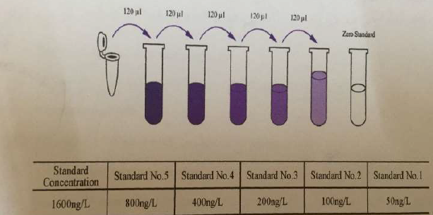
All samples, materials and reagents were left at room temperature for 30 minutes before use, then the standard concentrations were prepared by conducting a series of dilutions in five sterile tubes to obtain the concentrations of interleukin-22 (50-100-200-400-800) as in Figure 1.
Figure 1: Series Dilutions of the Standard Solution Substrate for IL-22
Then, by pipette, 50 μl of diluted standard solution tubes were transferred to the first five holes on the special plate (for each tube a hole used). The rest of the pits were mixed gently, the plate was covered with an adhesive cover and incubated for half an hour at a temperature of 37 0 C. The concentrated washing solution was diluted with distilled water (30 times), the cap was gently removed from the plate, then the plate pits were washed with the washing solution, and the process was repeated five times, turning the plate over Blotting paper to get rid of the remaining solution add 50 μl of HRP-Conjugate enzyme solution to all pits of the plate except for the control plank pits, return the adhesive cap and incubate as in step 4, wash the plate as in step 6, add 50 μl of staining solution A and 50 μl of staining solution B in the dark to all the pits and mix gently, then the plate was incubated for 15 minutes at 37°C, 50 μl of stop solution was added to all pits to finish the reaction, absorbance reading O.D. The samples were etched into a calibration plate using an ELISA device at a wavelength of 450 nm.
RESULTS AND DISCUSSION
Results of Culture Samples
200 samples of them gave bacterial growth when grown on the culture media as shown in the sample collection paragraph in the separation of materials and methods of work, at a rate of 80.0% of the total of 250 samples, median urine samples, while the remaining 50 samples, at a rate of 20.0%, did not give any bacterial growth as shown in the Table 1, which is a growth result completely identical to what was reached by the researcher[15], who obtained 160 positive growth samples with a percentage of 80.0% and 20% negative growth, which is a high growth result compared to what was reached by the researcher [13] who obtained On 86 samples positive for bacterial growth at a rate of 66.15% and 44 samples did not show bacterial growth at a rate of 33.85%, the lack of growth of some samples may be due to a viral or fungal cause or from anaerobic bacteria that we cannot isolate by the usual culture methods that were used in this study, which needs to Culture media and special conditions for development or as a result of the patient's use of random antibiotics led to the disappearance of pathogenic bacteria [16].
Table 1: Results of Transplantation of Samples Taken from Patients with Urinary Tract Infections
Percentage %
N.O
Implant results
80.0
200
positive growth
20.0
50
negative growth
100.0
250
total summation
Samples grown on culture media showed two types of growth: Gram positive, 37.5%, and Gram negative, 62.5%, as shown in Table 2. This result is close to what was found by [17] 43% of bacteria Positive bacteria and 57% negative bacteria and [18] in Tikrit, which recorded 39.5% of gram-positive bacteria and 57.5% of gram-negative bacteria, and as a result, the gram-negative bacteria of those infected had the highest percentage of gram-positive bacteria and this agrees with the study current.
Table 2: Number and Percentages of Gram-Positive and Gram-Negative Bacterial Isolates
Percentage %
N.O
isolate type
37.5
75
Positive for cream dye
62.5
125
Negative for gram dye
100.0
200
the total
Phenotypic and Microscopic Identification of Bacterial Isolates Causing Urinary Tract Infections
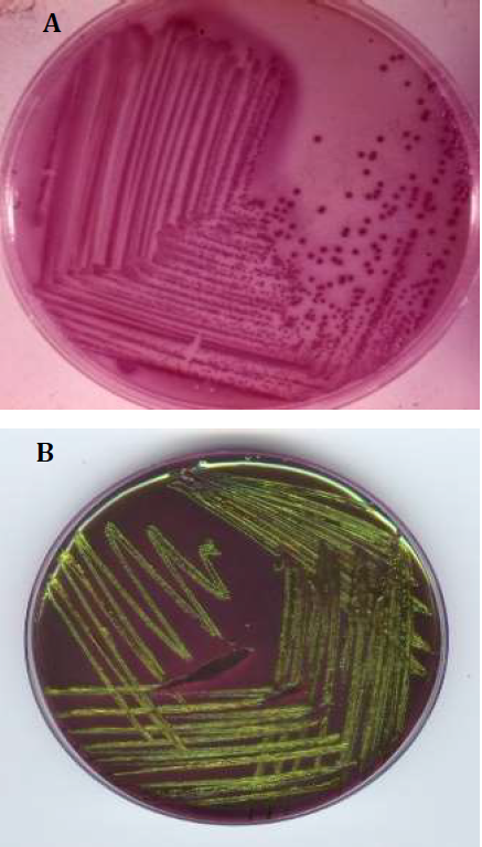
The results of phenotypic and microscopic tests that were performed on the growing bacteria (using Gram stain) showed that all Gram-negative bacteria grow on Maconkey medium, which does not allow Gram-positive bacteria to grow on it and are bacillary in shape. Escherichia coli colonies appeared in pink on MacConkey medium as a result of fermentation of lactose sugar (Figure 2), and its medium-sized colonies are regular, dry, and convex. 1) This bacteria is one of the natural flora found in the intestines of humans and animals, and one of the most common types of urinary tract infections [19].
Figure 2: Shows (A) E.Coli Colonies on Maconkey Medium And (B) Shows on EMB Mediu
Resistance of Bacterial iIolates to Antibiotics
The results of the bacterial isolates showed different types of resistance to the antibiotics used as shown in (Table 3) and (Figure 3), and the results were interpreted according to.
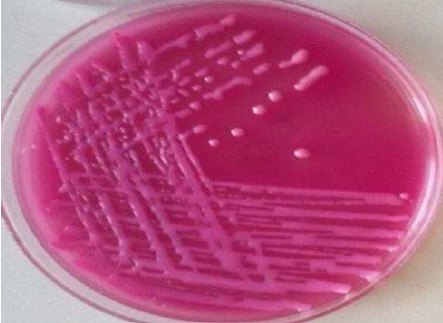
Figure 3: Shows Colonies of Klebsiella Bacteria on Maconkey Medium
Table 3: Sensitivity of Bacterial Species Isolated from Urinary Tract Infections to Antibiotics
E.coli isolates showed absolute resistance to CFM, and low resistance to both F and CN antigens, with different percentages (30,36.6) respectively, and very weak resistance to Me, AK antigens with ratios (11.6 and 8.3). As for E.coli isolates, they showed resistance with ratios (11.6 and 8.3). % (i.e. sensitive towards isolates with a percentage of 91.7%), and this agrees with all patel and its group 2019 with percentages (91.89%). As for the anti-F, the results of our study agreed with [20]by 34.1%, and our study did not agree with the researcher [13], where the proportion of her study was 89.4%, and the anti-CN of E.coli was resistant to this antibody by 30%, and this converges with the study of [15]where it was (and 37.5%), but it does not agree with (95.8%), but it does not agree with[17] with a percentage of (95.8%). % and 2.8%) respectively, while the anti-CFM matched the results of the researcher [13], where it was 89.74%. One of the important reasons for the resistance of E.coli bacteria to antibiotics may be due to changes in the permeability barrier, which leads to difficulty in reaching the target site and thus obstructing its work, which is a special feature in Gram-negative bacteria [21]. The resistance of these bacteria may be the result of a change in the composition of certain enzymes or the loss of their function and thus lead to negative changes in the site on which the antibiotic works, or the resistance results from enzymes that remove the toxicity of the antibiotic [22], as for the resistance of bacteria to lactam-antibiotics. β comes from bacterial production of β-lactamase enzymes that degrade the β-lactam ring [23].
S.aureus bacteria where the absolute resistance to the anti-CFM is 100%, and the resistance to anti-Me in our study is 36%, and this result is similar to the study of the researcher [24]where the result of her study was 28.1% and our study does not agree with the researcher[25] where This antagonist is sensitive to 100%, and anti-F is 10% resistant, and this does not agree with [26], where the resistance ratios in their studies were (72.2%). As for the anti-gentamicin, it was moderately resistant by 49%, and this agrees with the study [15]. The result of the resistance to the antigen was 51.4%, and our study does not agree with [25] with rates (14.7%). As for the anti-Amikacin, it is 25.5% resistant, and this agrees with [25] where it is 23.3% resistant, and we do not agree with [26], where it is 11.1%. As for Cefixime, it is absolutely resistant, and this corresponds to the study [26] by 100%, while K.pnumonia bacteria is isolated as it has absolute resistance to CFM. As for meropenen, it was 17% resistant (i.e. 83% sensitive) and this The result is similar to the study[24], where it was 100% sensitive.
Anti-Nitrofurantion showed a medium resistance of 57% and this is in agreement with the study of [15] and [26], where the resistance they had was (65% and 64.7%), respectively. We also used the antigen in our study, gentamicin, and it had a resistance of 22.8% and this agrees with the study [27]by 33.3%, but it does not agree with [15] by 89.7%, and Amikacin has a high sensitivity of 94.3% and this agrees with the study of [26] and a study [28] with a percentage of 100% for each of them, and an approach to the study [15], where it was sensitive with a percentage of 75.9%. Cefixime has absolute resistance, and this is similar to [26] and[15] with a ratio of 82.3 and 79.3, respectively. The most important of these mechanisms is its production of β-lactamase enzymes, as studies have proven that it possesses many types of these enzymes that destroy a wide range of β-lactamases . And Proteus.mirabilis bacteria have absolute resistance to the anti-CFM, and they are resistant to anti-Me by 20%, anti-F and CN are resistance by 33.3% each, while anti-Me is sensitive by 20% and here we agree with the study [24] where it is resistant by 25%, and the result of anti-F is completely identical to the study[26] with a percentage of 33.3%, anti-CN and in our study it agrees with the study of [13] where it is 25%, but anti-AK is sensitive in our study by 80% and this is close to a result [26] 100%. The reason for the resistance of these bacteria isolates to the antibiotic is due to several reasons, including the presence of mutations or plasmids that show the character of resistance towards the antibiotic, and the bacteria may have the ability to form a biofilm that has a role in this resistance [29] (Figure 4).
Figure 4: Susceptibility of Proteus.Mirabilis, S.Aureus and K.Pnumonia Isolated To Antibiotics
The results of the current study showed a high concentration of interleukin in the serum of patients with urinary tract infection compared to healthy controls, as its level in interleukin 22 reached its level in the infected (445.0±16.4 mg/dl) and in the healthy (135.3±18.4) and there are highly significant differences as the probability value reached p<0.001 as shown in (Table 4).
Table 4: Measurement of Interleukin IL22
Injury and control samples
Mean±SD
IL 22
injury samples
445.0± 16.4
control samples
135.3 ±18.4
p- value
23.01 **
p-Value
0,0008
Our study matched with the study of [30] where the levels of IL-22 were significantly elevated in their study in patients with Candida compared to control samples, and showed that the presence of IL-22 in urine is an important means in diagnosing Candida infection in the urinary tract.
CONCLUSION
The results showed that the prevalence of urinary tract infections in patients was of different ages ranging from (15-60) years, and females recorded higher infections than males, and it was found that the highest infection appeared in ages ranging from (31-45) years for both genders, and E.coli bacteria were the predominant bacteria over the rest of the bacterial species while the least bacterial types causing the disease were K. kristinae and E.aerogenes
The results showed a high percentage of IL-22 used in the study compared to control samples
Recommendation
Expanding the study of other types of cellular compounds, including chemical attraction and interleukins and determining their relationship with the various causes of urinary tract infections
Assessment of IL-22 levels in viral infections
REFERENCE
Stefaniuk, E. et al. “Etiology and antibiotic susceptibility of bacterial pathogens responsible for community-acquired urinary tract infections in Poland.” European Journal of Clinical Microbiology and Infectious Diseases, vol. 35, 2016, pp. 1363–1369.
Nicolle, L.E. “Urinary tract infection and pyelonephritis.” National Kidney Foundation Primer on Kidney Diseases, WB Saunders, 2014, pp. 412–419.
Foxman, B. “Urinary tract infection syndromes: Occurrence, recurrence, bacteriology, risk factors and disease burden.” Infectious Disease Clinics of North America, vol. 28, no. 1, 2013, pp. 1–13.
Levison, M.E. and D. Kaye. “Treatment of complicated urinary tract infection with an emphasis on drug-resistant gram-negative uropathogens.” Current Infectious Disease Reports, vol. 15, no. 2, 2013, pp. 109–115.
Olson, P.D. et al. “Androgens enhance male urinary tract infection severity in a new model.” Journal of the American Society of Nephrology, vol. 27, no. 6, 2016, pp. 1625–1634.
Schaeffer, A.J. and L.E. Nicolle. “Urinary tract infections in older men.” New England Journal of Medicine, vol. 374, no. 6, 2016, pp. 562–571.
Harper, M., and G. Fowlis. “Management of urinary tract infections in men.” Trends in Urology, Gynaecology and Sexual Health, 2007.
Nicolle, L. et al. “Uncomplicated urinary tract infection in women: Current practice and the effect of antibiotic resistance on empiric treatment.” Canadian Family Physician, vol. 52, no. 5, 2006, pp. 612–618.
Sim, J. Urinary tract infection. Nidus Information Services, 2001.
Eider, J. S. “Urinary tract infection.” Nelson Textbook of Pediatrics, 19th ed., Saunders, 2012, pp. 1829–1834.
Jones, C. and J. Kausman. “Newborn urinary tract infections.” Rickham’s Neonatal Surgery, Springer, 2018, pp. 1153–1160.
Ali, Z. S. and Y. AbdulRaheem. “Assessment of exclusive breastfeeding effect against urinary tract infection in infancy.” Iraqi Academic Scientific Journal, vol. 18, no. 3, 2019, pp. 204–210.
Ibrahim, M.I.K. Isolation and identification of bacteria causing urinary tract infection for women of childbearing age and their relationship to some immune factors. Master’s thesis, Tikrit University, 2020.
Leboffe, M.J. and B.E. Pierce. A photographic atlas of the microbiology laboratory. 4th ed., 2011.
Al-Zaidi, A.J.R. Determination of the type and concentration of active compounds in the fruits of Citrullus colocynthis and the effect of aqueous and alcoholic extracts on some bacterial isolates causing urinary tract infection. Master’s thesis, Tikrit University, 2020.
Kitagawa, K. et al. “International comparison of causative bacteria and antimicrobial susceptibilities of urinary tract infections between Kobe, Japan and Surabaya, Indonesia.” Japanese Journal of Infectious Diseases, vol. 71, no. 1, 2018, pp. 8–13.
Al-Hamdani, A.J.A. Study of the biological effect of Sidr and pomegranate extracts on ESKAP-resistant polybacterial group isolated from pregnant women with urinary tract infection. Master’s thesis, Tikrit University, 2019.
Ibrahim, E.E.D. A bacteriological study of urinary tract infections for pregnant women and those with high blood pressure and diabetes in the Tikrit region. Master’s thesis, Tikrit University, 2010.
Ahmad, W. et al. “Frequency of Escherichia coli in patients with community-acquired urinary tract infection and their resistance pattern against some commonly used antibacterials.” Journal of Ayub Medical College Abbottabad, vol. 27, no. 2, 2015, pp. 333–337.
Al-Obaidi, M.J.A. Microbiological and epidemiological study of urinary tract infections in children in Hawija district. Master’s thesis, Tikrit University, 2007.
Spanu, T. et al. “Occurrence of extended-spectrum beta-lactamase and other antimicrobial drug resistance.” Antimicrobial Agents and Chemotherapy, vol. 46, no. 1, 2002, pp. 196–202.
Kadir, M. et al. “Prevalence of urinary tract infection in patients with renal stones.” 2010.
Magdy, M.A. “Detection of extended-spectrum beta-lactamase-producing Klebsiella pneumoniae and Escherichia coli from environmental surfaces.” International Journal of Biological Chemistry, vol. 10, 2013, pp. 3923.
Hellan, S.F.A. Inhibitory effect of bioproducts extracted from some types of cyanobacteria on multi-antibiotic resistant bacterial species. Master’s thesis, Tikrit University, 2019.
Khalaf, N. M. Evaluation of the efficacy of some plant extracts on beta-lactamase-producing pathogenic bacteria isolated from skin infections. Master’s thesis, University of Diyala, 2020.
Hussein, M.A.R.H. The role of some immune molecules in urinary tract infections and gestational diabetes in pregnant women. Master’s thesis, Tikrit University, 2021.
Al Mazrouei, O.S.M. The cumulative effect of aqueous and alcoholic extracts of radish and ginger plants on some bacterial species isolated from urinary tract infections in the city of Tikrit. Master’s thesis, Tikrit University, 2017.
Darwish, S. N. Immunological changes of some indicators associated with bacterial types that cause urinary tract infection in pregnant women. Master’s thesis, Tikrit University, 2021.
Kwiecińska, J. et al. “Effects of ceftazidime and ciprofloxacin on biofilm formation in Proteus mirabilis rods.” The Journal of Antibiotics, vol. 66, 2013, pp. 593–597.
Ahmadikia, K. et al. “Increased urine interleukin-17 and interleukin-22 levels in patients with candidal urinary tract infection.” Iranian Journal of Kidney Diseases, vol. 12, no. 1, 2018, pp. 33.
Recommended Articles
Research Article
Serum IL-22, miRNA-146a Expression and Total Antioxidant Status as Integrated Biomarkers in Rheumatoid Arthritis
Khalid R. Kareem
Published: 13/08/2026
Download PDF
Research Article
Electrochemical Comparative Studies On The Redox Behavior Of Cu(II) And Cr(II) Before And After Interactions With Ciprofloxacin In Solutions
H. M. Naseem Akhtar,
M. Q. Ehsan
Published: 10/08/2021
Download PDF
Research Article
A Rare Case Report of Waugh Syndrome in an Eight Months Old Male
Pielly Hanna,
...
Bassem Abou Merhi
Published: 10/03/2021
Download PDF
Research Article
Study on Prevalence and Etiology of Early Neonatal Deaths in Jasin district, Melaka from 2014-2017
Sujat a Khobragade,
...
Yadneshwar Khobragade
Published: 30/05/2021
Download PDF
PO Box 101, Nakuru
Kenya.
Email: office@iarconsortium.org
Editorial Office:
J.L Bhavan, Near Radison Blu Hotel,
Jalukbari, Guwahati-India
Abbas, H. F., Al-Bayaty, A. J. & Khuder, H. S. (2022). Isolation and Identification Bacteria Causes Urinary Tract Infection and Testing Their Sensitivity to Some Antibiotics and Changes in the Level of Immune Proteins Resulting from Infection. International Academic Journal of Applied Bio-Medical Sciences, 3(2), 1-5.
MLA
Abbas, Haneen F., Ayyub J. Al-Bayaty and Huda S. Khuder. "Isolation and Identification Bacteria Causes Urinary Tract Infection and Testing Their Sensitivity to Some Antibiotics and Changes in the Level of Immune Proteins Resulting from Infection." International Academic Journal of Applied Bio-Medical Sciences 3.2 (2022): 1-5.
Chicago
Abbas, Haneen F., Ayyub J. Al-Bayaty and Huda S. Khuder. "Isolation and Identification Bacteria Causes Urinary Tract Infection and Testing Their Sensitivity to Some Antibiotics and Changes in the Level of Immune Proteins Resulting from Infection." International Academic Journal of Applied Bio-Medical Sciences 3, no. 2 (2022): 1-5.
Harvard
Abbas, H. F., Al-Bayaty, A. J. and Khuder, H. S. (2022) 'Isolation and Identification Bacteria Causes Urinary Tract Infection and Testing Their Sensitivity to Some Antibiotics and Changes in the Level of Immune Proteins Resulting from Infection' International Academic Journal of Applied Bio-Medical Sciences 3(2), pp. 1-5.
Vancouver
Abbas HF, Al-Bayaty AJ, Khuder HS. Isolation and Identification Bacteria Causes Urinary Tract Infection and Testing Their Sensitivity to Some Antibiotics and Changes in the Level of Immune Proteins Resulting from Infection. International Academic Journal of Applied Bio-Medical Sciences. 2022 Jul;3(2):1-5.


 Download PDF
 Download XML