Enzymes present in thermophiles are more stable than its mesophilic homologues [1]. Thermophilic enzymes are biotechnologically more advantageous than the mesophilic enzymes because they are easier to purify by heat treatment, have higher resistance to chemical denaturants, and can withstand higher substrate concentrations. Due to their higher rate of reaction and higher reaction temperatures they are also less susceptible to microbial contaminations. Organisms isolated from hot water springs can be a great source of thermostable enzymes which can be used in industries [2].
α-Amylase (E.C. 3.2.1.1.) is one of the most industrially and clinically essential enzymes which accounts for 30% of the world’s enzyme production. The chemical hydrolysis of starch in the starch processing industry at present has been successfully taken over by amylases [3]. For over 50 years α-amylases have been used in various industries such as starch processing, detergent, textile, Food, Paper pharmaceutical, ethanol, and alcoholic beverages [3-5]. Many Bacillus spp. and related genera Such as Bacillus licheniformis and Bacillus subtilis produce extracellular α-amylases that are widely used in industries [6-8]. Purity, biochemical characters, and kinetic properties of an enzyme are crucial factors in industrial application [9].
This research was carried out to find potential novel thermophilic α-amylase producing bacteria from Sri Lankan hot water springs to be utilized through synthetic biology approaches. In this article we discuss the process of isolation, biochemical characterization and identification of α-amylase producing Caldimonas manganoxidans MS1 from a Sri Lankan hot water spring and functioning characterization of its extracellular α-amylase after purification.
Experimental Section
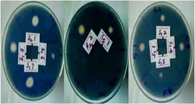
Isolation, Biochemical Characterization, and Identification of α-Amylase Producing Bacteria: Soil and water samples were collected using aseptic techniques from Nelumwewa (Polonnaruwa district) and Mahaoya (Ampara district) hot water springs, two of the hottest in Sri Lanka and were transported to the laboratory in ice and stored at 4°C until further studies. These samples were then inoculated into modified starch nutrient broth containing 0.5% (w/v) peptone, 0.2% (w/v) yeast extract, 0.5% (w/v) NaCl, 0.5g/L KCl, 0.5g/L MgSO4.7H2O, 0.04 g/L MnSO4, 0.3 g/L FeSO4, 0.87 g/L K2HOP4, 0.022 g/L CaCl2 and 1% (w/v) soluble potato starch as carbon source. The final pH was maintained at 6.9 and enriched the growth of thermostable α-amylase producers for 72 hours at 50°C [10]. The enriched samples were then inoculated into starch nutrient agar plates by streak plate method and spread plate technique and screened for α-amylase production using starch hydrolysis test. The α-amylase producing colonies were selected by flooding the plates with iodine solution (Figure 1) after incubating for 24 hours [11]. Potential candidates were selected by monitoring their ability to hydrolyze starch on agar plates and subjected to colony morphological studies and biochemical characterization. The best candidate was selected by measuring the amount of reducing sugars released over time by using starch as the carbon source in the nutrient broth by Dinitrosalicylic acid (DNS) method [12]. Isolated colonies were preserved as pure cultures. The most effective amylase producer was subjected to identification by 16S rRNA gene sequencing. PCR amplification was done using 27F 5' (AGA GTT TGA TCM TGG CTC AG) 3' and 1492R 5' (TAC GGY TAC CTT GTT ACG ACT T) 3' primers and the sequencing was carried out with 785F 5' (GGA TTA GAT ACC CTG GTA) 3' and 907R 5' (CCG TCAATT CMT TTR AGT TT) 3' primers. The sequences were assembled using BioEdit 7.2.6. CAP contig assembly program [13]and analyzed in NCBI BLASTN tool [14]and the Neighbor Joining phylogenetic tree was generated using BLAST pairwise alignments [15]. All the biochemical tests were carried out according to the Cowan and Steel's manual for the identification of medical bacteria [16].
Figure 1: Isolated Bacterial Colonies on Starch Nutrient Agar Screened By Iodine Test after 24 Hours of Incubation at 50 ºc
Culture Optimization and α-amylase Production from Caldimonas Manganoxidans MS1
The maximum production of α-amylase was obtained by optimizing the temperature and the media composition. The temperature optimization was carried out in modified starch nutrient broth. The bacteria were cultured at 40 °C, 50 °C and 60 °C and at every 3-hour interval the broth was assayed for α-amylase activity. The pH of the medium, optical density at 600 nm and cell count were also measured by plate count method. The culture media optimization was done using modified starch nutrient broth with peptone, yeast extract or soybean flour as nitrogen source. Effect of oxygen availability was measured using the same culture volume in two difference sized Erlenmeyer flasks (500mL and 250mL). Intracellular and extra cellular enzyme production was measured by analysing the pellet and supernatant separately.
Purification and Characterization of MS1 α-Amylase
The fermentation was carried out using media containing 10% soybean flour, 1% soluble potato starch, 0.5% NaCl enriched with 0.5g/L KCl, 0.5g/L MgSO4.7H2O, 0.04 g/L MnSO4, 0.3 g/L FeSO4, 0.87 g/L K2HOP4 and 0.022 g/L CaCl2 at 50 °C and pH 6.9 with high agitation. The enzyme was purified by centrifugation and the charge of the enzyme at pH 6.9 was determined by cellulose acetate paper electrophoresis. The protein was partially purified by ammonium sulphate fractionation followed by dialysis using 0.02M sodium phosphate buffer at pH 6.9 and purified to homogeneity by DEAE-Sephadex A-25 ion exchange chromatography. The purity of the enzyme was confirmed by native polyacrylamide gel electrophoresis (PAGE) and the mobility was estimated. The functional characterization was carried out for the effect of temperature (10 °C -90 °C range) at pH 6.9, effect of pH (3-10) using 0.02M citrate/ phosphate buffer (pH 3, 4 and 5), 0.02M sodium phosphate buffer (pH 6, 6.9 and 8) and 0.02M carbonate/ bicarbonate buffer (pH 9 and 10) at 50 °C and the effect of metal ions, surfactants, bleaching agents and chelators (1mM) at pH 6.9 and 50°C. The Km and Vmax values for the enzyme were calculated using Lineweaver-Burke, Eadie-Hofstee and Hanes-Woolf plots.
α-Amylase activity was calculated by assaying the amount of reducing sugar produced as maltose equivalents by Dinitrosalicylic acid (DNS) method [12].The protein content was assayed by Lowry’s method [17]. All the assays were performed in triplicates.
RESULTS AND DISCUSSION
Isolation, Biochemical Characterization, and Identification of α-Amylase Producing Bacteria.
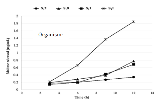
In preliminary screening process, growths were observed at a temperature range between 40 °C-60 °C in the starch nutrient broth. Altogether ten (10) different types of bacteria were isolated and four of them were selected for further studies (Figure 1). Isolate S51 produced 1.85 mg/mL of reducing sugars (as maltose) in 12 hours which was the highest among selected organisms. Results given in figure 2.
Figure 2: Starch Utilization Analysis by DNS Method
The organism S51 was isolated from a water sample obtained from Mahaoya hot water spring in Polonnaruwa district of Sri Lanka (N 7.55221, E 81.35302). During the sample collection the recorded temperature and pH were 57ºC and 7.2 respectively.
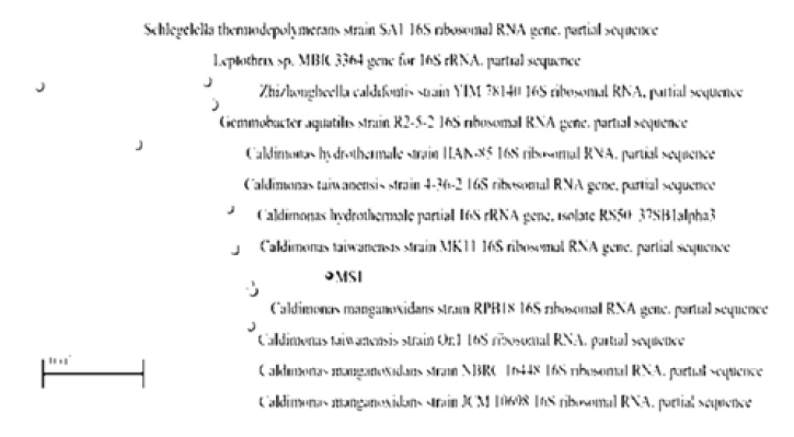
Comparison of almost full-length sequence of 16S rRNA gene (1498 bp) from S51 with available 16S rRNA sequences in GenBank revealed, S51 is a member of Comamonadaceae family of Burkholderiales order which comes under β-proteobacteria and showed identities of 98% to Caldimonas manganoxidans and Caldimonas taiwanensis, 97% to Caldimonas hydrothermale and Leptothrix sp. and 95% to Zhizhongheella caldifontis. The organism was named as Caldimonas manganoxidans MS1 and the assembled partial sequence of 16S rRNA gene was deposited to NCBI GenBank under the accession number MZ562348. The phylogenetic tree showing the position of Caldimonas manganoxidans MS1 within α and β proteobacteria given in Figure 3.
Figure 3: Neighbor Joining Phylogenetic Tree Showing the Position of Caldimonas Manganoxidans MS1 with Max Seq Difference of 0.75
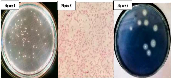
The Caldimonas manganoxidans MS1 colonies were circular, convex, small, creamy white and non-pigmented (Figure 4). In broth, no sheath was formed, and granular cell clumps were present. Biochemical characterization of S51 revealed it was a Gram-negative rod (Figure 5) which showed optimum growth at 50 °C and pH 6.9.
Figure 4: Caldimonas Manganoxidans MS1 (S51) Colonies on Starch Nutrient Agar after 24 Hours of Incubation at 50ºc and Ph 6.9
Figure 6: Caldimonas Manganoxidans MS1 (S51) Iodine Test after 24 Hours of Incubation at 50ºc and Ph 6.9
Based on preliminary screening (Figure 1), being Gram-negative and thermostable while effectively producing α-amylase (Figure 2, Figure 6) drove us to investigate the organism S51 (Caldimonas manganoxidans MS1). The isolated Caldimonas manganoxidans MS1 belong to moderate thermophiles. Organisms that grow optimally between 50 °C and 60 °C are classified as moderate thermophiles whereas extreme thermophiles show optimum growth between 60 °C and 80 °C and are widely distributed among the genera such as Bacillus, Clostridium, Thermoanaerobacter, Thermus, Fervidobacterium, Thermotoga and Aquifex. Many hyperthermophiles grow optimally between 80 °C and 110 °C such as Pyrococcus [18].
When comparing the biochemical properties given in table 1 to other Caldimonas spp., Takeda et al. [19] reported Caldimonas manganoxidans HS isolated from a hot water spring in Japan was able to utilize maltose, sucrose, D-glucose, citrate, and mannitol. It was not able to utilize myo-inositol, D-arabinose, D-mannose, lactose, L-rhamnose, raffinose, D-xylose, trehalose or D-salicin and was able to grow optimally at 50°C and pH 7-8.
Another Similar organism Caldimonas taiwanensis On1 was able to utilize glucose, mannitol, maltose, citrate, gelatin, D-trehalose. Indole positive, catalase positive and was oxidase negative and could not utilize, aesculin, arabinose, mannose, inositol, lactose, D-raffinose, D-rhamnose, sucrose, phenylalanine, arginine, alanine, and urea. They showed optimum growth at 55 °C and pH 7 [20].
Another closely related species, Caldimonas hydrothermale was reported by Bouraoui et al. [21] from a public thermal bath in south Tunisia. They showed positive reaction for aesculin hydrolysis, nitrate reduction, fermentation of D-maltose, D-cellobiose, mannose, lactose, D-raffinose, L-rhamnose, D-salicin, D-xylose, D-glucose, trehalose, and sucrose. Negative reaction was observed for urease, citrate utilization, fermentation of arabinose, L- xylose, inositol, inulin and dulcitol. They showed no gelatin liquefaction and no indole production. They Showed 3% salt tolerance. Catalase and oxidase tests were positive. No decarboxylation was recorded on lysine, arginine, and ornithine. Optimum growth was observed at 55 °C and pH 7. Based on available evidence we suggest the Caldimonas manganoxidans MS1 variant possesses distinctive biochemical properties to other closely related members of Caldimonas genus.
Culture Optimization and α-Amylase Production from Caldimonas Manganoxidans MS1
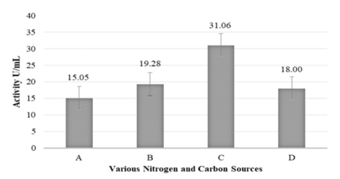
The organism showed production of 15 U/mL at 50 °C after 18 hours when modified starch nutrient broth was used with 1% starch. The α-amylase production was enhanced to 31 U/mL at 50 °C after 18 hours when 1% (w/v) soybean flour and 1% (w/v) starch were used (Figure 7).
Figure 7: Media Optimization Using (A) Peptone+Yeast Extract as Nitrogen Source and 1% Starch as Carbon Source (B) Peptone+Yeast Extract as Nitrogen Source and 2% Starch as Carbon Source (C) 10% Soybean Flour as Nitrogen Source and 1% Starch as Carbon Source (D) 10% Soybean Flour as Nitrogen Source and 2% Starch as Carbon Source
Temperature is a key factor for growth and enzyme production. In this study the effect of temperature was studied by culturing the organism at a range between 40-70 °C where at 70 °C no bacterial growth was observed. The highest cell count of 18x106 was observed at 50 °C while the cell counts at 40 °C and 60 °C were 9.3x10⁶ and 3.7x10⁶ respectively.
Growth and enzyme production declined after increasing the temperature; this can be because of decreased solubility of oxygen at high temperatures [22]. The decreased activity in the later phase of growth could be due to catabolite repression by glucose released from starch hydrolysis and proteolysis of α-amylase [23].
In this study we have used soybean flour to enhance the α- amylase production which resulted in 106% increase compared to the 15 U/mL activity achieved where peptone, yeast extract and 1% starch (modified starch nutrient broth) was used. Meanwhile, 2% starch showed a slight decrease in enzyme production. This may be due to higher substrate concentration leads to feed forward inhibition of enzyme production (Figure 6). Similarly, soybean meal has shown increased amylase production in Bacillus sp. according to Lasa and Berenguer [1]. In the study by Alrumman et al. [23] the enzyme production of Bacillus axarquiensis reached a maximum of 64.5 U/mL with the potato wastewater containing 0.5% starch, 0.4% yeast extract, 0.04% CaCl2.H2O and 0.05% KH2PO4. The optimization of the potato wastewater medium with addition of starch, nitrogen, phosphate, and calcium led to approximately a 4-fold increase in the production of α-amylase compared to starch broth medium. This suggests that use of agriculture wastes provides a good, cheap substrate for α- amylase production over costly synthetic nutrient media which is economically efficient.
Submerged fermentation we have used provides greater opportunity for enzyme production by allowing greater control over the temperature, nutrient contents and oxygen level by aeration and agitation than solid state fermentation [24].
Caldimonas manganoxidans MS1 showed a low intracellular α-amylase activity. Intracellular α-amylase have been reported in Streptococcus bovisand Escherichia coli[25-26]. Although the role of intracellular α-amylases are not clear, Brooker & McCarthy [27] suggested that it plays a key role in rapid cell growth.
Caldimonas manganoxidans MS1 produces extracellular α-amylase. Culturing on starch nutrient broth supplemented with salt solution, the culture supernatant showed an extracellular activity of 13 U/mL with a specific activity of 180 U/mg and the cell pellet after sonication showed an intracellular activity of 4.4 U/mL with a specific activity of 9 U/mg. Oxygen availability studies revealed that culture in 500mL Erlenmeyer flask produced 31 U/mL while 250 mL flask produced only 23 U/mL of enzyme suggesting higher enzyme production when more was oxygen available.
Purification and Characterization of MS1 α-Amylase
Ammonium Sulphate Fractionation: During the preliminary studies, 40% saturated precipitate gave the highest α-amylase activity while 90% saturated precipitate showed the highest specific activity. Both saturations were used for further purification. Throughout all the fractions 20-90% the pellet collected showed α- amylase activity. This may be because the media used for fermentation contained 10% soybean flour which contains considerable amounts of proteins which could possibly interfere with the precipitation of the target protein. In this case we purified two samples of bacterial supernatants, one saturated up to 40% and the other up to 90%. In both cases we obtained a significant amount of enzyme. There is also a possibility that the Caldimonas manganoxidans MS1 possess isoforms of α- amylase. Numerous studies suggest that ammonium sulphate precipitation can occur at different saturations depending on the enzyme. Bano et al. [28] purified α-amylase from Bacillus subtilis KIBGE-HAS at 40% saturation. Streptomyces aureofaciens 77 amylase was precipitated upon 50-70% saturation which gave the maximum yield[29]. Moreover Geobacillus sp. NMS2 α- amylase has been purified at 90% saturation [10].
Cellulose Acetate Paper Electrophoresis
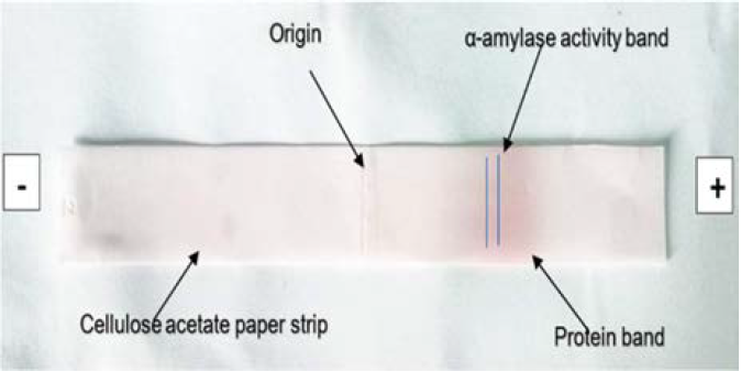
MS1 α-amylase is negatively charged at pH 6.9 hence the protein band moved towards the anode. The α-amylase band was located around 3-3.5cm from the origin (Figure 8).
Figure 8: Cellulose Acetate Electrophoresis
DEAE-Sephadex A-25 Ion-exchange Chromatography
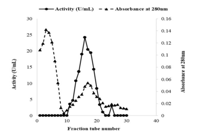
The purified MS1 α-amylase showed an activity of 19 U/mL with a specific activity of 467 U/mg with 18-fold purification and the recovery was 49%. Due to the α- amylase being negatively charged, an anion exchange column, DEAE-Sephadex A-25 was selected for further purification. From the pooled fractions obtained by DEAE-Sephadex A-25 chromatography (Figure 9), the homogeneity was confirmed by a single band in native polyacrylamide gel electrophoresis (Figure 10).
Figure 9: Α-Amylase Activity and Absorbance at 280nm of Column Eluted Fractions
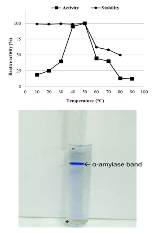
Figure 10: Single Protein Band Seen in Native Polyacrylamide Gel Electrophoresis Confirms the Homogeneity of Purified MS1 Α-Amylase
In comparison, different techniques have been used by researchers to purify α-amylases from crude samples. Purification of extracellular α-amylase from Bacillus subtilis KIBGE HAS was carried out by ultrafiltration, ammonium sulphate precipitation and gel filtration chromatography and the enzyme was purified to homogeneity with a purification fold of 96.3 with specific activity of 13011 U/mg [28]. The Bacillus methylotrophicus strain P11-2 α-amylase was purified through DEAE-FF anion exchange, and Superdex 75 10/300 GL gel filtration chromatography. The purified α-amylase showed a specific activity of 330.7 U/mg that corresponds to 13.1-fold purification [30].
Native Polyacrylamide Gel Electrophoresis (PAGE)
The gel showed a sharp single band 1.0 cm from the origin which indicated that the enzyme was homogenously purified. The mobility (Rf ) of the protein band was 0.286.
Effect of Temperature on MS1 α-amylase Activity
Temperature is a crucial factor for enzyme stability. Enzymes show maximum activity at its optimum temperature. The purified α-amylase of Caldimonas manganoxidans MS1 during the study showed an optimum activity at 50 °C and pH 6.9. Activity increased with the temperature up to 50 °C and then declined with further increases. Thermal stability is important in certain industries where the enzyme must retain its activity even in elevated temperatures that are used in different processes. The MS1 α-amylase is highly stable over the range of 10 °C -50 °C, where the relative activities were more than 98%, moreover the enzyme retained 62% of its activity at 60°C and 49% of its activity even at 80 °C after incubation for one hour. Studies on Bacillus licheniformis by Vaseekaran et al. [31] showed the enzyme retained 69% of its activity at 80 °C after 30minutes and 59% after one hour. Similarly, Geobacillus sp. IIPTN is stable over a range of 80 °C -120 °C [32]. Even though the optimum temperature is lower than certain Bacillus spp, The MS1 enzyme is highly stable over a range of temperatures.
Effect of pH on MS1 α-amylase Activity
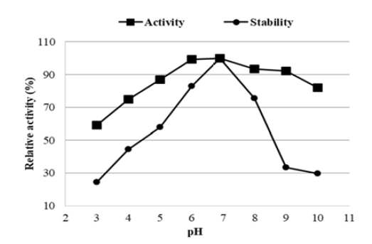
Activity profile at different pH of the purified MS1 α-amylase revealed that the enzyme is highly stable in the range of pH between 5-9 where the relative activities were within 87- 92% with the optimum at pH 6.9. Enzyme’s pH stability profile revealed the enzyme to be highly stable at pH 6.9 and retained its 58%, 83%, 75% of activities at pH 5, 6 and 8 respectively after one hour of incubation. At pH 3, 9 and 10 relative activities were reduced to 24%, 33% and 30% respectively (Figure 11). The study suggests that the enzyme is suitable to be used in the pH range between 6-8. Another similar species Caldimonas taiwanensis an amylolytic bacteria showed an optimum activity at pH 7 [20]. Different studies have reported that most of the Bacillus spp. α-amylases have shown maximum activity in an acidic to neutral range [33]. Bacillus licheniformis EMS6 α- amylase isolated by Adnan [34] was active in the range of pH 4.5-9 with the optimum at pH 7. Bacillus sp. WA21 revealed that enzyme α-amylase retained more than half of its activity at 85ºC and between pH 5-9 [4].
Figure 11: Effect of Ph on MS1 Α-Amylase at 50 ºc
Effect of Metal Ions, Surfactants, Chelating Agents, and Bleaching Agents on MS1 α-amylase Activity
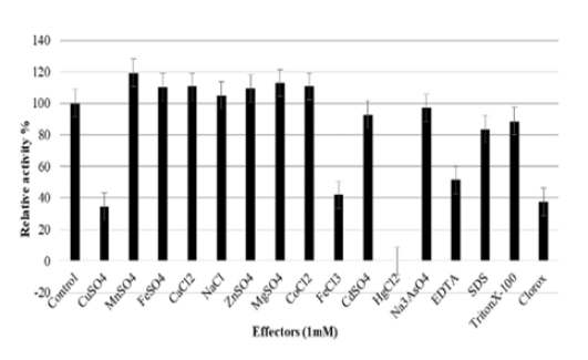
In our study the enzyme activity was enhanced in the presence of Ca2+, Mg2+, Mn2+, Fe2+, Co2+, Zn2+ and Na+ whileFe3+, Cu2+, AsO43- Cd2+ and Hg2+ inhibited the enzyme activity at 1mM concentration (Figure 12). Presence of Hg2+ led to complete inactivation of the enzyme. It has been reported that Hg, Au, and Ag bind to amino acids Cys or His, Pt binds to Met or His and Pb and lanthanides bind to Asp or Glu[35]. Presence of Mn2+ showed the highest relative activity of 119% where in the presence of Cu2+, Fe3+ it showed a relative activity of 34.5% and 42% respectively. Similarly, Halobacillus sp. MA-2 amylasewas inhibited by Cd2+ and Cu2+ [36]. Zn2+ showed inhibitory activity in Bacillus sp. ANT-6, Inhibitions by Fe3+ were observed in Bacillus subtilis 65, Bacillus sp. WN11 and in E. coli. Bacillus subtilis 65 enzymes are also inhibited by Cu2+, Mn2+, Zn2+ andCd2+ [37-39] In most research studies Mn2+ and Zn2+ inhibit the enzyme, In the study by Arikan [40] Bacillus sp. A3-15 α-amylase was inhibited by ZnCl2 and NaCl. But in contrast, Mn2+, Zn2+ and Na+ showed to enhance the activity of MS1 α-amylase at 1mM concentrations.
α-Amylases are metalloenzymes. Binding of ligands such as metal ions increase the stability according to Schellman’s binding theory, where high affinity binding of ligands shifts the folding-unfolding equilibrium towards the folded state[35]. Most of the amylases have Ca2+in their structure. According to Deb et al. [41] Bacillus amyloliquefaciens P-001 amylase activity was enhanced by Ca2+, Mg2+ and Fe2+ while Cu2+ and Mn2+inhibited the enzyme. Similarly, Bacillus cereus MS6 α -amylase was enhanced by Ca2+ and Mg2+ [42]. In Aspergillus oryzae EI 212 it was reported that Ca2+ inhibited the activity while EDTA treatment did not affect the activity[43]. In Bacillus sp. RM16, Hassan et al. [44] reported that the enzyme was calcium independent. Interestingly with MS 1 α-amylase, the enhanced enzyme activity with Mn2+ may be due to organism itself being able to metabolize Manganese. With regards to increased activity in presence of both Mn2+ and Zn2+, we suggest since both ions are di-valent they have been utilized as enzyme co-factors which helped to enhance the activity. This is a unique feature of our MS1 α- amylase whereas in most cases Mn2+ and Zn2+ inhibited α -amylase activity.
Figure 12: Effect of Metal Ions, Surfactants, Chelating Agents, And Bleaching Agents On MS1 Α-Amylase at 50 ºc and Ph 6.9
Chelating agents like EDTA and surfactants such as TritonX-100 and SDS also inhibited the activity of the enzyme in our study. In the presence of EDTA the relative activity was 51.5% and with SDS and TritonX-100 the enzyme retained 83% and 88% of its initial activity respectively after 1 hour. Addition of Clorox which is a commercial solution of NaOCl (Sodium hypochlorite) reduced the relative activity to 37%. Chelating agents remove metal ions from the structure leading to instability [45].Removal of calcium from specific ion-binding sites in the protein structures reduced the melting temperatures tremendously for Bacillus amyloliquifaciens and Bacillus licheniformis and removal of Calcium leads to increased susceptibility to proteolytic degradation [46-47].
Kinetic studies of MS1 α-amylase
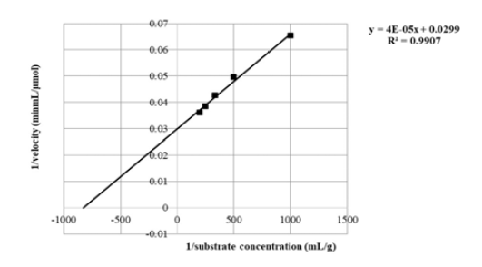
Km and Vmax values for Caldimonas manganoxidans MS1 α- amylase obtained through Lineweaver–Burke (Figure 13), Eadie-Hofstee and Hanes-Woolf plots were 1.33mg/mL and 33 μmol/min/mL; 1.30 mg/ml and 34 μmol/min/mL; 1.4mg/ml and 35 μmol/min/mL respectively.
Figure 13: Lineweaver -Burke Plot for MS1 Α-Amylase
In comparison to Km and Vmax values for Caldimonas manganoxidans MS1 (Figure 12), characterization of extracellular α-amylase of Bacillus subtilis KIBGE-HAS showed that the enzyme had a Km and Vmax value of 2.68 mg/mL and 1773 U/mL, respectively[28]. According to Talekar and Patil [48], Bacillus strearothermophilus NCIM 2922 showed a Km and Vmax of 1.7mg/mL and 112.8 μmol/min respectively. The lower of Km of Caldimonas manganoxidans MS1 suggests higher affinity towards soluble starch substrate. Having a lower Km and Vmax compared to above mentioned Bacillus spp. suggest the Caldimonas manganoxidans MS1 α-amylase is more efficient.
CONCLUSION
The biochemical properties suggest that the organism Caldimonas manganoxidans MS1 identified in this study is a novel variant of existing strains of Caldimonas manganoxidans and MS1extracellular α-amylase showed novel functional properties in contrast to already studied similar enzymes. The broad thermostability and stability at a wide pH range is an advantage for various applications. The enzyme activity was affected by surfactants like SDS and TritonX-100 to a lower extent, which implies that the enzyme is suitable for the detergent industry. The enzyme shows high specificity to soluble potato starch. The enzyme activity was enhanced by Mn2+, Ca2+, Mg2+, Fe2+, Na+, Co2+ and Zn2+and inhibited by Hg2+, Cu2+, Fe3+, Cd2+ and AsO43-. The functional characterization of the enzyme suggested the enzyme has novel properties compared to already studied α -amylases. Further optimization of metal ion concentrations to obtain maximum activity will be useful for industrial applications.
The production level of extracellular metabolites like enzymes in most of the hyperthermophiles seems to be too low to be used directly in industrial applications. Hence, molecular cloning of corresponding genes in mesophilic hosts can be used to overcome this problem for large scale enzyme production. The enzyme could be immobilized by various methods for repeated use. Protein engineering can be used to modify this protein to increase the thermostability and the pH tolerance, which will be useful for industrial applications.
Acknowledgment
Authors would like to acknowledge the financial assistance from the Department of Biochemistry and Molecular Biology of Faculty of Medicine, University of Colombo and support from the academic and non-academic staff.
REFERENCE
Lasa, I. and J. Berenguer. “Thermophilic enzymes and their biotechnological potential.” Microbiologia, vol. 9, no. 2, 1993, pp. 77–89.
Li, W.F. et al. “Structural features of thermozymes.” Biotechnology Advances, vol. 23, no. 4, 2005, pp. 271–281.
Gupta, R. et al. “Microbial α-amylases: A biotechnological perspective.” Process Biochemistry, vol. 38, no. 11, 2003, pp. 1599–1616.
Asad, W. et al. “Extracellular enzyme production by indigenous thermophilic bacteria: Partial purification and characterization of α-amylase by Bacillus sp. WA21.” Pakistan Journal of Botany, vol. 43, no. 2, 2011, pp. 1045–1052.
Souza, P.M.D. and P.D.O.E. Magalhães. “Application of microbial α-amylase in industry: A review.” Brazilian Journal of Microbiology, vol. 41, no. 4, 2010, pp. 850–861.
El-Banna, T.E. et al. “Production and immobilization of alpha-amylase from Bacillus subtilis.” Pakistan Journal of Biological Sciences, vol. 10, no. 12, 2007, pp. 2039–2047.
El-Banna, T. et al. “Optimization and immobilization of α-amylase from Bacillus licheniformis.” Catrina: The International Journal of Environmental Sciences, vol. 3, no. 1, 2008, pp. 45–53.
El-Fallal, A. et al. “Starch and microbial α-amylases: From concepts to biotechnological applications.” Carbohydrates: Comprehensive Studies on Glycobiology and Glycotechnology, 2012, pp. 459–488.
Sivaramakrishnan, S. et al. “α-Amylases from microbial sources: an overview on recent developments.” Food Technology and Biotechnology, vol. 44, no. 2, 2006, pp. 173–184.
Mathew, C.D. and S. Rathnayake. “Isolation and characterization of alpha amylase isolated from a hot water spring in Sri Lanka.” International Journal of Microbiological Research, vol. 5, no. 4, 2014, pp. 50–61.
Kaur, P. and A. Vyas. “Characterization and optimal production of alkaline-amylase from Bacillus sp. DLB 9.” African Journal of Microbiology Research, vol. 6, no. 11, 2012, pp. 2674–2681.
Bernfeld, P. “Amylases, alpha and beta.” Methods in Enzymology, vol. 1, 1955, pp. 149–158.
Hall, T. “BioEdit: A user-friendly biological sequence alignment editor and analysis program for Windows 95/98/NT.” Nucleic Acids Symposium Series, vol. 41, 1999, pp. 95–98.
Zhang, Z. et al. “A greedy algorithm for aligning DNA sequences.” Journal of Computational Biology, vol. 7, nos. 1–2, 2000, pp. 203–214.
Altschul, S.F. et al. “Basic local alignment search tool.” Journal of Molecular Biology, vol. 215, no. 3, 1990, pp. 403–410.
Barrow, G. and R.K.A. Feltham, editors. Cowan and Steel’s manual for the identification of medical bacteria. Cambridge University Press, 2004.
Lowry, O. et al. “Protein measurement with the Folin phenol reagent.” Journal of Biological Chemistry, vol. 193, no. 1, 1951, pp. 265–275.
Bertoldo, C. and G. Antranikian. “Starch-hydrolyzing enzymes from thermophilic archaea and bacteria.” Current Opinion in Chemical Biology, vol. 6, no. 2, 2002, pp. 151–160.
Takeda, M. et al. “Caldimonas manganoxidans gen. nov., sp. nov., a poly (3-hydroxybutyrate)-degrading, manganese-oxidizing thermophile.” International Journal of Systematic and Evolutionary Microbiology, vol. 52, no. 3, 2002, pp. 895–900.
Chen, W.M. et al. “Caldimonas taiwanensis sp. nov., an amylase-producing bacterium isolated from a hot spring.” Systematic and Applied Microbiology, vol. 28, no. 5, 2005, pp. 415–420.
Bouraoui, H. et al. “Caldimonas hydrothermale sp. nov., a novel thermophilic bacterium isolated from a Roman hot bath in south Tunisia.” Archives of Microbiology, vol. 192, no. 6, 2010, pp. 485–491.
Campbell, L.L. and B. Pace. “Physiology of growth at high temperatures.” Journal of Applied Bacteriology, vol. 31, no. 1, 1968, pp. 24–35.
Alrumman, S.A. et al. “Isolation of thermoalkalophilic α-amylase-producing bacteria and optimization of potato wastewater medium.” Advances in Life Science and Technology, vol. 20, 2014, pp. 41–51.
Renge, V.C. et al. “Enzyme synthesis by fermentation method: a review.” Scientific Reviews and Chemical Communications, vol. 2, no. 4, 2012, pp. 585–590.
Satoh, E. et al. “Purification, characterization, and nucleotide sequence of an intracellular maltotriose-producing alpha-amylase from Streptococcus bovis 148.” Applied and Environmental Microbiology, vol. 63, no. 12, 1997, pp. 4941–4944.
Whitehead, T.R. and M.A. Cotta. “Identification of intracellular amylase activity in Streptococcus bovis and Streptococcus salivarius.” Current Microbiology, vol. 30, no. 3, 1995, pp. 143–148.
Brooker, J.D. and J.M. McCarthy. “Gene knockout of the intracellular amylase gene by homologous recombination in Streptococcus bovis.” Current Microbiology, vol. 35, no. 3, 1997, pp. 133–138.
Bano, S. et al. “Purification and characterization of novel α-amylase from Bacillus subtilis KIBGE HAS.” AAPS PharmSciTech, vol. 12, no. 1, 2011, pp. 255–261.
Ibrahim, A.N. et al. “Precipitation and purification of amylase enzyme produced by Streptomyces aureofaciens 77.” Archives of Pharmacal Research, vol. 13, no. 1, 1990, pp. 28–32.
Xie, F. et al. “Purification and characterization of a novel α-amylase from Bacillus methylotrophicus strain P11-2.” Process Biochemistry, vol. 49, no. 1, 2014, pp. 47–53.
Vaseekaran, S. et al. “Isolation and identification of a bacterial strain producing thermostable α-amylase.” Tropical Agricultural Research, vol. 22, no. 1, 2011, pp. 1–11.
Dheeran, P. et al. “Characterization of hyperthermostable α-amylase from Geobacillus sp. IIPTN.” Applied Microbiology and Biotechnology, vol. 86, no. 6, 2010, pp. 1857–1866.
Krishnan, T. and A.K. Chandra. “Purification and characterization of α-amylase from Bacillus licheniformis CUMC305.” Applied and Environmental Microbiology, vol. 46, no. 2, 1983, pp. 430–437.
Adnan, F. “Kinetics and thermodynamic studies of alpha amylase from Bacillus licheniformis mutant.” Pakistan Journal of Botany, vol. 42, no. 5, 2010, pp. 3507–3516.
Sugahara, M. et al. “Effect of heavy atoms on the thermal stability of α-amylase from Aspergillus oryzae.” PLoS One, vol. 8, no. 2, 2013, e57432.
Amoozegar, M.A. et al. “Production of amylase by newly isolated moderate halophile Halobacillus sp. strain MA-2.” Journal of Microbiological Methods, vol. 52, no. 3, 2003, pp. 353–359.
Burhan, A. et al. “Enzymatic properties of a novel thermostable, thermophilic, alkaline and chelator-resistant amylase from an alkaliphilic Bacillus sp.” Process Biochemistry, vol. 38, no. 10, 2003, pp. 1397–1403.
Hayashida, S. et al. “Production and characteristics of raw-potato-starch-digesting α-amylase from Bacillus subtilis 65.” Applied and Environmental Microbiology, vol. 54, no. 6, 1988, pp. 1516–1522.
Mamo, G. et al. “A highly thermostable amylase from a newly isolated thermophilic Bacillus sp. WN11.” Journal of Applied Microbiology, vol. 86, no. 4, 1999, pp. 557–560.
Arikan, B. “Highly thermostable, thermophilic, alkaline, SDS- and chelator-resistant amylase from a thermophilic Bacillus sp.” Bioresource Technology, vol. 99, no. 8, 2008, pp. 3071–3076.
Deb, P. et al. “Production and partial characterization of extracellular amylase enzyme from Bacillus amyloliquefaciens P-001.” SpringerPlus, vol. 2, no. 1, 2013, pp. 1–12.
Mohammed, M.A.A.Z. et al. “Identification and characterization of novel halophilic Bacillus cereus Ms6.” Advances in Environmental Biology, vol. 5, no. 5, 2011, pp. 992–999.
Kundu, A.K. and S. Das. “Production of amylase in liquid culture by a strain of Aspergillus oryzae.” Applied Microbiology, vol. 19, no. 4, 1970, pp. 598–603.
Hassan, S.A. et al. “Purification and biochemical characterization of a Ca²⁺-independent, thermostable and acidophilic α-amylase.” African Journal of Biotechnology, vol. 10, no. 32, 2011, pp. 6082–6089.
Riordan, J. “The role of metals in enzyme activity.” Annals of Clinical & Laboratory Science, vol. 7, no. 2, 1977, pp. 119–129.
Fitter, J. and S. Haber-Pohlmeier. “Structural stability and unfolding properties of thermostable bacterial α-amylases.” Biochemistry, vol. 43, no. 30, 2004, pp. 9589–9599.
Machius, M. et al. “Crystal structure of calcium-depleted Bacillus licheniformis α-amylase at 2.2 Å resolution.” Journal of Molecular Biology, vol. 246, no. 4, 1995, pp. 545–559.
Talekar, S. and J. Patil. “Production and characterization of thermostable alpha amylase by Bacillus stearothermophilus NCIM 2922.” Journal of Cell and Tissue Research, vol. 12, no. 1, 2012, pp. 3037–3042.
Recommended Articles
Research Article
Serum IL-22, miRNA-146a Expression and Total Antioxidant Status as Integrated Biomarkers in Rheumatoid Arthritis
Khalid R. Kareem
Published: 13/08/2026
Download PDF
Research Article
Electrochemical Comparative Studies On The Redox Behavior Of Cu(II) And Cr(II) Before And After Interactions With Ciprofloxacin In Solutions
H. M. Naseem Akhtar,
M. Q. Ehsan
Published: 10/08/2021
Download PDF
Research Article
A Rare Case Report of Waugh Syndrome in an Eight Months Old Male
Pielly Hanna,
...
Bassem Abou Merhi
Published: 10/03/2021
Download PDF
Research Article
Study on Prevalence and Etiology of Early Neonatal Deaths in Jasin district, Melaka from 2014-2017
Sujat a Khobragade,
...
Yadneshwar Khobragade
Published: 30/05/2021
Download PDF
PO Box 101, Nakuru
Kenya.
Email: office@iarconsortium.org
Editorial Office:
J.L Bhavan, Near Radison Blu Hotel,
Jalukbari, Guwahati-India
Farook, R. S. M., Witharana, C. & Mathew, D. (2022). A Novel Thermostable Alpha Amylase Producing Caldimonas Variant From A Hot Water Spring In Sri Lanka. International Academic Journal of Applied Bio-Medical Sciences, 3(2), 1-8.
MLA
Farook, Raushan S. M., Chamindri Witharana and Deepal Mathew. "A Novel Thermostable Alpha Amylase Producing Caldimonas Variant From A Hot Water Spring In Sri Lanka." International Academic Journal of Applied Bio-Medical Sciences 3.2 (2022): 1-8.
Chicago
Farook, Raushan S. M., Chamindri Witharana and Deepal Mathew. "A Novel Thermostable Alpha Amylase Producing Caldimonas Variant From A Hot Water Spring In Sri Lanka." International Academic Journal of Applied Bio-Medical Sciences 3, no. 2 (2022): 1-8.
Harvard
Farook, R. S. M., Witharana, C. and Mathew, D. (2022) 'A Novel Thermostable Alpha Amylase Producing Caldimonas Variant From A Hot Water Spring In Sri Lanka' International Academic Journal of Applied Bio-Medical Sciences 3(2), pp. 1-8.
Vancouver
Farook RSM, Witharana C, Mathew D. A Novel Thermostable Alpha Amylase Producing Caldimonas Variant From A Hot Water Spring In Sri Lanka. International Academic Journal of Applied Bio-Medical Sciences. 2022 Jul;3(2):1-8.


 Download PDF
 Download XML