This study aimed to the identification of Fifty isolates of Iraqi Carp fish (Cyprinus carpio) collected from the ponds and farms in different locations of Karbala governorate during the summer of 2021 from June to September. Biochemical identification was completed by using the VITEK 2 compact system, depending on several biochemical and physiological test responses. Six groups of rats (6 rats per group) were used to determine the lethal dose of 50 (LD50). All rats infected (virulent) group were injected with (8.4 ⤫ 108 CFU/ml) of virulent Aeromonas veronii. The pathogenesis study showed a significant increase in the experimental (Virulent) group compared with the control group. All rats in the Virulent group suffered from severe clinical signs of diseases and died within 3-4 days. Post mortem examination & bacterial isolation after injection it general congestion and hyperemia of multiple organs, the small intestine mildly congested and hemorrhagic areas, Organs collected were culturally positive for A. vironii.
Keywords
Aeromonas Veronii
Identification
Pathogenesis
Rats
INTRODUCTION
Aeromonas are gram-negative, facultative anaerobic bacterium. These bacteria have been isolated from different aquatic environments such as freshwater, wastewater, marines, and lakes [1]. Aeromonas were studied for a long time before as an important part of microbiology, but not so long ago it has been detected as the main group of pathogens that cause disease for both animals and humans [2-3].
Fish are generally indirectly linked with surrounding aquatic conditions; hence, they are always affected by different risks such as the infections of microbes, especially, the opportunistic pathogen Aeromonas bacteria [4]. The carp fish (Cyprinus carpio) are usually exposed to bacteria invasion, especially Aeromonas. The interaction of this fish with Aeromonas is highly critical under the stress status, which includes human involvement in fishing, sorting, and carrying of fish as well as the aquatic conditions [5]. Aeromonads septicemia which is caused by pathogenic species of Aeromonas is a critical fish illness, which sorely affects the aquaculture of the world [6-7].
Carp fish considers an important transmits of this bacteria to humans [8]. Many species of these bacteria such as A. Hydrophilia and A.veronii are related to cases of sepsis, wound infections, and enteritis in humans, and are causing a higher risk for immunocompromised persons [9]. Previous studies show that there are different factors involved in the pathogenicity of this bacteria such as secretion systems, extracellular enzymes, toxins, iron-binding systems, surface polysaccharides, adhesion, and motility [10-13].
MATERIALS AND METHODS
The bacteria were isolated from the fish carp (Cyprinus carpio) and were collected from ponds and farms located in the Euphrates river, Karbala, Iraq. A total number (of 40) carp fish were collected randomly from these farms with an average weight (of 500 to 1250)g during summer 2021 from June to September. The samples (skin, gills, and viscera) were bundled in sterilized bags and moved promptly in a cooled case with sterile conditions to the microbiology lab of the college of veterinary medicine, Al-qasim Green University in order to dissect and study their morphological characteristics of them, and then they were transferred to the Public Health Laboratory in Karbala city for bacterial isolation and identification.
Isolation of bacteria was done based on the general principles of culturing by using a spread plating procedure. The samples were inoculated on the nutrient agar medium by spreading technique [14] at 37°C for 48 hr [15]. Biochemical identification of the isolate A. veronii was completed by using the VITEK 2 compact system, which depends on 62 biochemical and physiological test responses. The VITEK 2 compact system is a completely computerized process that gives bacterial detection via biochemical examination by using colorimetry. This system is doing all the processes that are needed for this microbe identification and permits dynamic examination by perusing every test in 15 min [16].
Aeromonas veronii bacteria were inoculated in Brain heart infusion broth at 37 Cº for 24 hrs. The suspension was washed and centrifuged for 15 min under sterilized conditions. The bacteria were re-suspended in the phosphate buffer saline (PBS) with pH 7.2. A series with a dilution factor of 10 (Tenfold dilution) were done.
Bacterial plate count is the method used to count A. veroni in each diluent to find out the viable A. veronii [17]. 0.1ml of diluent was put on the agar surface and spread by a sterile glass rod, then incubated at 37C for 24hrs. The colonies were counted and viable A. veronii has been determined and the diluents were selected, which have these concentrations for injecting the rats intraperitoneally 2x101 C.F.U/ml, 2x10² C.F.U/ml, 2x10³ C.F.U/ml, 2x10⁴ C.F.U/ml, 2x10⁵ C.F.U/ml, 2x10⁶ C.F.U/ml.
28 Both male rats were selected and divided into 7 groups, each group containing 4 rats and adapting for 2 weeks before starting the experiment. The weight of each selected rat was 180 to 220g. The first six groups of these seven rats groups were injected with 0.5 mL of diluents intraperitoneally and the latest group was considered a control group. The LD50 was estimated according to [18].
Sixteen rats were selected with a weight of 180 to 220g and randomly divided into two groups (n = 8), which were the virulent group that was injected with live bacterial strain, and the control group that was injected by PBS.
Statistical Analysis
Statistical analyses were carried out by using SPSS 17.0. Turkey's tests were used for performed multiple comparisons between treatments. Differences were counted to be statistically significant when p<0.05 by analysis of variance (ANOVA).
RESULTS
Six isolates were suspected to be related to Aeromonas among the fifty obtained isolates (Table 1).
Table 1: Bacterial Isolates From Carp Fish
Site
No. of sample
No. of isolating
No. of Aeromonas. spp
Skin
40
19 = 47.5%
3 = 15.7%
Gills
15 = 37.5%
1 = 6.6%
Viscera
16 = 40%
2 = 12.5%
Total
50
6 = 12%
These isolates were identified depending on their cultural, morphological characteristics, and biochemical identification.
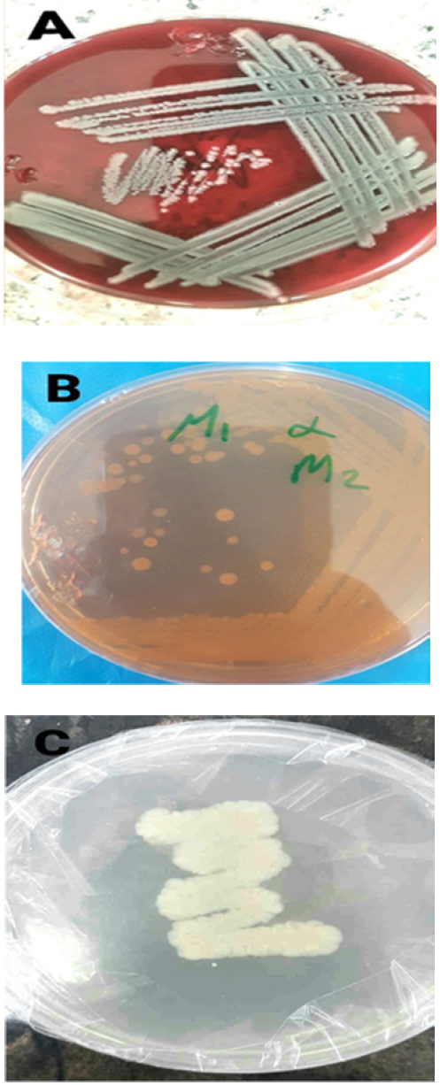
The colonies that appeared on the Blood agar are (1-3mm) in diameter, large, convex, smooth, rounded, and β- hemolytic colonies which gave their grey color (Figure 1A), while on Macconkey agar, the colonies of Aeromonas isolates are colorless and non-lactose fermenters (Figure 1B). The colonies on nutrient agar were creamy white color (Figure 1C).
Figure 1: A.Veronii Appeared Smooth, Convex, Rounded Colonies: A- Β-Hemolytic Colonies Which Showed a Grey Color on Blood Agar. B- The Colonies of A.Veronii Are Colorless and Non-Lactose Fermenters on Macconkey Agar. C- Creamy White Color Colonies Observed on Nutrient Agar
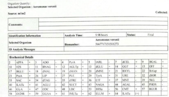
Biochemical identification was completed by using the VITEK 2 compact system, which depends on several biochemical and physiological test responses, resulted in Aeromonas veronii .
The result of Vitek2 compact system has showed the bio-number (5667717151501273) as affirmed diagnosis for A. veronii isolate (Figure 2)
Figure (2): Calculate the bio-number in Vitek 2 compact system (+): the test Positive. (-): the Test Negative. (5667717151501273): the Bio-number
Ten hours after the injection of the virulent bacteria group, symptoms of tiredness, nausea, trembling, lack of appetite, irregular movement and complete paralysis of the hind limbs were observed. These included decreased infected rats' diet, weary attitude, and depression. The rats of the infected group suffered from other patho-clinical signs in the first five days following injection, in which all rats exhibited listlessness, huddling, and ruffled hair coat, while there were no significant symptoms that appeared in the control groups.
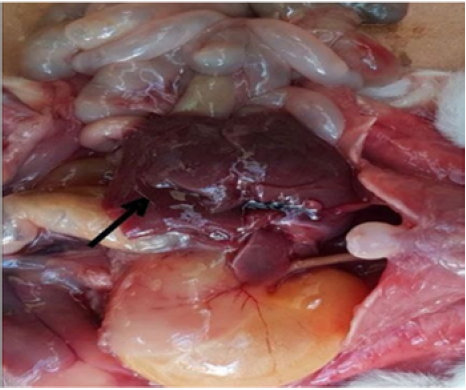
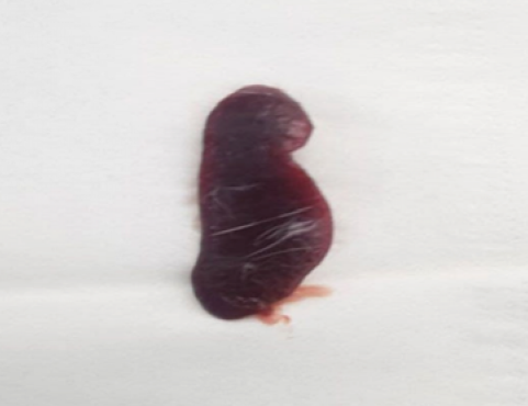
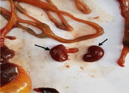
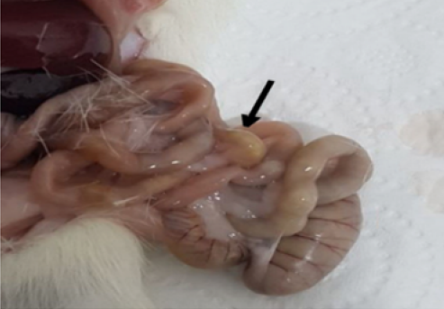
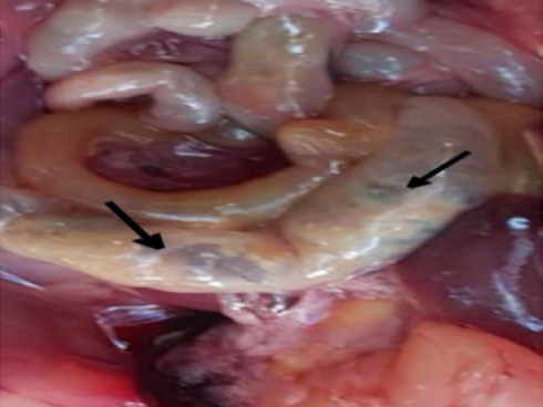
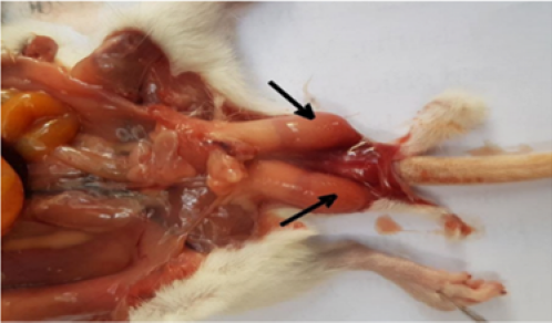
Hyperemia was observed in the organs of infected rats, especially the liver (Figure 3), heart, spleen (Figure 4), and kidneys (Figure 5), with pale areas. The lung was characterized by a gray color with a hard texture, and the small intestines were characterized by mildly congested, and hemorrhagic areas were observed (Figure 6). The large intestine showed congestion in different areas (Figure 7). Few hemorrhagic spots were observed in testes (Figure 8).
Figure 3: The Liver Displayed Hemorrhage Areas and Diffuse Focal Necrosis
Figure 4: Hyperemia was Observed in the Spleen
Figure 5: Hyperemia was Observed in the Kidney
Figure 6: Mildly Congested and Hemorrhagic Areas in the Small Intestine
Figure 7: The Congestion of the Large Intestine and Filled With Feces
Figure 8: Few hemorrhagic spots were observed in testes
DISCUSSION
The colonies of all Aeromonads species appear with almost the same morphological characteristics on the media, these results are agreed with [19] who showed that the aeromonads colonies observe with gray color and β-hemolytic colonies on the blood agar, palled (non-lactose fermenters) on MacConkey agar and shining with creamy colonies on nutrient and tryptic soy (TS) agars. Furthermore, A.veronii are positive for catalase and oxidase [20,3] and they have tolerated NaCl concentrations in the range of 0.3 to 5% [9].
Al-Qaese [21] reported that A. caviae was virulent to rat with an LD50 value of (1.0 x 10⁸ CFU) per rat, which was lower than the A. veronii in the same model animals of this study. While [22] results found that the LD50 of mice injected intraperitoneally with A.veronii (SL7231-1) was 4.51 × 10⁹ CFU/ml, which was higher than the LD50 ( 8.4 ⤫ 108) of virulent A.veronii recorded in this study. This variety of LD50 because of species different or host studied. Li et al. [22] study showed a significant variability of the pathogenicity of A.veronii strains and the pathogenicity of A. veronii are in positive correlation with the virulence gene number, thus, A. veronii that carry a high number of virulence genes are more virulent.
The pathogenicity of this bacteria is moderate, and this depends on dose-volume and the strength of the immune system. In conclusion, we proved that the pathogenicity of A.veronii is moderate in rats.
The rat's patho-clinical signs of the infected group agreed with [23] who observed that the mice that were intraperitoneally injected with Aeromonas hydrophelia showed diminished appetite, gloominess, and weary behavior. Furthermore, [23] found that these clinical signs are more evident in the mice that were injected intraperitoneally than those with oral administration. The experimental study showed a significant increase in the Virulent group compared with the control group. As a result, the model rodents infected with different species of Aeromonas observed a high similarity of clinical signs. In addition, these results are in agreement with Chen et al. [24] who showed that the BALB/c mice models were intraperitoneally injected with A.veronii, A. caviae and A. dhakensis observed similar clinical symptoms in infected rats in this study.
The post-mortem examination was in accordance with results obtained by [23] who shows that the model mice that were injected intraperitoneally with A. hydrophelia observed a mildly congested intestine, high congestion in the large intestine, and lesions with hemorrhage signs in the liver, spleen, and lung tissues.
So, let us conclude by Aeromonas veronii has been linked to sepsis, and enteritis in humans, as shown in path clinical signs of infected group rats, and compared to the control group. also, recommend Studying the ability of bacteria to transfer through wounds to the bloodstream and the virulence factors.
THANK everyone for bringing their expertise and experience in the research and constructive interaction to make it a success.
REFERENCE
Rashid, M. et al. “Isolation and identification of Aeromonas hydrophila from silver carp and its culture environment from the Mymensingh region.” Journal of Bangladesh Agricultural University, vol. 11, no. 2, 2013, pp. 373–376.
Janda, J.M. and S.L. Abbott. “The genus Aeromonas: taxonomy, pathogenicity, and infection.” Clinical Microbiology Reviews, vol. 23, 2010, pp. 35–73.
Martin-Carnahan, A. and S.W. Joseph. “Order XII. Aeromonadales ord. nov.” Bergey’s Manual of Systematic Bacteriology, edited by D.J. Brenner et al., Williams & Wilkins, 2005, pp. 556–578.
Beaz-Hidalgo, R. et al. “Comparison of phenotypical and genetic identification of Aeromonas strains isolated from diseased fish.” Systematic and Applied Microbiology, vol. 33, 2010, pp. 149–153.
Austin, B. and D.A. Austin. Bacterial fish pathogens: disease of farmed and wild fish. 6th ed., Springer International Publishing, 2016.
Jayavignesh, V. et al. “Biochemical characterization and cytotoxicity of Aeromonas hydrophila isolated from catfish.” Archives of Applied Science Research, vol. 3, no. 3, 2011, pp. 85–93.
Kavitha, K. et al. “Emerging ulcer disease in threatened murrel Channa aurantimaculata.” Journal of Research in Animal Sciences, vol. 2, no. 1, 2014, pp. 87–93.
Praveen, P.K. et al. “Incidence of Aeromonas spp. infection in fish and chicken meat and its public health hazards.” Veterinary World, vol. 9, no. 1, 2016, pp. 6–11.
Parker, J.L. and J.G. Shaw. “Clinical microbiology and disease of Aeromonas spp.” Journal of Infection, vol. 62, 2011, pp. 109–118.
Ottaviani, D. et al. “Putative virulence properties of Aeromonas strains isolated from food, environmental, and clinical sources in Italy.” International Journal of Food Microbiology, vol. 144, 2011, pp. 538–545.
Tomás, J.M. “The main Aeromonas pathogenic factors.” ISRN Microbiology, 2012, pp. 1–22.
Sun, J. et al. “Virulence properties of Aeromonas veronii isolated from diseased gibel carp.” International Journal of Molecular Sciences, vol. 17, no. 4, 2016, pp. 496.
Hossain, S., et al. “Virulence properties and multidrug resistance profiles of motile Aeromonas spp. isolated from zebrafish.” Letters in Applied Microbiology, vol. 67, 2018, pp. 598–605.
Spanggaard, B. et al. “Microflora of the rainbow trout intestine: traditional and molecular identification.” Aquaculture, vol. 182, 2000, pp. 1–15.
Eddy, S.D. and S.H. Jones. “Microbiology of summer flounder fingerling production at a marine fish hatchery.” Aquaculture, vol. 211, 2002, pp. 9–28.
Ligozzi, M. et al. “Evaluation of the VITEK 2 system for identification and antimicrobial susceptibility testing of gram-positive cocci.” Journal of Clinical Microbiology, vol. 40, 2002, pp. 1681–1686.
Quinn, P.J. et al.Clinical veterinary microbiology. 6th ed., Mosby, 2004.
Reed, L.J. and H. Muench. “A simple method of estimating fifty percent endpoints.” American Journal of Hygiene, vol. 27, 1938, pp. 493–497.
Mzula, A. et al. “Phenotypic and molecular detection of Aeromonas infection in farmed Nile tilapia in Tanzania.” Heliyon, vol. 5, no. 8, 2019, pp. e02220.
Erdem, B. et al. “Virulence factors and antibiotic resistance in motile Aeromonas isolated from fish.” Turkish Journal of Biology, vol. 34, 2010, pp. 453–462.
Al-Qaese, B.I. “Pathological effects of Aeromonas caviae and clinical chemistry changes in rats.” Iraqi Journal of Veterinary Medicine, vol. 27, no. 1, 2003, pp. 61–77.
Li, T. et al. “Aeromonas veronii infection in commercial freshwater fish and public health risk.” Animals, vol. 10, no. 4, 2020, pp. 608.
Kim, K.T. et al. “Enhanced virulence of Aeromonas hydrophila induced by stress and serial passaging in mice.” Animals, vol. 11, no. 2, 2021, p. 508.
Chen, P.L. et al. “Virulence diversity among bacteremic Aeromonas isolates.” PLoS One, vol. 9, no. 11, 2014, pp. e111213.
Recommended Articles
Research Article
Serum IL-22, miRNA-146a Expression and Total Antioxidant Status as Integrated Biomarkers in Rheumatoid Arthritis
Khalid R. Kareem
Published: 13/08/2026
Download PDF
Research Article
Electrochemical Comparative Studies On The Redox Behavior Of Cu(II) And Cr(II) Before And After Interactions With Ciprofloxacin In Solutions
H. M. Naseem Akhtar,
M. Q. Ehsan
Published: 10/08/2021
Download PDF
Research Article
A Rare Case Report of Waugh Syndrome in an Eight Months Old Male
Pielly Hanna,
...
Bassem Abou Merhi
Published: 10/03/2021
Download PDF
Research Article
Study on Prevalence and Etiology of Early Neonatal Deaths in Jasin district, Melaka from 2014-2017
Sujat a Khobragade,
...
Yadneshwar Khobragade
Published: 30/05/2021
Download PDF
PO Box 101, Nakuru
Kenya.
Email: office@iarconsortium.org
Editorial Office:
J.L Bhavan, Near Radison Blu Hotel,
Jalukbari, Guwahati-India
Radhi, M. M., Hadi, S. H. & Jawad, T. A. (2022). Microbiological Identification for Aeromonas Veronii and Its Experimental Pathogenesis of Necropsy Findings In Rats. International Academic Journal of Applied Bio-Medical Sciences, 3(1), 1-5.
MLA
Radhi, Mustafa M., Sabaa H. Hadi and Thikra A. Jawad. "Microbiological Identification for Aeromonas Veronii and Its Experimental Pathogenesis of Necropsy Findings In Rats." International Academic Journal of Applied Bio-Medical Sciences 3.1 (2022): 1-5.
Chicago
Radhi, Mustafa M., Sabaa H. Hadi and Thikra A. Jawad. "Microbiological Identification for Aeromonas Veronii and Its Experimental Pathogenesis of Necropsy Findings In Rats." International Academic Journal of Applied Bio-Medical Sciences 3, no. 1 (2022): 1-5.
Harvard
Radhi, M. M., Hadi, S. H. and Jawad, T. A. (2022) 'Microbiological Identification for Aeromonas Veronii and Its Experimental Pathogenesis of Necropsy Findings In Rats' International Academic Journal of Applied Bio-Medical Sciences 3(1), pp. 1-5.
Vancouver
Radhi MM, Hadi SH, Jawad TA. Microbiological Identification for Aeromonas Veronii and Its Experimental Pathogenesis of Necropsy Findings In Rats. International Academic Journal of Applied Bio-Medical Sciences. 2022 Jan;3(1):1-5.


 Download PDF
 Download XML