Contents
Download PDF
pdf Download XML
422 Views
468 Downloads
Share this article
Research Article | Volume 3 Issue 1 (Jan-June, 2022) | Pages 1 - 6
Bacteriological Profile of Isolates from the Patients admitted in Intensive Care Units in a Tertiary Care Hospital
 ,
 ,
 ,
 ,
1
Medical Officer, National Health Mission, Shimla, Himachal Pradesh, India
2
Civil Hospital, Theog, District Shimla, Himachal Pradesh, India
3
Department of Microbiology, IGMC, Shimla, Himachal Pradesh, India
Under a Creative Commons license
Open Access
Received
Jan. 3, 2022
Revised
Jan. 9, 2022
Accepted
Feb. 19, 2022
Published
March 10, 2022
Abstract

Background: The profile of organisms causing ICU infections varies widely one country to the other, one hospital to another and even amongst different ICUs of the same hospital.The present study attempts to know the bacteriological profile of bacterial isolates from ICU patients in our institution. Material and Methods: The present study was conducted in the department of Microbiology Indira Gandhi Medical College and Hospital where in the clinical samples received from various ICUs were analysed for bacteriological profile. The duration of the study was from 14th January 2019 to 13th January 2020. Results: The age distribution of the patients whose samples were sent was age ranged from 1day to 90 years. Numbers of males were more compared to females. A total of 2458 samples were processed out of which 766 (31.16%) were found to be culture positive, sterile were 1463 (59.52%) and contaminants were found in 229 (9.32%). Among all isolates gram negative bacteria were most common, followed by Candida spp. and gram positive bacteria. Positivity rate was maximum from general ICU 58.36% followed by sick neonatal care unit 25.06% and lowest positivity rate was seen from respiratory ICU 16.66%.The percentage of gram negative bacteria were maximum from HDU (80%), gram positive bacteria were found to be maximum from CTVS-ICU (27.27%) and Candida were maximum from pediatric ICUs (83.08%). In pediatric ICUs highest percentage of Candida spp. were isolated from NICU (64.28%) followed by PICU (60.14%) and SNCU (60%).From GICU Klebsiella pneumoniae was the most common isolate followed by Non fermenter group of organism. Klebsiella pneumoniae was also the commonest isolate from HDU, SNCU and NICU. Whereas Non fermenter group of organism was common in RICU and CTVS ICU. In PICU Escherichia coli was the most common isolate.Conclusion: A periodic surveillance of the ICUs is thus essential as many of the pathogens are not incubating in the patient at the time of admission but are transmitted by the hands of the treating physicians and other inanimate objects that are used during invasive procedures.

Keywords
INTRODUCTION

Intensive care unit (ICU) is a specialized setting in hospitals, that offers close monitoring and personalized care for the sick patients [1].


Even though ICU represents only 5% of total hospital beds, they account for almost 20-25% of all health care associated infections.2The rate of ICU infections worldwide is 23.7 infections per 1,000 patient days, with the rate of nosocomial infections being 5-30% amongst the ICU patients [3]. 


Patient’s own indigenous flora of the skin, mucous membrane, gastrointestinal tract, or respiratory tract which may invade the patient’s body during surgical or invasive procedures. The exogenous sources are commonly acquired in the ICU by other patients, health care workers, visitors and inanimate objects including patients bed surface, equipments [4].


The profile of organisms causing ICU infections varies widely one country to the other, one hospital to another and even amongst different ICUs of the same hospital [5]. The present study attempts to know the bacteriological profile of bacterial isolates from ICU patients in our institution. This study aims to improve patient’s outcome and infection control practices in the hospital.


Aims and Objectives

To identify the bacteriological profile of bacterial isolates in the samples received from different intensive care units (HDU, GICU, CTVS-ICU, PICU, RICU, NICU, SNCU

MATERIALS AND METHODS
  • Study Setting: The present study was conducted in the department of Microbiology, Indira Gandhi Medical College and Hospital Shimla

  • Study Design: It was a descriptive cross sectional study

  • Study period: The study period was of one year from 14th January 2019 to 13th January 2020

  • Selection of cases: In this study all clinical samples received in the department of Microbiology for bacterial culture and susceptibility testing from patients admitted in the intensive care units of this institution during the study period


Samples received in the department of Microbiology and processed using standard procedures were of Bloodm,Sterile Body fluids (cerebrospinal fluid and pleural fluid), Pus samples, Sputum,Swabs (throat and wound) ,Respiratory secretions,Urine and Central line catheter tip.


The demographic profile of the patient whose samples were included in the study was also recorded.


Processing of Samples

The samples of blood and other sterile body fluids received in BACTEC blood culture vials were first cultured using the BD BACTEC™ FX system for five days. The positive blood culture vials were further processed within 8hrs. For obtaining pure growth for further processing, the positive blood culture vials were inoculated on blood agar and MacConkey agar plates and incubated aerobically at 37oC overnight for 18-24 hrs. The positive cultures detected in the BD BACTEC™ FX system were subjected to identification and antibiotic susceptibility testing by BD Phoenix™ FX Automated Microbiology system. Negative report was given only after 5days of incubation in BD BACTEC ™ FX automated system.


Body fluid such as pus, sputum, respiratory secretions, pleural fluid were received in sterile containers, inoculated onto blood agar and MacConkey agar plates, and were incubated at 37o C for 24 hrs. Cerebrospinal fluid was received in sterile container and inoculated on blood agar and chocolate agar plates and incubated at 370 C for 24 hrs.Uncentrifuged urine but well-mixed urine was inoculated by a calibrated loop onto plates of cystine lactose electrolyte deficient (CLED) agar. Throat swab was inoculated onto blood agar plates and incubated at 370 C for 24 hrs.


Morphology and Characteristics

The pure growth obtained after 24 hours of incubation on solid media was identified on the basis of colony characters, gram staining and preliminary biochemical reactions. When pure growth is not obtained on primary plating then it was subcultured on blood agar and MacConkey agar to get pure growth.


Preliminary Identification of Bacteria


  • Colony characters: The growth of the organism was observed on Blood Agar and MacConkey Agar. The characters of the colony were studied like size, shape, surface, edge, elevation, consistency, colour of the colony, odour, and effect on media

  • Gram Staining: Gram staining of the growth was done using modified Huckers method. Based on the gram staining of organism whether gram positive or gram negative, the panels for antimicrobial sensitivity were selected

  • Preliminary Biochemical Reactions: Preliminary biochemical reactions such as catalase, coagulase and oxidase were performed to identify the organism.

The isolates were then processed in BD Phoenix™ FX automated system for further identification. The final report indicating organism identified was communicated to respective ICUs and also recorded in the performa


Quality Control

The quality check was performed using standard strains as per the manufacturer’s instructions.


Exclusion Criteria

The samples where BD Phoenix™ FX automated system gave inconclusive results or failed to identify the isolates were excluded from the study.


Statistical Analysis

The Data collected was entered in Excel spreadsheet and accuracy of data entered was checked by cross-verification of the data entered. Categorical variables were expressed as frequencies and percentages.

RESULTS

The present study was conducted in the department of Microbiology Indira Gandhi Medical College and Hospital where in the clinical samples received from various intensive care units were analysed for bacteriological profile and antibiotic susceptibility pattern. The duration of the study was from 14th January 2019 to 13th January 2020. The observations were as follows.


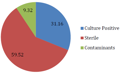
The clinical samples were received from general ICU, neonatal ICU, paediatric ICU, HDU, respiratory ICU, sick neonatal care unit and CTVS-ICU. The bacterial isolates were studied in detail in the present study and Candida spp. was also documented. In the present study 2458 samples were processed out of which 766 (31.16%) were found to be culture positive, sterile were 1463 (59.52%) and contaminants were found in 229 (9.32%) Figure 1. 


Figure 1: Culture Positivity among Clinical Samples


The number of isolates in all culture positive specimens was 840, as more than one isolates were also obtained in some specimens.


Among all isolates gram negative bacteria were most common, followed by Candida spp. and gram positive bacteria.


In the present study positivity rate was maximum from general ICU 58.36% followed by sick neonatal care unit 25.06% and lowest positivity rate was seen from respiratory ICU 16.66% Table 1-3.


Table 1: Distribution of Clinical Specimens from Different ICUs

IsolateNo. of IsolatePercentage
Gram negative bacteria47756.78%
Gram positive bacteria9711.54%
Candida spp.26631.66%
Total840100%


Table 2: Pattern of Organisms Isolated In All Clinical Specimens

IcusNumber Of Specimens N = 2458Culture Positive N = 766Percentage Positivity
GICU55032158.36%
HDU2006030%
RICU1202016.66%
CTVS ICU1002121%
PICU63012920.47%
SNCU75018825.06%
NICU1082725%


Table 3: Distribution of Isolates from Different ICU

ICU’s

NO. OF ISOLATES

n = 840

GRAM NEGATIVE BACTERIA

n = 477

GRAM POSITIVE BACTERIA

n = 97

CANDIDA SPP.

n = 266

GICU356 (42.38%)277 (77.80%)40 (11.23%)39 (10.95%)
HDU75 (8.92%)60 (80%)13 (17.33%)2 (2.66%)
RICU21 (2.50%)15 (71.42%)4 (19.04%)2 (9.52%)
CTVS ICU22 (2.61%)14 (63.63%)6 (27.27%)2 (9.09%)
PICU138 (16.42%)37 (26.81%)18 (13.04%)83 (60.14%)
SNCU200 (23.80%)64 (32%)16 (8%)120 (60%)
NICU28 (3.33%)10 (35.71%)0%18 (64.28%)


The percentage of gram negative bacteria were maximum from HDU (80%), gram positive bacteria were found to be maximum from CTVS-ICU (27.27%) and Candida were maximum from pediatric ICUs (83.08%). In pediatric ICUs highest percentage of Candida spp. were isolated from NICU (64.28%) followed by PICU (60.14%) and SNCU (60%).


The age and sex wise distribution as categorized into various groups is depicted in Table 4-5.


In the present study, from GICU Klebsiella pneumoniae was the most common isolate followed by Non fermenter group of organism. Klebsiella pneumoniae was also the commonest isolate from HDU, SNCU and NICU. Whereas Non fermenter group of organism was common in RICU and CTVS ICU. In PICU Escherichia coli was the most common isolate.


Table 4: Age Sex Distribution of Patients with Positive Culture

Age

Gender

NICU

SNCU

PICU

GICU

HDU

RICU

CTVS ICU

0-7 days early neonates

M

12

62

25


F

9

41

13


7-28 days late neonate

M

3

46

16


F

2

22

17


29 days-1 yrs infant

M


18


F


9


1-5 yrs preschool

M


12


F


4


6-12 yrs school

M


3


1


F


3


13-19yrs adolescent

M


3

1


1

F


4


20-30yrs

M


6

2

1


F


3


31-40yrs

M


15

6

2


F


7

2


41-50

M


20

7

1

2

F


12

5

1

3

51-60

M


21

6

1

3

F


15

2

2

2

61-70

M


11

4

2

2

F


7

1

2

2

71-80

M


10

1

2

2

F


5

2

2

1

≥81 yrs 

M


3


2

1

F


1


Total


26

171

128

137

39

18

19


Table 5: ICU Wise Distribution of Clinical Isolates

ORGANISMGICUHDURICUCTVS ICUPICUSNCUNICUTotal
Klebsiella Pneumoniae7818-25302135
Non Fermentergroup Of Organism51844810287
Escherichia Coli331443108274
Pseudomonas Aeruginosa3582-42152
Acinetobacter Baumannii3553-33-49
Staphylococcus Aureus1432386-36
CONS521266-22
Klebsiella Oxytoca11-1-24119
Enterococcus Faecium124---2-18
Enterobacter Aerogenes93-1-2116
Enterobacter Cloacae93-12-116
Enterococcus Faecalis931-2--15
Stenotrophomonas Maltophila5-1121-10
Citrobacter Koseri4----1-5
Pseudomonas Pseudoalkaligens2----2-4
Citrobacter Freundi11-1---3
Proteus Mirabilis2--1---3
Β Haemolytic Streptococcus-1-1-1-3
Serratia Marcescence1---1--2
Proteus Vulgaris1------1
Salmonella Choleraesuis-----1-1
Enterococcus Casseliflavus-----1-1
Α Haemolytic Streptococcus----1--1
Streptococcus Pneumonia----1--1
Total317731920558010574
DISCUSSION

Infection is a major factor that determines the clinical outcome among patients requiring intensive care unit (ICU) support. The causes of infection within an ICU are multi‑factorial. The consequences depend upon the source involved, organisms associated, underlying morbidity, timeliness and appropriateness of the treatment and the intervention received [6].In the present study Eight hundred and forty (840) bacterial and Candida isolates were reported in this study form various clinical specimens that were received and processed as routine in the department of Microbiology.


The demographic profile of the ICU patients in the present study revealed that the number of males admitted in different ICUs were greater than females, (male: female ratio being 1.67:1). Which is similar to that reported by Iwuafor A.et al. Sarvepalli A. et al. and Turtkan M. et al. [7-9]. The reason for male predominance in this study could be due to more health seeking behaviour of males.


In the present study the overall culture positivity was found to be 31.16%. This was found to be similar to a study conducted by Patil P. et al. [1] The positivity rate was found to be more than that reported by Chelazzi et al. (9.3%) and Nangino et al. (8.9%) [10-11]. The positivity rate was less than that reported by Vincent J.et al. (51%) and Babbar P. et al. 44.8% [12-13]. The probable reason for the lower culture positivity rate in the present study can be due to prior use of antibiotics, improper sample collection and transportation and the samples were also not processed for anaerobic bacteria and filamentous fungi.


Majority of the pathogens causing infections in the patients admitted to the ICUs in the present study, were gram negative bacteria (56.66%) followed by gram positive bacteria (11.42%). The results from this study are similar to few recent studies where gram negative bacteria were most commonly isolated [12,14,15]. However according to global studies gram positive bacteria were found to be more compared to gram negative bacteria [16-18]. This predominance of gram negative bacteria in the present study can be explained by the fact that the prevalence of gram negative bacteria is higher in developing countries as compared to developed countries [2]. 


In the present study maximum number of samples were obtained from SNCU, but the overall culture positivity was found to be maximum from GICU (58.36%). Similar results were seen in the studies conducted by Patil P. et al. [1] and Jawady A. et al. [15]. In contrary to the present study Gupta S. et al. and Mahaluca A. et al. documented maximum positivity rate from NICU and PICU [16,19]. The culture positivity rate with respect to different ICUs is variable in different studies across the country and the world. 


In the present study gram negative bacteria were found to be maximum from HDU (80%), gram positive bacteria were maximum from CTVS-ICU (27.27%) and Candida were maximum in NICU (64.28%). The predominant gram negative bacteria isolated were Klebsiella pneumoniae and Non fermenter group of organism followed by Candida spp. and gram positive bacteria such as Staphylococcus aureus and coagulase negative Staphylococcus. The findings are in consistent with other studies from India such as by Sahu et al. Patil P.et al. and Pattanayak C. et al. and Jawady A. et al. [1,15,20,21]. Several studies from other parts of the world show predominance of gram positive bacteria from ICUs [7,16,18]. The predominance of gram negative bacteria may be due to their wide prevalence in the hospital environment, and most of them represent a wide range of normal flora. 


The most common bacteria isolated in the present study were Klebsiella pneumoniae followed by Non fermenter group of organism. Klebsiella pneumoniae was also found to be the most common isolate from blood, endotracheal secretions, pus and drain. Whereas in urine Enterococcus faecium and Escherichia coli were found to be predominant isolates. Klebsiella pneumoniae was also found to be predominant isolate from ICU infections in several other studies [22-24]. Unlike the present study Pradhan et al. found Acinetobacter baumannii as predominant pathogen from endotracheal secretions and Pseudomonas spp. from urine. Richards M. et al. found CONS as predominant isolate from blood, Pseudomonas aeruginosa from respiratory secretions, Escherichia coli from urine and Pseudomonas aeruginosa from pus. Jain et al. found Citrobacter koseri from blood, Escherichia coli from urine, Pseudomonas aeruginosa from sputum and Staphylococcus aureus from pus [25-26]. Klebsiella pneumoniae was found to be predominant isolate in this study this can probably be explained by the fact that the members of Enterobacteriaceae are found as normal commensal flora of human body, which make them one of the most important endogenous bacteria causing infections in immune-compromised patients. Increased colonization of Enterobacteriaceae organisms in the respiratory tract of patients on prolonged mechanical ventilation also contributes to higher infections due to these bacterias in ICUs [27].


Non fermenter gram negative bacteria have emerged as important pathogen in the hospitals particularly in ICUs, probably due to wide spread and injudicious use of antibiotics. These bacteria are normally inhabitants of water and soil. In the hospital environment they may be isolated from instruments, hospital linen and also from skin. Most of these are MDR pathogen. Hence pose a challenge to the treating physician. In the present study Non fermenter gram negative bacteria were next common group isolated from several clinical specimens. They were increasingly reported from endotracheal secretions. Pseudomonas and Acinetobacter baumannii were predominant Non fermenter bacteria isolated from endotracheal secretions. Pseudomonas and Acinetobacter baumannii are reported as predominant isolates from various ICUs in literature [28-29].

CONCLUSION

Multidrug resistant bacteria are a growing problem in the intensive care units which is leaving the clinicians with only fewer options to treat critically ill patients. The increasing multidrug resistant pathogens and the immunocompromised nature of the patients lead to increased mortality and morbidity in the ICUs and also the cost of treatment worldwide. 


Present study covers all the samples received from different ICUs in our institution and provide an insight in to early initiation of antimicrobial therapy and influence the outcome of patients admitted to these ICUs. A periodic surveillance of the ICUs is thus essential as many of the pathogens are not incubating in the patient at the time of admission but are transmitted by the hands of the treating physicians and other inanimate objects that are used during invasive procedures.

REFERENCE
  1. Patil, P.S. and R. Ashok. “Bacteriological profile and antimicrobial susceptibility pattern in intensive care unit of tertiary care hospital, Aurangabad.” International Journal of Clinical and Biomedical Research, July 2017, pp. 26–30.

  2. Chaudhry, D. and B. Prajapat. “Intensive care unit bugs in India: how do they differ from the Western world?” Journal of the Association of Chest Physicians, vol. 5, no. 1, January 2017, pp. 10.

  3. Wikaningtyas, P. et al. “Profile of antibiotic resistance and usage pattern in ICU of a private hospital in Bandung, Indonesia.” International Journal of Pharmacy and Pharmaceutical Sciences, vol. 7, no. 2, February 2015, pp. 160–162.

  4. Sastry, A.S. Essentials of Medical Microbiology. 1st ed., Jaypee Brothers Medical Publishers, 2016, pp. 81–83.

  5. Zaveri, J.R. et al. “A study on bacteriological profile and drug sensitivity and resistance pattern of isolates of patients admitted in intensive care unit of a tertiary care hospital in Ahmedabad.” National Journal of Medical Research, vol. 2, no. 3, July 2012, pp. 330–334.

  6. Bhattacharya, S. and A.S. Mondal. “Clinical microbiology in the intensive care unit: strategic and operational characteristics.” Indian Journal of Medical Microbiology, vol. 28, no. 1, January 2010, pp. 5–10.

  7. Iwuafor, A.A. et al. “Incidence, clinical outcome and risk factors of intensive care unit infections in the Lagos University Teaching Hospital, Lagos, Nigeria.” PLOS One, vol. 11, no. 10, October 2016, e0165242.

  8. Sarvepalli, A.K. and P.K. Dharana. “Clinical profile, bacterial profile and outcomes of acute bacterial meningitis in a tertiary care hospital: one-year study.” International Journal of Advances in Medicine, vol. 4, March 2017, pp. 502–507.

  9. Turktan, M. et al. “Community-acquired infections among refugees leading to intensive care unit admissions in Turkey.” International Journal of Infectious Diseases, vol. 58, May 2017, pp. 111–114.

  10. Chelazzi, C. et al. “Epidemiology, associated factors and outcomes of ICU-acquired infections caused by Gram-negative bacteria in critically ill patients: an observational, retrospective study.” BMC Anesthesiology, vol. 15, 2015, pp. 125–131.

  11. Nangino, G.D. et al. “Financial impact of nosocomial infections in intensive care units of a philanthropic hospital in Minas Gerais.” Revista Brasileira de Terapia Intensiva, vol. 24, 2012, pp. 357–361.

  12. Vincent, J.L. et al. “International study of the prevalence and outcomes of infection in intensive care units.” JAMA, vol. 302, no. 21, December 2009, pp. 2323–2329.

  13. Babbar, P. et al. “Healthcare-associated infections in intensive care units: a pilot study in a tertiary care public hospital in India.” Journal of Prevention and Infection Control, vol. 5, no. 1, 2019, pp. 1.

  14. Aly, S.A. et al. “Bacterial catheter-associated urinary tract infection in the intensive care unit of Assiut University Hospital.” Al-Azhar Assiut Medical Journal, vol. 14, no. 2, April 2016, pp. 52.

  15. Al-Jawad, Z.A., and H.M. Al-Habib. “Antibiogram profiles of bacterial isolates from intensive care units in Mosul Teaching Hospitals.” Rafidain Journal of Science, vol. 23, no. 1, January 2012, pp. 52–59.

  16. Mahaluca, F.A. et al. “Antibacterial resistance pattern of Gram-positive bacteria isolated in the ICU from a reference hospital in Southern Mozambique.” Journal of Bacteriology and Parasitology, vol. 9, 2018, pp. 348.

  17. van Vught, L.A. et al. “Incidence, risk factors, and attributable mortality of secondary infections in the intensive care unit after admission for sepsis.” JAMA, vol. 315, no. 14, April 2016, pp. 1469–1479.

  18. Orsini, J. et al. “Microbiological profile of organisms causing bloodstream infection in critically ill patients.” Journal of Clinical Medicine Research, vol. 4, no. 6, December 2012, pp. 371–376.

  19. Gupta, S. and B. Kashyap. “Bacteriological profile and antibiogram of blood culture isolates from a tertiary care hospital of North India.” Tropical Journal of Medical Research, vol. 19, no. 2, July 2016, pp. 94–99.

  20. Pattanayak, C. et al. “A study on antibiotic sensitivity pattern of bacterial isolates in the intensive care unit of a tertiary care hospital in Eastern India.” International Journal of Basic and Clinical Pharmacology, vol. 2, no. 2, March 2013, pp. 153–159.

  21. Sahu, M.K. et al. “Incidence and microbiological profile of nosocomial infections and their antibiotic resistance patterns in a high-volume cardiac surgical intensive care unit.” Annals of Cardiac Anaesthesia, vol. 19, no. 2, April 2016, pp. 281.

  22. Deep, A. et al. “Clinical and microbiological profile of nosocomial infections in the pediatric intensive care unit (PICU).” Indian Pediatrics, vol. 41, no. 12, December 2004, pp. 1238–1246.

  23. Afroz, Z. et al. “Bacteriological profile of gram-negative nosocomial isolates from intensive care units and their antibiogram in a tertiary care hospital of South India.” IP International Journal of Medical Microbiology and Tropical Diseases, vol. 4, 2018, pp. 243–250.

  24. Pokhrel, B. et al. “Bacteriological profile and antibiotic susceptibility of neonatal sepsis in neonatal intensive care unit of a tertiary hospital in Nepal.” BMC Pediatrics, vol. 18, no. 1, December 2018, pp. 1–8.

  25. Richards, M.J. et al. “Nosocomial infections in pediatric intensive care units in the United States.” Pediatrics, vol. 103, no. 4, April 1999, pp. e39–e39.

  26. Jain, A.K. et al. “Microbiological profile of infection in intensive care unit and their antimicrobial susceptibility pattern with special reference to metallo β-lactamases and AmpC.” International Journal of Medical Science and Public Health, vol. 7, 2018, pp. 229–234.

  27. Moolchandani, K. et al. “Antimicrobial resistance surveillance among intensive care units of a tertiary care hospital in Southern India.” Journal of Clinical and Diagnostic Research, vol. 11, no. 2, February 2017, pp. DC01–DC07.

  28. Global Alliance for Infections in Surgery. “Let’s be careful with non-fermenting Gram-negative bacteria including Pseudomonas aeruginosa and Acinetobacter baumannii.” Global Alliance for Infections in Surgery, 2018.

  29. Bonomo, R.A. and D. Szabo. “Mechanisms of multidrug resistance in Acinetobacter species and Pseudomonas aeruginosa.” Clinical Infectious Diseases, vol. 43, suppl. 2, September 2006, pp. S49–S56.

  30.  

Recommended Articles
Research Article
Electrochemical Comparative Studies On The Redox Behavior Of Cu(II) And Cr(II) Before And After Interactions With Ciprofloxacin In Solutions
Published: 10/08/2021
Download PDF
Research Article
A Rare Case Report of Waugh Syndrome in an Eight Months Old Male
...
Published: 10/03/2021
Download PDF
Research Article
Study on Prevalence and Etiology of Early Neonatal Deaths in Jasin district, Melaka from 2014-2017
...
Published: 30/05/2021
Download PDF
Research Article
Clinicopathological Correlation in Cases of Acute Appendicitis: An Overview
...
Published: 05/04/2025
Download PDF
Chat on WhatsApp
Flowbite Logo
PO Box 101, Nakuru
Kenya.
Email: office@iarconsortium.org

Editorial Office:
J.L Bhavan, Near Radison Blu Hotel,
Jalukbari, Guwahati-India
Useful Links
Order Hard Copy
Privacy policy
Terms and Conditions
Refund Policy
Shipping Policy
Others
About Us
Team Members
Contact Us
Online Payments
Join as Editor
Join as Reviewer
Subscribe to our Newsletter
+91 60029-93949
Follow us
MOST SEARCHED KEYWORDS
Copyright © iARCON International LLP . All Rights Reserved.