Eighty patients were collected from Yarmouk Hospital, located in Karkh district, Baghdad, Iraq and paper aim to know and assessment between gonadotropin levels and inhibin B levels in fertile women for obese women The ELISA device was relied upon in the evaluation and analysis of the results, in addition to the reliance on the statistical analysis program in evaluating the results, where the relationship was extracted in addition to the mean value and standard regression and An inverse relationship was found between the variables that link gonadotropin levels and inhibin B with obesity.
Obesity is characterized by an excessive amount of fatty tissue in the body. BMI is directly related to adipose tissue mass and is currently used to diagnose obesity, with a BMI (outside of pregnancy) being 30 kg/m2.
Adipose tissue is an active organ of the endocrine glands. With an excess of adipose tissue, the level of leptin increases and the level of adiponectin decreases, which leads to insulin resistance (IR). Obesity is also often associated with hyperandrogenism. These and various other hormonal changes cause ovulation.
It is still not entirely clear whether obesity in itself is a factor in increasing the risk of adverse pregnancy outcomes or predisposing to the development of other conditions that increase this risk.
Negative pregnancy outcomes are often associated with carbohydrate metabolism disorders, which are found in a significant proportion of obese women. However, in obese women with normal glucose tolerance, the probability of a complicated pregnancy continues to increase.
Various mechanisms of dysregulation of the metabolic, vascular and pro-inflammatory effects of adipose tissue in relation to other organs and systems are believed to be involved in the pathogenesis. This assumption is supported by the fact that the incidence of some pregnancy complications increases with the development of obesity
Obesity is an increasingly prevalent health risk and causes many female reproductive disorders In fact, overweight women have a higher incidence of impaired menstrual cycle, ovulation, and infertility than other women of childbearing age Although altered pulsatile gonadotropin secretion is a well-defined mechanism in obese patients it is not known whether obesity and/or hyperinsulinemia may have independent effects on ovarian hormone secretion especially in women with a normal menstrual cycle and normal appearance of the ovaries, in the absence of hyperandrogenism. Furthermore, there is no information available about the potential effects of obesity and/or hyperinsulinemia on the number of ovarian follicles in the early follicular phase of obese women with normal cycles.
Recently, have attracted the interest of many researcher , Inhibin are heterodimeric gonadotropin peptides containing an α-subunit and a βA subunit (inhibin b) or βB subunit (inhibin B) that have been proposed to negatively regulate follicle-stimulating hormone (FSH) 1 secretion, although there is no direct evidence for this phenomenon Locally, act by promoting the growth of follicles, thus reflecting the growth reserve of the small antral follicle Interestingly, in girls with anorexia nervosa, weight gain, with increases in adipose tissue and leptin levels, was paralleled by increases in inhibin B levels.
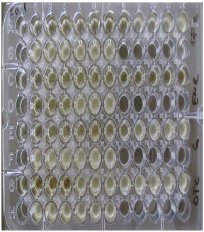
80 patients were collected from Yarmouk Hospital, located in Karkh district, Baghdad, Iraq, where obese women were collected and the effect of the disease on gonadotropin and inhibin B levels in fertile women was known and for determine FSH done depend on ELISA FSH which kit consists of an immunosorbent assay closely linked to the enzyme by relying on the sandwich principle, where the wells are tight and closed, then comes the role of the incubator, which incubates the samples that contain FSH, then the conjugate is washed in addition to that after adding the substrate solution, the developed color intensity is proportional to the concentration of FSH in the sample and about Inhibin b was analyzed by relying on Elisa inhibin b, where sodium dodecyl sulfate was used by heating to 100 ° C, then following the next step, which is incubating the samples, depending on microtiter plates Coated with a monoclonal antibody to the B subunit of inhibin (Figure 1).

Figure 1: Microtiter Plates
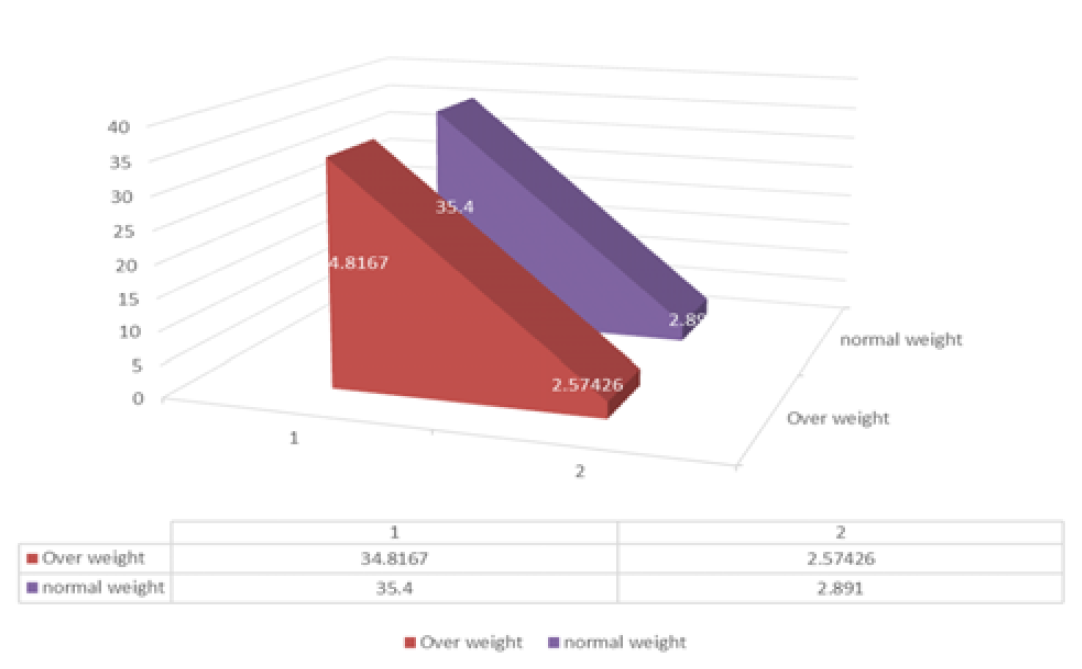
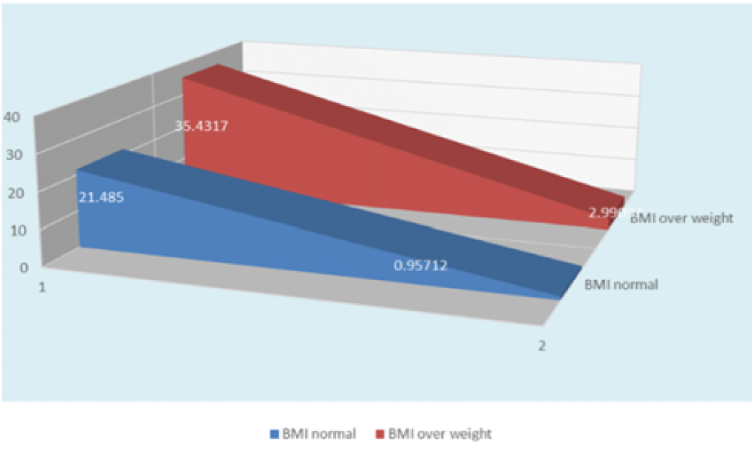
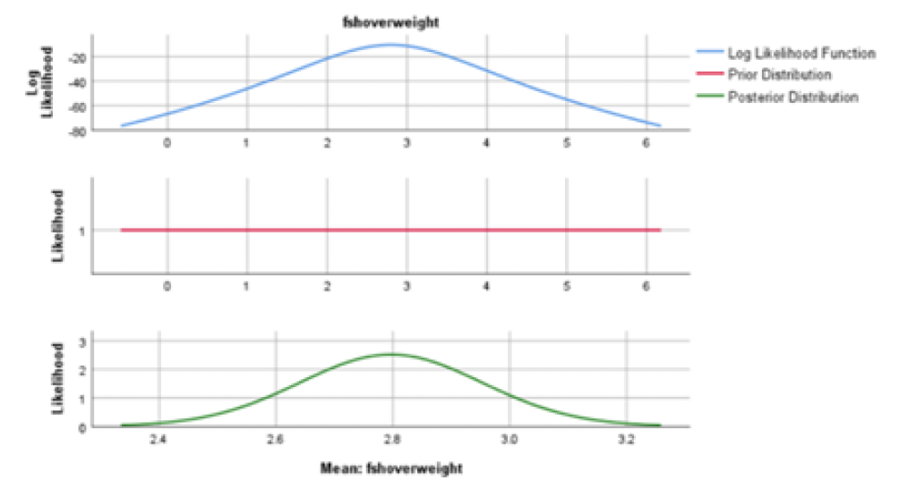
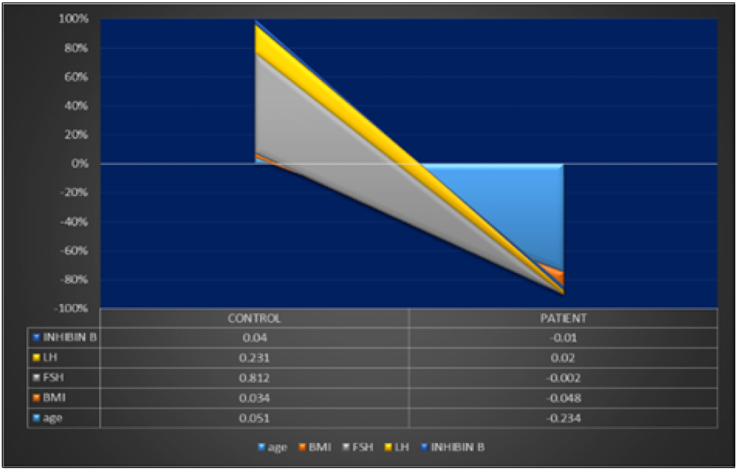
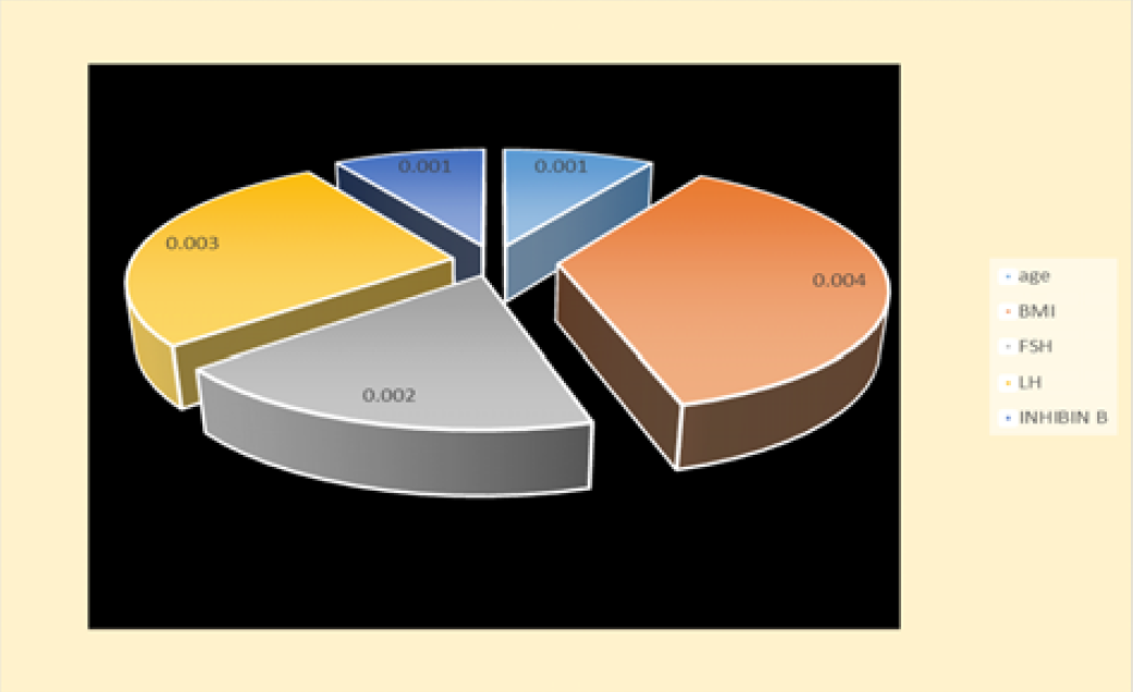
With age, the number of follicles in a woman's ovary decreases and the synthesis of inhibin B decreases. In early perimenopause, the level of inhibin B in the follicular phase of the cycle decreases earlier than the level of estradiol and inhibin A. The onset of menopause (Figure 2-4). In the postmenopausal period, the hormone is detected at a very low concentration (less than 5 pg / ml) or not at all. Elevated concentrations of inhibin B in postmenopausal women require the exclusion of granulosa cells or mucinous ovarian carcinoma, for which this hormone is a specific marker. With the above-mentioned ovarian tumors, it is possible to increase the concentration of inhibin B by more than 60-fold. Joint tests for CA 125 and inhibin B together with additional instrumental research methods make it possible to identify the tumor and control the treatment process (Table 1-4 and Figure 5-6).

Figure 2: Explain Mean and SD of Ages

Figure 3: Explain Curve of BMI

Figure 4: FSH Mean Posterior
Table 1: Descriptive Statistics of Ages
| Descriptive Statistics | |||||
| T | N | Minimum | Maximum | Mean | Std. Deviation |
| Over weight | 60 | 30.00 | 39.00 | 34.8167 | 2.57426 |
| normal weight | 20 | 30.00 | 40.00 | 35.4000 | 2.89100 |
Table 2: Mean and SD of BMI
| Descriptive Statistics | N | Minimum | Maximum | Mean | Std. Deviation |
| BMI Normal Weight | 20 | 19.90 | 22.90 | 21.4850 | 0.95712 |
| BMI Over Weight | 60 | 30.20 | 39.90 | 35.4317 | 2.99097 |
Table 3: Final Result of Overweight
| Statistics | Age Overweight | BMI Overweight | FSH Overweight | Lh OverweightP | Inhibin b Over | |
| N | Valid | 60 | 60 | 60 | 60 | 60 |
| Missing | 2 | 2 | 2 | 2 | 2 | |
| Mean | 34.8167 | 35.4317 | 2.7967 | 2.1033 | 33.8167 | |
| Median | 35.0000 | 36.3000 | 2.7000 | 2.1000 | 34.0000 | |
| Mode | 33.00 | 38.20 | 2.10 | 1.80a | 33.00a | |
| Std. Deviation | 2.57426 | 2.99097 | 1.20084 | 0.84492 | 15.02934 | |
| Range | 9.00 | 9.70 | 4.00 | 3.10 | 50.00 | |
| Minimum | 30.00 | 30.20 | 0.90 | 0.80 | 9.00 | |
a: Multiple modes exist. The smallest value is shown
Table 4: Final Result of Normal Weight
| Statistics | Normal Weight | Bmi Normalweight | Fsh Normalweight | Lh Normalweight | Inhibin B Normal Weight | |
| N | Valid | 20 | 20 | 20 | 20 | 20 |
| Missing | 42 | 42 | 42 | 42 | 42 | |
| Mean | 35.4000 | 21.4850 | 9.9235 | 6.8485 | 92.987 | |
| Median | 35.5000 | 21.4000 | 9.8500 | 7.1000 | 87.5000 | |
| Mode | 33.00a | 21.30 | 8.90a | 6.10a | 80.00 | |
| Std. Deviation | 2.89100 | 0.95712 | 1.51274 | 1.47441 | 15.20699 | |
| Range | 10.00 | 3.00 | 6.23 | 4.70 | 61.00 | |
| Minimum | 30.00 | 19.90 | 6.67 | 4.10 | 60.00 | |
a: Multiple modes exist. The smallest value is shown

Figure 5: Correlation between Parameters

Figure 6: p Value between Parameters
Quantification of inhibin B and other sex hormones can be useful in the study of ovarian reserve (follicular reserve / ovarian reserve) - the number of eggs and follicles capable of growing and developing in the ovaries. The more follicles in the ovary, the higher the level of inhibin B. And with natural depletion (due to age-related changes) or premature depletion of the ovary, the ability to fully ovulate, normal pregnancy and normal conception of the fetus is lost. In this regard, the level of inhibin B is often determined to assess the possibility of a normal pregnancy for a woman of a more mature age and / or to predict the effectiveness and feasibility of assisted reproductive technologies (in vitro fertilization - IVF, insemination with donor sperm).
Inhibin is an important protein hormone that has two parts, alpha and beta. It should be noted that there are two types of beta, which are beta-A, which gives inhibin A, and beta-B, which is inhibin B. The gonads secrete this hormone, especially the granulosa cells in the ovaries in women, the Sertoli cells in the testicles in men, and the placenta during pregnancy. The function of this hormone is.
Inhibition of the secretion of follicle-stimulating hormone (FSH) by the pituitary glands, a hormone responsible for growth processes, sexual puberty, the development of the female reproductive system, and the reproductive process Presents data on the consequences of obesity on the female reproductive system and the offspring of overweight or obese mothers, such as infertility, miscarriage, premature birth, stillbirth, congenital malformations and prematurity, as well as the high risk of caesarean section. Obesity accompanies PCOS, which impairs metabolism and increases the risk of depression and eating disorders. Maternal obesity and hyperglycemia can affect fetal formation through epigenetic mechanisms without affecting the nucleotide sequence. Moreover, metabolic risks increase
Hales, C.M. et al. “Prevalence of obesity among adults and youth: United States, 2015–2016.” NCHS Data Brief, no. 288, 2017, pp. 1–8.
Wang, Y.C. et al. “Health and economic burden of the projected obesity trends in the USA and the UK.” The Lancet, vol. 378, no. 9793, 2011, pp. 815–825.
Tzeng, C.R. et al. “Cluster analysis of cardiovascular and metabolic risk factors in women of reproductive age.” Fertility and Sterility, vol. 101, no. 5, 2014, pp. 1404–1410.
Wei, S. et al. “Obesity and menstrual irregularity: associations with SHBG, testosterone, and insulin.” Obesity, vol. 17, no. 5, 2009, pp. 1070–1076.
Wise, M.R. et al. “Obesity and endometrial hyperplasia and cancer in premenopausal women: a systematic review.” American Journal of Obstetrics and Gynecology, vol. 214, no. 6, 2016, pp. 689–e1.