Bacteriological Profile and Antibiotic Sensitivity Pattern In Early Onset Sepsis and Late-Onset Sepsis and comparison of ampicillin gentamicin combination with regard to 3 rd generation cephalosporins for empirical treatment initiation
Pankaj Katoch
1
1
1Medical officer(Specialist), Department of Microbiology, IGMC, Shimla, India
Background: Diagnosis of bloodstream infection in neonates is quite difficult due to varied symptoms, thus empirical therapy guided by knowledge of causative agents and their local antibiotic susceptibility profile is a critical step to improve therapeutic outcomes in such cases. So we planned to study the bacteriological profile and antibiotic susceptibility pattern in neonatal sepsis to compare the efficacy of ampicillin and gentamicin combination as compared to 3rd generation cephalosporin's so that empirical antibiotics treatment could be tailored to treat neonatal sepsis. Methods: It was a prospective observational study conducted throughout one and a half years from January 2015 to June 2016 in the Department of Paediatrics and Microbiology, Indira Gandhi Medical College and Hospital, Shimla (INDIA). Blood culture samples of neonates suspected of having EOS or LOS were sent to the Microbiology department where they were inoculated into BACTEC TM Peds plus/F which was then inserted into the BD BACTEC fluorescent series instrument for incubation. The antibiotic sensitivity testing was done by disc diffusion as per CLSI guidelines. Zone sizes were measured and interpreted by BD PHOENIX AUTOMATED AST machine according to CLSI standards 2016. Results: Bacteria were isolated from 183 samples and 162 samples were negative out of the listed 345neonates. These 183 neonates were enrolled as cases in the study. Out of 183 cases, 70 cases were inborn and 113 were outborn. The positive blood culture isolation rate was 48.3% and 56.5% in inborn and outborn respectively. There were 102 males and 81 females in the study. The blood culture isolation rate was 53%. Bacteria were isolated from 37 samples of suspected EOS neonates with a positive isolation rate of 48% and 146 samples of suspected LOS with a positive culture isolation rate of 54.5%. Conclusion: The prevailing pattern of causative etiological agents and their sensitivity pattern is very important as it helps in the selection of specific and effective antibiotic(s) for treatment of the index case. It also helps to formulate an institutional policy regarding the selection of antibiotics at the time of admission in neonates admitted with suspected sepsis. This helps in preventing the misuse of antibiotics and the emergence of antibiotic resistance.
Keywords
Classroom Instruction
Education
Freedom
Pedagogy
Philosophy of Man
INTRODUCTION
Sepsis is one of the leading causes of loss of lives in neonates the world over accounting for 3 million deaths worldwide of neonatal sepsis. It is an even more alarming problem in developing countries like India 1700cases/100000 neonates die each year from sepsis.1Globally septicemia accounts for about (23%) quarter of all deaths, with neonatal septicemia having a fatality rate of 15% of all deaths. One of the effective strategies in lowering mortality and morbidity due to neonatal sepsis is prompt diagnosis and proper selection of antimicrobial agents. Neonatal sepsis has nonspecific signs and symptomatology and culture sensitivity reports are received very late [1-2].
There is a gap of the very valuable period between the presentation of neonatal sepsis and reporting of the blood culture and sensitivity reports. So prevailing pattern of causative etiological agents and its sensitivity pattern is very important as it helps in the selection of specific and effective antibiotic(s) for treatment of the index case.
It also helps to formulate an institutional policy regarding the selection of antibiotics at the time of admission in neonates admitted with suspected sepsis. This helps in preventing the misuse of the antibiotics and emergence of antibiotic resistance. This also helps to identify the emerging antimicrobial resistance. So we planned to study the bacteriological profile and antibiotic sensitivity pattern in neonatal sepsis, so empirical antibiotics treatment could be started promptly to treat neonatal sepsis and prevent any further complications [3].
MATERIALS AND METHODS
It was a prospective observational study conducted throughout one and a half years from January 2015 to June 2016 in the Department of Paediatrics and Microbiology, Indira Gandhi Medical College and Hospital, Shimla.
Inclusion Criteria
All the neonates were admitted with suspected sepsis
Patients who consented to the study of their neonates
Exclusion Criteria
Parents not willing to enroll their neonates in the study
Neonates admitted to the other hospital before the admission our hospital
All the newborns who were admitted to the newborn unit of the Department of Paediatrics with suspected sepsis and meeting the criteria for enrolment were enrolled in the study. Blood culture samples were taken observing standard precautions and procedures from all enrolled cases. The sample was sent to the Microbiology department where they were inoculated into BACTEC TM Peds plus/F which was then inoculated into the BD BACTEC fluorescent series instrument for incubation. Each bottle contained a sensor that detects the increase in CO2 produced by the growth of microorganisms. The bottle sensor monitors every 10 minutes for an increase in its fluorescence, which was proportional to the amount of CO2 present. A positive reading indicates the presumptive presence of viable microorganisms in the bottle. A positive bottle was sub-cultured on blood agar and MacConkey agar plates. Following the subculture on solid media from each positive bottle a smear was prepared for gram staining from that blood culture bottle. The Gram-stained smear was examined for the presence of microorganisms and a presumptive report conveyed to departments of Paediatrics.
The Blood agar and Mac Conkey agar plates were incubated aerobically at 37ºc for 24 to 48 hrs and then observed for the growth of bacteria. All bacterial isolates were identified using standard biochemical identification methods which included catalase, oxidase, coagulase, bile solubility, Sugar fermentation, indole, methyl red, citrate utilization, urease, and nitrate reduction test for identification to the genus or species level. The antibiotic sensitivity testing was done by disc diffusion as per CLSI guidelines .23 Zone sizes were measured and interpreted by BD PHOENIX AUTOMATED AST machine according to CLSI standards. The data was analyzed for the blood culture positivity rate, bacteriological profile, and sensitivity pattern in early and late-onset sepsis.
Statistical Analysis
The data will be analyzed using statistical analysis-Epiinfo7.The data collected will be entered into a spreadsheet. The data will be checked for any missing values and completed. Analysis in terms of demographic variables, positivity in the processed samples, type of species prevalent, will be done using statistical software Epi-info version 7(7.1.1.0).
RESULTS
389 neonates were admitted to the neonatal unit of our hospital with suspected sepsis between January 2015 to June 2016. Out of which parents of 345 neonates consented to take part in the study. Out of which 186 were male and 159 were female neonates. The male to female ratio was 1.17:1. 77 neonates were <3days of age and were listed as suspected early-onset sepsis (EOS) and 268.neonates were>3 days and were listed as suspected late-onset sepsis (LOS). Out of these145 newborns were inborn i.e. who were delivered in our hospital and were suspected of sepsis while 200 newborns were outborn i.e. who were referred from other hospitals with clinical suspicion of sepsis. (Table 1)
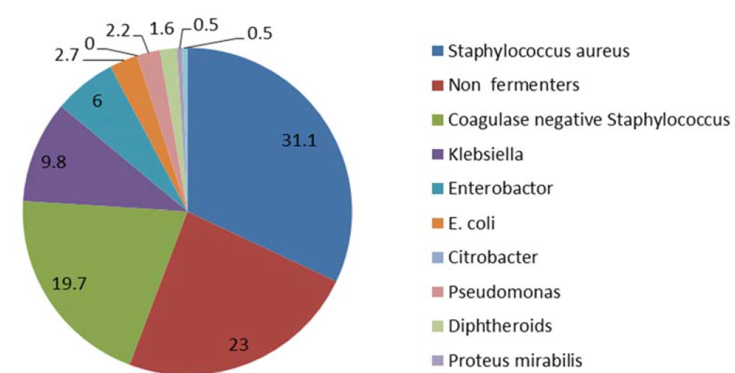
Bacteria were isolated from 183 samples and 162 samples were negative out of listed 345neonates. These 183 neonates were enrolled as cases in the study. Out of 183 cases, 70 cases were inborn and 113 were outborn. The blood culture isolation rate was 48.3% and 56.5% in inborn and outborn respectively. There were 102 males and 81 females in the study. The culture positivity rate was 53%. Bacteria were isolated from 37 samples of suspected EOS neonates with a positivity rate of 48% and 146 samples of suspected LOS with a positivity rate of 54.5%. Gram-positive bacteria were isolated from the 97 cases and gram-negative bacteria were grown in 86 cases. The most common isolate was Staphylococcus aureus in 38(31.1%) followed by non- fermenters in 42(23%) cases. Details of various isolates among 183 cases have been given in Table 2.
The positive isolation rate was 48.3% and 56.5% in inborn and outborn respectively. There were 102 males and 81 females. The blood culture isolation rate was 53%. Bacteria were isolated from 37 samples of suspected EOS neonates with a blood culture positivity rate of 48% and 146 samples of suspected LOS with a positivity rate of 54.5%. Gram-positive bacteria were isolated from the 97 cases and gram-negative bacteria were grown in 86 cases. The most common isolate was Staphylococcus aureus in 38(31.1%) followed by non-fermenters in 42(23%) cases. Gram-positive bacteria were isolated more from positive blood cultures as compared to gram-negative bacteria. The therapeutic outcome of neonates both with EOS and LOS caused by sensitive isolates was relatively good as compared to those with resistant isolates.
On sensitivity patterns, most of the isolates were sensitive to ampicillin and gentamicin combination and a little less proportion was sensitive to third-generation cephalosporins. Other antibiotics showed sensitivity as imipenem, meropenem, vancomycin, and linezolid as expected from 2 nd line drugs. (Table 3-9)
Table 1: Socio-Demiographic Profile of All the Neonates Were Admitted With Suspected Sepsis
Age
Sex
Place of delivery
Gram staining
EOS
LOS
Male
Female
Inborn
Out born
positive
negative
Suspected
77
268
186
159
145
200
NA
NA
Confirmed
37
146
102
81
70
113
97
86
Table 2: Distribution of bacterial isolates from blood culture
Bacteria
Number (%age)
Staphylococcus aureus
57(31.1%)
Non fermenters
42 (23.0%)
Coagulase negative Staphylococcus
36(19.7%)
Klebsiella
18(9.8%)
Enterobactor
11(6.0%)
E. coli
5(2.7%)
Citrobacter
5.27 %
Pseudomonas
4 (2.2%)
Diphtheroids
3(1.6%)
Proteus mirabilis
1(0.5%)
Enterococcus
1(0.5%)
Total
183(100%)
Table 3: Showing Antibiotic Sensitivity Pattern of Staphylococcus Aureus Isolates
Sensitivity
Amika
Amoxiclav
Ampisal
Ampicillin
Aztreon
Cipro
Clinda
Cotri
Eryth
Genta
Levo
Lino
Netli
Tobra
Vanco
Sensitive
20
30
52
36
11
25
26
21
27
25
23
11
9
9
11
Resistant
37
37
5
21
0
32
31
36
30
32
32
0
2
2
0
Total
57
57
57
57
11
57
10
57
57
57
57
11
11
11
11
Table 4: Showing Antibiotic Sensitivity Pattern of Coagulase-Negative Staphylococci
Amika
Amoxiclav
Ampisal
Ampicill
Azithromy
Aztreon
Ceftazidime
Cipro
Clinda
Cotri
Eryth
Genta
Levo
Lino
Piper
Tetra
Tige
Vanco
Sensitive
15
17
18
20
16
18
12
36
18
20
15
21
15
18
11
16
13
18
Resistant
10
19
4
16
9
0
24
23
4
16
21
15
21
0
7
20
5
0
Total
36
36
22
36
25
18
36
13
22
36
36
36
36
18
18
36
18
18
Table 5: Showing Antibiotic Sensitivity Pattern of Citrobacter
Ampicill
Cipro
Cotri
Eryth
Genta
Lino
Vanco
Sensitive
2
2
1
0
3
5
4
Resistant
3
3
4
5
2
0
1
Total
5
5
5
5
5
5
5
Figure 1: Distribution of Bacterial Isolates from Blood Culture
Table 6: Showing Antibiotic Sensitivity Pattern of E.Coli
Sensitivity
Amika
Amoxiclav
Ampisal
Ampicill
Imip
Aztreon
Cefopodo
CFZ/CLV
Ceftri
CFZ
Cipro
cefepime
Cotri
Doxy
Colis
Genta
Piper+Taz
Sensitive
2
1
4
2
2
2
2
1
3
2
1
2
1
1
2
3
3
Resistant
3
4
1
3
0
0
0
1
2
3
4
3
4
4
0
2
2
Total
5
5
5
5
2
2
0
2
5
5
5
5
5
5
2
5
5
Table 7: Showing Antibiotic Sensitivity Pattern of Klebsiella
Sensitivity
Amika
Ampisal
Ampicill
Imip
Aztreon
Cefopodo
CFZ/CLV
Ceftri
CFZ
Cipro
Cefepime
Cotri
Colis
Genta
Mero
PB
Piper+Taz
Sensitive
3
5
3
2
2
2
4
3
3
2
2
0
9
6
6
3
2
Resistant
15
13
15
7
7
16
14
15
15
16
16
18
0
12
3
6
7
Total
18
18
18
9
9
18
18
18
18
18
18
18
9
18
9
9
9
Table 8: Showing Antibiotic Sensitivity Pattern of Enterobacter Spp
Sensitivity
Amika
Ampicillin
Ampisal
Imp
Aztreon
Cefopodo
Ceftri
CFZ
Cipro
Cotri
Colis
Genta
Mero
PB
Piper+Taz
Sensitive
2
3
3
4
4
2
4
3
2
2
5
3
4
4
4
Resistant
9
8
2
1
1
3
7
8
9
9
0
8
1
1
7
Total
11
11
5
5
5
5
11
11
11
11
5
11
5
5
11
Table 9: Showing Antibiotic Sensitivity Pattern of Non-Fermenters
Sensitivity
Amika
Ampicillin
Imip
Aztreon
Cefopodo
Ceftri
CFZ
Cipro
cefepime
Genta
Piper+Taz
Sensitive
12
18
8
7
4
18
14
13
15
17
19
Resistant
30
24
2
3
6
24
28
29
27
25
23
Total
42
42
10
10
10
42
42
42
42
42
42
DISCUSSION
Clinical signs and symptoms in neonates though useful have limited specificity as compared to bacteriological culture and sensitivity of blood samples. The common isolates in our studies were Staph. aureus, Non-fermentors, CONS, Klebsiella,E.Coli the findings are consistent with those of the above-mentioned topic. It also points out a very important finding that infections by these agents cause a bigger threat to child survival in developing countries and should be taken seriously. The findings of our study suggest that empirical therapy with ampicillin and gentamycin may be started as soon as possible to minimize morbidity and mortality in both EOS and LOS [4-5].
In a study carried out on neonatal sepsis in India by Joshi et al., in Pune [3-5].It was reported that out of 100 cases of neonatal sepsis 25% of cases were blood culture positive. Among them, gram-negative bacteria constituted the most common isolates which included P. aeruginosa (38.3%),K pneumoniae (30.4%), E.coli ( 15.5% ), and Acinetobacterspp (7.8%).Acinetobacter and Citrobacter sepsis have been reported previously among newborns from Southeast Asia by ZA Bhutta in a study done in Karachi. Case death rates of 42-61% were reported among neonates with Citrobacter and Acinetobacter sepsis.4In a study done by Brook I in Washington it was reported that important causative agents causing bacteremia and meningitis in children under the age of one-month-old were Group B Streptococcus, E.coli, Listeria monocytogenes, S. pneumonia, Haemophilus influenzae, S. aureus, Neisseria meningitides, and Salmonella species [3-6].
CONCLUSION
Neonatal septicemia is a very important cause of loss of life in neonates. Prompt and proper clinical diagnosis as well as starting empirical treatment as soon as possible are the most important to reduce neonatal deaths. In neonatal deaths, it is as critical in a given setting as pathogens causing bacterial sepsis differ in antibiotic sensitivity patterns in various settings. Clinical signs and symptoms in neonates though useful have limited specificity as compared to bacteriological culture and sensitivity of blood samples. The common isolates in our studies were Staph. aureus, Non-fermentors, CONS, Klebsiella,E.Coli the findings are consistent with those of the above-mentioned topic. It also points out a very important finding that infections by these agents cause a bigger threat to child survival in developing countries and should be taken seriously. The findings of our study suggest that empirical therapy with ampicillin and gentamicin may be started as soon as possible to minimize morbidity and mortality in both EOS and LOS. Hence our study concludes that ampicillin and gentamicin combination given together showed a better gram-positive and gram-negative cover as compared to cephalosporins given alone or in combination for empirical therapy in suspected cases of septicaemia.
REFERENCE
Stam, J. et al. “Antibiotic use in infants in the first year of life in five European countries.” Acta Paediatrica, vol. 101, no. 9, September 2012, pp. 929–934. doi:10.1111/j.1651-2227.2012.02728.x.
Clinical and Laboratory Standards Institute. Performance Standards for Antimicrobial Susceptibility Testing. Twenty-Fourth Informational Supplement, vol. 34, no. 1, CLSI, January 2014, M100–S24.
Joshi, S. et al. “Mixed Salmonella infection—a case report.” Indian Journal of Medical Microbiology, vol. 20, no. 2, 2002, pp. 113–114.
Bhutta, Z.A. “Enterobacter sepsis in the newborn—a growing problem in Karachi.” Journal of Hospital Infection, vol. 34, no. 3, 1996, pp. 211–216.
Brook, I. “Unexplained fever in young children: how to manage severe bacterial infection.” BMJ, vol. 327, no. 7423, 2003, pp. 1094–1097.
Yurdakök, M. “Antibiotic use in neonatal sepsis.” The Turkish Journal of Pediatrics, vol. 40, no. 1, 1998, pp. 17–33.
Recommended Articles
Research Article
Electrochemical Comparative Studies On The Redox Behavior Of Cu(II) And Cr(II) Before And After Interactions With Ciprofloxacin In Solutions
H. M. Naseem Akhtar,
M. Q. Ehsan
Published: 10/08/2021
Download PDF
Research Article
A Rare Case Report of Waugh Syndrome in an Eight Months Old Male
Pielly Hanna,
...
Bassem Abou Merhi
Published: 10/03/2021
Download PDF
Research Article
Study on Prevalence and Etiology of Early Neonatal Deaths in Jasin district, Melaka from 2014-2017
Sujat a Khobragade,
...
Yadneshwar Khobragade
Published: 30/05/2021
Download PDF
Research Article
Clinicopathological Correlation in Cases of Acute Appendicitis: An Overview
Rahul Rai,
...
Tushar
Published: 05/04/2025
Download PDF
PO Box 101, Nakuru
Kenya.
Email: office@iarconsortium.org
Editorial Office:
J.L Bhavan, Near Radison Blu Hotel,
Jalukbari, Guwahati-India
Katoch, P. (2021). Bacteriological Profile and Antibiotic Sensitivity Pattern In Early Onset Sepsis and Late-Onset Sepsis and comparison of ampicillin gentamicin combination with regard to 3 rd generation cephalosporins for empirical treatment initiation. International Academic Journal of Applied Bio-Medical Sciences, 2(2), 1-5.
MLA
Katoch, Pankaj. "Bacteriological Profile and Antibiotic Sensitivity Pattern In Early Onset Sepsis and Late-Onset Sepsis and comparison of ampicillin gentamicin combination with regard to 3 rd generation cephalosporins for empirical treatment initiation." International Academic Journal of Applied Bio-Medical Sciences 2.2 (2021): 1-5.
Chicago
Katoch, Pankaj. "Bacteriological Profile and Antibiotic Sensitivity Pattern In Early Onset Sepsis and Late-Onset Sepsis and comparison of ampicillin gentamicin combination with regard to 3 rd generation cephalosporins for empirical treatment initiation." International Academic Journal of Applied Bio-Medical Sciences 2, no. 2 (2021): 1-5.
Harvard
Katoch, P. (2021) 'Bacteriological Profile and Antibiotic Sensitivity Pattern In Early Onset Sepsis and Late-Onset Sepsis and comparison of ampicillin gentamicin combination with regard to 3 rd generation cephalosporins for empirical treatment initiation' International Academic Journal of Applied Bio-Medical Sciences 2(2), pp. 1-5.
Vancouver
Katoch P. Bacteriological Profile and Antibiotic Sensitivity Pattern In Early Onset Sepsis and Late-Onset Sepsis and comparison of ampicillin gentamicin combination with regard to 3 rd generation cephalosporins for empirical treatment initiation. International Academic Journal of Applied Bio-Medical Sciences. 2021 Jul;2(2):1-5.


 Download PDF
 Download XML