The goal of this study was to determine the antioxidant, antimicrobial and anti-inflammatory capacity of oven-dried methanolic extract (OME) of Moringa oleifera (M. oleifera) leaves. OME showed promising total phenolic content (2.6783 ± 0.0555 mg/g, 0.2678 ± 0.0055 mg/g, 0.0586 ± 0.0037 mg/g) and total flavonoid content (0.8079 ± 0.0050 mg/g, 0.0808 ± 0.0005 mg/g, 0.0140 ± 0.0018 mg/g) with tested concentration of 100 mg/mL, 10 mg/mL and 2 mg/mL. Ferric reducing activity (5.6515 ± 0.0983 mM Fe2+, 2.0751 ± 0.0410 mM Fe2+, 0.6015 ± 0.0128 mM Fe2+) and DPPH radical scavenging activity (87.99 ± 0.24%, 16.21 ± 0.17%, 5.82 ± 0.80%) of OME was in correlation with phytochemical compounds. OME also showed antimicrobial activity on E. coli and S. aureus which is comparable to commercial antibiotic (Streptomycin). The IC50 of OME in proteinase inhibition was 2.57 ± 0.02 mg/mL. Overall, OME could be a potential remedy in the management of oxidative stress and microbial infections.
Moringa oleifera (M. oleifera) is known as a multipurpose tree as each part of the plant has its medical value. It has been used as traditional remedies, cosmetic oil, water coagulant and forage for livestock [1]. M. oleifera leaves is the most investigated part of the plant due to the rich sources of phytochemicals demonstrating antioxidant, anti-inflammatory, antimicrobial and antidiabetic activity [2-3]. The mechanisms of these therapeutic properties are closely related to their antioxidant capacity among others [4]. The interest in plants with antioxidant, anti-inflammatory and antimicrobial has been considerably increased as a result of commercial drugs resistance and their side effects. The resistance may due to the indiscriminate use of commercial drugs while undesirable side effects might lead to emergence of uncommon diseases [5-7]. For instance, non-steroidal anti-inflammatory drugs (NSAIDs) resistance in dysmenorrhea mentioned by Oladuso et al. Verma et al. [8] also reported the increasing antibiotic resistance in bacteria by NSAIDs. Hypermutable strains bacteria have higher potential of drug resistance even with very low concentrations of antimicrobials [9]. This phenomenon has given scientists the impetus to discover novel and alternative compounds from medicinal plants.
There are many extraction methods to recover the bioactive compounds from plants. Apart from the duration and temperature, the extraction solvent is one of the factors which interfere with the phytochemical contents and bioactivities of plants [4,10]. Since the bioactive compounds have different chemical properties and polarities which may not be soluble in a particular solvent. The most suitable solvents would be the aqueous mixtures consisting of polar solvents such as methanol, ethanol and acetone. Polar solvents have been proven to increase the solubility of polyphenols from plant matrices [10]. The notion of the best extraction solvent depends on the targeted compounds and varies from one plant to another. It is therefore important to determine the extraction solvent which will be the most suitable for bioactive compounds of M. oleifera leaves [4]. Hence, the objective of this study was to determine the effects of the extraction solvent on phytochemical contents and bioactivity of M. oleifera fresh leaves extracts as well as the relationship between the phytochemical content with antioxidant, antimicrobial and anti-inflammatory activities.
M. Oleifera Leaves and Reagents
Fresh M. oleifera leaves were collected from Taman Bukit Tiram, Johor Bahru, Malaysia. Methanol, hydrochloric acid (HCl), sodium acetate trihydrate, gallic acid, Folin-Ciocalteu reagent, perchloric acid and glacial acetic acid were purchased from Merck. 2,4,6-Tri(2-pyridyl)-s-triazine (TPTZ) was purchased from Acros Organics. Bovine serum albumin (BSA) was purchased from Vivantis. Trypsin was purchased from amersco. Tris base was purchased from Fisher. Iron (III) chloride hexahydrate was purchased from Qrec. Iron (II) sulphate was purchased from Systerm. 2,2-diphenyl-1-picrylhydrazyl (DPPH) and tryptone were purchased from Sigma-Aldrich. Sodium carbonate, Dimethyl sulfoxide (DMSO), agar, quercetin was purchased from VChem. Aluminium chloride 6-hydrate and sodium chloride were purchased from Bendosen. Yeast extract was purchased from Scharlab. Potassium acetate was purchased from HmbG. Escherichia coli ATCC10536 and Staphylococcus aureus ATCC29737 were kind gifts from the Assoc. Prof. Dr. Nik Ahmad Nizam Nik Malek under Faculty of Science, UTM.
Sample Preparation
Fresh and young M. oleifera leaves were harvested and oven dried (50
) to a constant mass before pulverized into powder. Oven-dry M. oleifera leaves powder (1 g) was extracted using 80% methanol (50 mL) according to Ali et al., (2013) with modification. The mixture was stirred (100 – 120 rpm) at room temperature for 6 hours. The extract was filtered from the residues by using Whatman No. 1 filter paper. The residues were re-extracted with the 25 mL methanol. Both extracts were combined and concentrated by evaporating the solvent under reduced pressure at 50

using rotary evaporator. The concentrated extracts were mixed with 50% DMSO to make 100 mg/mL and diluted to 2, 6, 10 mg/mL. Oven-dry M. oleifera leaves methanolic extract (OME) were stored at – 80
until used.
Determination of Polyphenol Content Total Phenolic Content
Total phenolic content (TPC) in each extract was determined as previously described [11], with minor modifications. Briefly, 100
L of extract was mixed with 1 mL of Folin-Ciocalteu reagent. After 5 min incubation, 800
of sodium carbonate was added and allowed to stand in darkness for 60 min. The absorbance of the reaction mixture was measured at 650 nm. The calibration curve was established using gallic acid (50 – 250
g/mL). TPC was expressed as gallic acid equivalent (GAE) in mg/g of extract.
Total Flavonoid Content
Total flavonoid content (TFC) of each extract was investigated using aluminium chloride colorimetry method described by Formagio et al., (2014) with slight modifications. In brief, 200
L of extract was mixed with 600
L of 95% ethanol, 40
L of 10% aluminium chloride, 40
L of 1 M potassium acetate and 1.12 mL ultrapure water. After incubation in darkness for 30 min, the absorbance of the reaction mixture was measured at 415 nm. The calibration curve was established using quercetin (100 – 500
g/mL). TFC was expressed as quercetin equivalent (QCE) in mg/g of extract.
Antioxidant Activity
Frap: Ferric reducing activity of the extracts was determined with the Ferric Reducing Antioxidant Power (FRAP) assay (Pulido et al., 2000), with modification. Briefly, a mixture consisting of 30
L of OME, 90
L of ultrapure water (UPW) and 900
L of FRAP reagent was incubated at 37
for 30 min. Absorbance of the mixture was read at 593 nm. FRAP values are presented in mM Fe2+ equivalents, calculated from a standard curve prepared with 0.5 to 4.0 mM FeSO4 . 7H2O.
Dpph Radical Scavenging Assay
DPPH radical scavenging activity was performed as described by Sanchez-Moreno et al. [12], with minor modification. In brief, 20
L of extract was mixed vigorously with 1.48 mL of DPPH solution. After 20 min incubation at 37
in darkness, the absorbance of the reaction mixture was measured at 517 nm. The percentage inhibition of radicals was calculated as shown below

Where Abscontrol is the absorbance of 50% DMSO with DPPH solution while Abssample is the absorbance of extract with DPPH solution.
Antimicrobial Assay
Bacterial Cultures: Strains Escherichia coli (E. coli) ATCC10536 and Staphylococcus aureus (S. aureus) ATCC29737 were used in this assay. Both strains were sub cultured from the stock on to Luria broth (LB) agar for 24 hours prior to testing.
Agar Well Diffusion
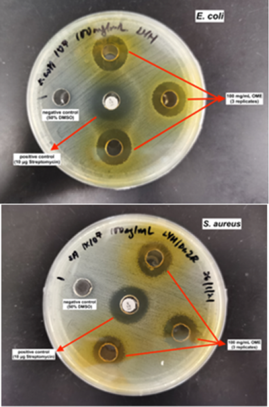
The antimicrobial ability of OME was investigated with agar well diffusion method by referring to Dahiya and Purkayastha [13]. A few colonies of each strain of bacteria were inoculated in phosphate buffer saline and diluted to 1 X 10⁷ CFU/mL according to the McFarland standard (0.5). The bacteria culture was spread on LB agar plates with cotton swab moistened with the bacterial suspension. Five wells of 8 mm diameter were punched into the agar medium and filled with 100
L (100 mg/mL) of OME. One of the wells that filled with 50% DMSO was served as negative control while standard antibiotic discs of streptomycin (10
g) was used as positive control. After 24 hours incubation at 37
, the diameters of the inhibition zones were measured in mm.
Proteinase Inhibition Assay
The anti-inflammatory capacity of the extract was determined using proteinase inhibition method from Naz et al. [14] with modification. The reaction mixture contained 100
L OME and 100
L 0.06 mg/mL trypsin (in 20 mM, pH 7.4 Tris HCl buffer) was incubated at 37
for 5 minutes. Then, 100
L 4% BSA was added before further incubation for 20 minutes. A volume of 200
L 70% perchloric acid was used to stop the reaction. The mixture was centrifuged at 3000 rpm for 10 minutes and measured at 220 nm. The calculation of percentage of inhibition is shown below:

Note
OA : Absorbance of blank solution without enzyme
OB : Absorbance of blank solution with enzyme
SA: Absorbance of sample solution without enzyme
SB : Absorbance of sample solution with enzyme
Statistical Analysis
Analyses were performed in triplicates and data are presented as mean ± standard errors (SE). Statistical analysis was performed by using SPSS Statistics software (version 26). Data were analyzed by the one way ANOVA test for the comparison of multiple concentration and p-value of <0.05 was considered statistically significant.
Effect of Solvent on Extraction Yield and Polyphenol Contents
Extraction is always the main procedure to recover and isolate phytochemicals from plant materials. However, the efficiency of extraction is generally influenced by the chemical structure of phytochemicals, extraction method, as well as the extraction solvent used. The effect of polarity of solvent in the extraction yield and antioxidant activity of phytochemical compounds in the plant sample has been proven [14,10]. In this study, methanol (polar solvent) were used to extract the M. oleifera leaves. The high percentage yield of OME (in Table 1) implied that the plant crude extract favours the highly polar solvent. Sultana et al. [15] also reported that methanolic extract had higher yield than ethanolic extract. Compounds other than phenolics such as protein and carbohydrate which have a higher solubility in methanol may also contribute to higher yield [16]. Moreover, high extraction yield could also be the influence of oven drying of the leaves. Dadi et al. [17] found that oven dried M. stenopetala leaves gave higher yield compared to room dried leaves. According to Vongsak et al. [18], dried M. oleifera leaves had higher extraction yield than fresh leaves. Oven dry could remove the excess water in the leaves within a short period of time. The shorter drying time could promote lower relative humidity [17]. Oven dry is more preferable than air dry because it has shorter drying time, its closed condition retains more bioactive compounds than sun drying and it could enhance the sensory attributes (colour and texture) [19]. The result implies that polarity of solvent and moisture of leaves will influence the extraction yield.
Table 1: Percentage of Yield, TPC and TFC of OME Based on Three Concentrations
Concentration (mg/mL) | Extraction yield (%) | TPC (mg GAE/g) | TFC (mg QCE/g) |
OME | 9.66 |
|
|
100 |
| 2.8163 | 0.7255 |
10 |
| 0.2686 | 0.0771 |
2 |
| 0.0589 | 0.0136 |
From Table 1, both TPC and TFC extracted showed a concentration-dependant manner. A correlation between TPC and TFC was observed from the result where the increase of TFC value is directly proportional to the TPC. This implies that flavonoids may be the dominating phenolic group in M. oleifera leaves. Based on Povolo [20], oven-dried M. oleifera leaves (158 mg GAE/g) had higher TPC compared to freeze-dried (77.5 mg GAE/g) while methanolic extract of M. oleifera leaves gave higher TPC and TFC than ethanolic extract was determined by Sultana et al. [20]. Vyas et al. [21] reported that TPC of shaded dried methanolic extract M. oleifera leaves was the highest among petroleum ether, chloroform and water. However, methanolic extract resulted in higher TPC while ethanolic extract with higher TFC were reported by Nobossé et al. [4]. The discrepancy in the results may be due to the concentration of extraction solvent used, the temperature of oven dry and the age of the leaves harvested. Aqueous methanol was used in this study instead of absolute methanol because the increase in the concentration of aqueous will enhance the solubility of phenolic compounds in the plant [22,10].
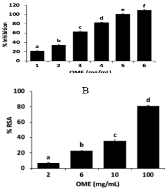
According to Nobossé et al. [4], phenolic compounds in the plant extract contribute to significant bioactivities.The antioxidant capacity of a plant is positively correlated with its phytochemical content [20,4,10]. Since the natural phytochemicals are complex in the crude extract, two different modes of antioxidant assays had been carried out to assess the antioxidant capacity of OME more accurately. The reducing power of OME was investigated from the colour changes, whereby the straw colour of test solution changes to shades of blue which indicates the reduction of Fe3+ complex to Fe2+ form. The colour change is depending on the presence of antioxidants in the extract. Besides, the radical scavenging activity of OME was determined through the transferring of electron from the antioxidant to DPPH radical, which results in the noticeable discolouration from purple to yellow. The stable free radical can be read with an absorption band at 517 nm [23]. Figure 1 shows the reducing power and DPPH scavenging activity of OME extract in a concentration-dependant manner. Both antioxidant results are supported by the oven-dry Scurrula ferruginea methanolic leaf extract from Justine et al. [24]. The study from Khodja et al. [19] mentioned that oven drying could preserve the nutritional quality and bioactive compounds in the leaves as it guarantees the removal of water, which contribute to the degradation of phenolics by enzymatic action and chemical oxidation. Also, the leaf extracted with aqueous methanol (80%) resulted in the highest scavenging activity and reducing power among the 100% methanol, 80% and 100% acetone. However, methanol extraction of shade dried Albizia odoratissima leaf possessed an intermediate FRAP and DPPH scavenging activity, which is lower than ethyl acetate but higher than hexane and chloroform extraction [25]. This variation might be due to plant species as well as different drying and extraction method. The phenolic content and various antioxidant compounds in crude extract are highly contributed by the antioxidant capacity of the plant [4]. Oven-dry method was once again observed to demonstrate higher total phenolic content in the study of Povolo [20].
Negative control showed no inhibition. The results were consistent with that reported by Abo El-Fadl et al. who observed that methanolic extract of oven-dry M. oleifera leaves had inhibition zones with diameter 18.2±1.2 mm (E. coli) and 16.1±0.63 mm (S. aureus). However, this data differed from Abdalla et al. [26] who pointed no antibacterial activity of the extraction of M. oleifera leaves using 20, 40 and 60% methanol against E. coli. The leaves extracted with 40 and 60% methanol showed the same diameter of inhibition zones against S. aureus. The variation in the strength of antibacteria of the extract might due to different plant sources and variety strains of bacteria used. Abdallah and Ali [27] reported that ethanolic extract of M. oleifera leaves had stronger antimicrobial activity than aqueous extract. The diameter of inhibition zones contributed by commercial antibiotic used, streptomycin was 20.3 ± 0.3 and 18.3 ± 0.3 mm against E. coli and S. aureus respectively.
Proteinase Inhibition Activity of OME
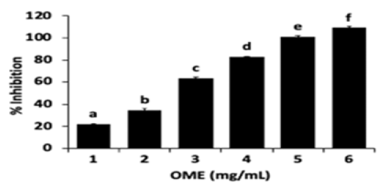
Excessive formation of reactive oxygen species (ROS) will lead to oxidative stress, leading to induction of cell damage that can culminate in cell death Antioxidants network play an important role in balancing the formation of ROS, however, the balance is somehow shifted to production of ROS even when undergoing the antioxidant therapy [28]. The accumulation of ROS will also induce inflammation. Therefore, there is a correlation between anti-inflammatory activity with the antioxidant capacity. Proteinases play an important role in arthritic reactions. Neutrophils are known to be the rich source of serine proteinases involved in inflammatory response [14]. BSA was used as substrate and trypsin was used as a serine protease enzyme to cut the peptide linkage of BSA. Based on Figure 2-3, OME showed significant anti-proteinase inhibition activity at different concentration. The activity was increasing in a concentration dependent manner. It possessed the highest inhibition activity (109.23 ± 1.56%) at the concentration of 6 mg/mL. IC50 value of OME was observed at 2.57 ± 0.02 mg/mL. Cold maceration extraction of M. oleifera leaves using distilled water was reported to have 29.35% inhibition at the concentration of 100
g/mL [29]. Padmalochana [30] reported that the IC50 of ethanol and acetone extract of M. oleifera leaves was 274.43 ± 0.73 and 322.10 ± 1.34
g/mL respectively. Shaded dried aqueous and methanolic extract of M. oleifera leaves showed better inhibition of proteinase (IC50 = 182.6 and 199.3
g/mL respectively) than other extraction solvents (n-hexane, ethyl acetate and butanol) [31]. By comparison, methanolic extraction of M. oleifera leaves gave better anti-inflammatory activity than other extraction solvents. The discrepancy between the results might be due to the difference in concentration of extraction solvent, drying method and substrate used in the assay. Studies on other plants also exhibited significant proteinase inhibitory activity. Methanolic extract of Niebuhria apetala leaves showed maximum inhibition activity (58%) at 500
g/mL [32]. Moreover, methanolic extract of Adansonia digitate leaves, Flueggea leucopyrus leaves and Vitex negundo leaves were reported to have stronger protease inhibition compared to chloroform and water extract [33]. However, aqueous extract of Aloe vera, Cannabis sativa, Datura stramanium and Calotropis gigantea exhibited stronger proteinase inhibition compared to methanolic extract [34]. Therefore, a suitable extraction method should be applied to a particular plant species which could affect the anti-inflammatory activity of the plant.

Figure 1: (A) Ferric Reducing Activity and (B) DPPH Radical Scavenging Activity of OME in Each of the Concentration. Each Bar Represents Mean
Standard Errors (N = 3). Bars Denoted by Different Letters are significantly Different (p <0.05) according to SPSS’s One Way ANOVA Test

Figure 2: Agar Well Diffusion Tests of 100 Mg/Ml OME on
CFU/Ml E. coli and S. Aureus

Figure 3: Proteinase Inhibition Activity of OME. Each Bar Represents Mean
Standard Errors (N = 3). Bars Denoted by Different Letters Are Significantly Different (p< 0.05) According To SPSS’s One Way ANOVA Test
The present study demonstrated the OME promising antioxidant capacity in different mechanism. OME also possessed antimicrobial as well as anti-inflammatory activities which are comparable to the standard drugs such as streptomycin (antibiotic) and piroxicam (NSAID). Further biological research could be focused on other antioxidant and anti-inflammatory assays in order to explore different mechanism of bioactive compounds in OME. Nevertheless, OME is potentially to be served as antioxidant remedies for pharmaceutical industries to treat the free-radical-associated oxidative damage. It could be processed to become commercial drugs which target the inflammation in different condition as well as the alternative antimicrobial drugs for treatment of infectious diseases. OME formulation could be a consideration as multifunctional formulation is a new trend of the cosmetic market.
Conflict of Interests
The authors have not declared any conflict of interests.
Acknowledgements
Dr. Zaidah Rahmat, Universiti Teknologi Malaysia
Universiti Teknologi Malaysia contract research grant
Chris Leong, Chris Leong Method R&D Sdn. Bhd, vote no. 4C276
Vergara-Jimenez, M. et al. “Bioactive components in Moringa oleifera leaves protect against chronic disease.” Antioxidants, vol. 6, no. 4, 2017, p. 91.
Yan, G. et al. “UPLC-Q-Orbitrap-MS2 analysis of Moringa oleifera leaf extract and its antioxidant, antibacterial and anti-inflammatory activities.” Natural Product Research, vol. 34, no. 14, 2020, pp. 2090–2094.
Natsir, H. et al. “Inhibitory activities of Moringa oleifera leaf extract against α-glucosidase enzyme in vitro.” Journal of Physics: Conference Series, vol. 979, no. 1, March 2018, p. 012019.
Nobossé, P. et al. “Effects of age and extraction solvent on phytochemical content and antioxidant activity of fresh Moringa oleifera L. leaves.” Food Science & Nutrition, vol. 6, no. 8, 2018, pp. 2188–2198.
Ghasemian, M. et al. “Review of anti-inflammatory herbal medicines.” Advances in Pharmacological Sciences, vol. 2016, 2016.
Fitriana, W.D. et al. “Antioxidant activity of Moringa oleifera extracts.” Indonesian Journal of Chemistry, vol. 16, no. 3, 2016, pp. 297–301.
Moyo, B. et al. “Antimicrobial activities of Moringa oleifera Lam leaf extracts.” African Journal of Biotechnology, vol. 11, no. 11, 2012, pp. 2797–2802.
Verma, T. et al. “Non-steroidal anti-inflammatory drugs, acetaminophen and ibuprofen, induce phenotypic antibiotic resistance in Escherichia coli: roles of marA and acrB.” FEMS Microbiology Letters, vol. 365, no. 22, 2018, fny251.
Reygaert, W.C. “An overview of the antimicrobial resistance mechanisms of bacteria.” AIMS Microbiology, vol. 4, no. 3, 2018, pp. 482–501.
Do, Q.D. et al. “Effect of extraction solvent on total phenol content, total flavonoid content and antioxidant activity of Limnophila aromatica.” Journal of Food and Drug Analysis, vol. 22, no. 3, 2014, pp. 296–302.
Baba, S.A. and S.A. Malik. “Determination of total phenolic and flavonoid content, antimicrobial and antioxidant activity of a root extract of Arisaema jacquemontii Blume.” Journal of Taibah University for Science, vol. 9, no. 4, 2015, pp. 449–454.
Sánchez-Moreno, C. et al. “A procedure to measure the antiradical efficiency of polyphenols.” Journal of the Science of Food and Agriculture, vol. 76, no. 2, 1998, pp. 270–276.
Dahiya, P. and S. Purkayastha. “Phytochemical screening and antimicrobial activity of some medicinal plants against multi-drug resistant bacteria from clinical isolates.” Indian Journal of Pharmaceutical Sciences, vol. 74, no. 5, 2012, pp. 443–450.
Naz, R. et al. “Antimicrobial activity, toxicity and anti-inflammatory potential of methanolic extracts of four ethnomedicinal plant species from Punjab, Pakistan.” BMC Complementary and Alternative Medicine, vol. 17, no. 1, 2017, p. 302.
Sultana, B. et al. “Effect of extraction solvent/technique on the antioxidant activity of selected medicinal plant extracts.” Molecules, vol. 14, no. 6, 2009, pp. 2167–2180.
Zieliński, H. and H. Kozłowska. “Antioxidant activity and total phenolics in selected cereal grains and their different morphological fractions.” Journal of Agricultural and Food Chemistry, vol. 48, no. 6, 2000, pp. 2008–2016.
Dadi, D.W. et al. “Influences of different drying methods and extraction solvents on total phenolic and flavonoids and antioxidant capacity of Moringa stenopetala leaves.” Journal of Pharmacognosy and Phytochemistry, vol. 7, no. 1, 2018, pp. 962–967.
Vongsak, B. et al. “Maximizing total phenolics, total flavonoids contents and antioxidant activity of Moringa oleifera leaf extract by the appropriate extraction method.” Industrial Crops and Products, vol. 44, 2013, pp. 566–571.
Khodja, Y.K. et al. “Conventional method and microwave drying kinetics of Laurus nobilis leaves: effects on phenolic compounds and antioxidant activity.” Brazilian Journal of Food Technology, vol. 23, 2020.
Povolo, C. Extraction and Characterization of Chemicals from Vegetal Matrices and Assessment of Their Properties for Nutraceutical and Cosmetic Applications, 2018.
Vyas, S. et al. “Comparative analysis of phenolic contents and total antioxidant capacity of Moringa oleifera Lam.” Pharmacognosy Journal, vol. 7, no. 1, 2015, pp. 44–51.
Mujtaba, A. et al. “Effect of solvents on extraction yield, total flavonoid, total phenolic contents, DPPH scavenging activity and antibacterial potential of three apricot cultivars.” Transylvanian Review, vol. 18, no. 10, 2016, pp. 1662–1676.
Hseu, Y.C. et al. “Antioxidant activities of Toona sinensis leaves extracts using different antioxidant models.” Food and Chemical Toxicology, vol. 46, no. 1, 2008, pp. 105–114.
Justine, V.T. et al. “Effect of drying methods and extraction solvents on phenolic antioxidants and antioxidant activity of Scurrula ferruginea leaf extracts.” Sains Malaysiana, vol. 48, no. 7, 2019, pp. 1383–1393.
Banothu, V. et al. “Phytochemical screening and evaluation of in vitro antioxidant and antimicrobial activities of the indigenous medicinal plant Albizia odoratissima.” Pharmaceutical Biology, vol. 55, no. 1, 2017, pp. 1155–1161.
Abdalla, A.M. et al. “Evaluation of antimicrobial activity of Moringa oleifera leaf extracts against pathogenic bacteria isolated from urinary tract infected patients.” Journal of Advanced Laboratory Research in Biology, vol. 7, no. 2, 2016, pp. 47–51.
Abadallah, M.S. and M. Ali. “Antibacterial activity of Moringa oleifera leaf extracts against bacteria isolated from patients attending General Sani Abacha Specialist Hospital Damaturu.” J. Allied Pharm. Sci., 2019, pp. 61–66.
Poljsak, B. et al. “Achieving the balance between ROS and antioxidants: when to use the synthetic antioxidants.” Oxidative Medicine and Cellular Longevity, 2013.
Kambizi, L. et al. “Proteinease inhibition, membrane stabilization, antioxidant and phytochemical evaluations of leaves, seeds and calyces of four selected edible medicinal plants.” Cogent Chemistry, vol. 3, no. 1, 2017, 1314064.
Padmalochana, K. “Anti-inflammatory activity and phytochemical analysis of Moringa oleifera ethanol and acetone leaves extract.” Journal of Drug Delivery and Therapeutics, vol. 8, no. 6-s, 2018, pp. 269–273.
Saleem, A. et al. “Antioxidant, anti-inflammatory and antiarthritic potential of Moringa oleifera Lam.” South African Journal of Botany, vol. 128, 2020, pp. 246–256.
Rajesh, A. et al. “Anti-inflammatory activity of methanol extract of Niebuhria apetala in vitro models.” Asian Journal of Pharmaceutical and Clinical Research, vol. 12, 2019, pp. 278–281.
Modi, C.M. et al. “Comparative evaluation of in vitro anti-inflammatory activity of different extracts of selected medicinal plants from Saurashtra region, Gujarat, India.” International Journal of Current Microbiology and Applied Sciences, vol. 8, no. 5, 2019, pp. 1686–1698.
Rastogi, S. et al. “In vitro study of anti-inflammatory and antioxidant activity of some medicinal plants and their interrelationship.” In Vitro, vol. 11, no. 4, 2018, pp. 2455–3891.