The protective effects of aqueous leaf extracts of Jatropha curcas (JC) and Hibiscus sabdariffa (HS) against hepatic toxicity resulting from lead acetate exposure were studied. The protective potentials of JC and HS were assessed by monitoring selected liver function and anti-oxidant indices. Sixty male Wistar rats were separated into ten equal groups. The first and second groups represented the normal and Lead acetate (PbA) (50mg/kg b.w) control groups respectively while groups 3 and 4 were given 100mg/kg JC and 200 mg/kg JC respectively. Groups 5 and 6 received 250 mg/kg HS and 500 mg/kg HS respectively. Groups 7-10 were given PbA (50mg/kg) and 100 mg/kg JC, 200 mg/kg JC, 250mg/kg HS, 500mg/kg HS respectively. Rats were orally administered their relevant doses for 28 days. Blood samples were collected from heart puncture at the end of the experiment (28 days) for hepatic function analysis. Results revealed that Pb exposure increased the gamma glutamyl transferase, total bilirubin, aminotransferase activities (AST and ALT), and malondialdehyde (MDA) levels and caused histological changes in the liver of the exposed rat groups. The concomitant treatment of PbA with aqueous extracts of JC and HS led to a significant protection from the deleterious effects of lead acetate.
Lead (Pb) is a toxic metal that induces a broad range of physiological, biochemical and neurological dysfunctions in humans as well as in animal model. It is a well-known multi-organ toxicant which damages the liver, kidney, reproductive system and other physiological organs [1]. Lead serves no useful purpose in the human body. Its presence in the body can lead to toxic effects, regardless of age, gender or exposure pathway [2]. It has been reported in previously published literature that lead exerts its toxic effect mainly by its ability to mimic divalent ions such as Ca2+, Mg2+, Fe2+ in cells and also induce generation of reactive oxygen species which disrupts the delicate prooxidant/antioxidant balance that exists in mammalian cells [3-4].
The liver is a vital organ which plays a key role in the biotransformation and detoxification of biotoxicants [5]. Mudipalli [6] evaluation of humans exposed to lead revealed that liver tissue is the largest repository of lead per g of wet tissue (33%) among the soft tissues. Lead toxicity has been associated with the disruption of the normal anatomical organization of hepatic lobules, loss of the distinctive cord-like structure of functional liver cells, hyperchromatic hepatocytes with occasional vacuolations and congestion of sinusoids [7]. Significant increase in free radical generation, activities of liver transaminase (alanine aminotransferase and aspartate aminotransferase) and total bilirubin have also been observed in experimental animals exposed to lead compounds [8].
Chelation therapy has been used as the classical treatment method for mitigating lead toxicity. However, the increasing side effects associated with chelators (especially synthetically derived) have paved the way for the resurged interest in the screening of natural plant products as preventive agents in attenuating the oxidative damage caused by heavy metals such as lead. The present study was designed to investigate the role of aqueous leaf extracts of two Nigerian medicinal plants against hepatotoxicity in male rats.
J. curcas belongs to Euphorbia family and has strong potentials for use both medicinally and nutritionally. Asuk et al. [9] reviewed the phytochemical and proximate composition of the leaf, stem and root parts of J. curcas and they reported that the JC leaves had appreciable amounts of polyphenols and flavonoids. Polyphenols and flavonoids are known to exercise anti-oxidative activities, protection against allergies, inflammation, platelet aggregation and hepatic toxicity [10-11]. H. sabdariffa is a plant with well-established neutraceutical properties. It has reportedly shown biological activities such as cancer prevention and liver protection [12-13].
These biological activities have been attributed to the high content of anthocyanins in the plant [14]. Prenesti et al. [15] found that the total antioxidant power of hibiscus decoctions were high and proposed that hibiscus beverages could act as a protection against free-radical damage. Although the hepato-protective effects of JC and HS extracts have been previously reported, there is still limited information available on their efficacy in lead induced hepatic damage. Hence, this study was designed to investigate the ameliorative potentials of JC and HS on hepatotoxicity, oxidative stress and liver tissue alterations in rats exposed to lead.
Chemicals
Lead acetate (Pb (CH3CO2)2) and all other chemicals used in the study were obtained from Sigma-Aldrich Corporation (St. Louis, Missouri, USA).
Plant material
The leaves of J. curcas were obtained from Abuja park of University of Port Harcourt, Rivers state, Nigeria while the H. sabdariffa calyces were obtained from a local market in Obior/Akpo Local Government Area of Rivers state, Southern Nigeria. The plant materials were washed using double distilled water and then dried at room temperature for 48 to 92 h and then grounded into powder and stored at room temperature until use. Plant aqueous extraction was done by cold maceration as previously described by Nagappan [16].
Animals care handling and Experiment Design
A total of 60 male adult albino rats of varying weights (90 – 200g), respectively bred in the animal house of the Department of Physiology, University of Port Harcourt were obtained for this study. The rats were divided into ten (10) groups and housed in polypropylene cages. They were maintained under ambient temperature and hygienic condition as prescribed by the committee for the purpose of control and supervision on experiments on animals (CPCSEA). Before the initiation of the experiment the rats were left to acclimatize for a period of seven (7) days to natural environmental conditions of 12 hours light and 12 hours dark cycle with ambient temperature of (25 ± 2) ºC. During the period of acclimatization and through the experiment the rats were fed with rat feed pellets and clean drinking water ad libitium. The research lasted for 28 days. After a period of adaptation, the animals were divided into ten experimental groups of 6 animals each according to their weight:
Group 1: Received distilled water only (normal control), no lead acetate and plant extracts were included in their diet
Group 2: Received oral administration of 50 mg/kg lead acetate PbA throughout the research (toxic control)
Groups 3-4: Received oral administration of 100 and 200 -mg/kg JC extracts respectively
Groups 5-6: Received oral administration of 250 and 500 -mg/kg HS extracts respectively
Groups 7-8: Received PbA (50mg/kg) + 100 and 200 -mg/kg JC extracts respectively
Groups 9-10: Received PbA (50mg/kg) + 250 and 500 -mg/kg HS extracts respectively
The concentrations of the aqueous JC and HS extracts in this study were selected to respectively represent one-tenth and one-twentieth portion of their LD50 concentration as previously established in literature. Mishra [17] and Dangambo et al. [18] reported the LD50 concentration of aqueous leaves extract of J. curcas to be above 2500mg/kg while that of H. sabdariffa has been estimated to be above 5000mg/kg for rats [19-21].
At the end of the experiment, the animals were fasted overnighted. The blood samples were collected, some of which was centrifuged to obtain the serum which was kept frozen at -20 ºC until used for biochemical analysis of AST, ALT, ALP and total bilirubin. The other portion of the blood samples were placed in Lithium heparin bottles after which they were centrifuged to obtain plasma used for the analysis of oxidative stress markers (Catalase, Superoxide dismutase, Glutathione and Malondialdehyde). Pieces of tissues from liver were immediately kept in 10% of formalin fixative to study histological alterations.
Biochemical analysis
Serum Biochemical Parameters: Enzymes marker were measured according to classic methods [22]. Total bilirubin, alkaline phosphatase (ALP), Gamma glutamyl transferase (GGT) and transaminases (AST and ALT) activities were estimated using an automatic clinical chemistry analyzer (Mindray BS800, Shenzen, China).
Oxidative stress markers
Determination of Superoxide Dismutase (SOD): SOD was estimated by the method that involved inhibition of superoxide-dependent reduction of tetrazolium dye methyl thiazolyl tetrazolium (MTT) to its formazan. The activity of superoxide dismutase was determined by the method of Misra and Fridovich [23].
Determination of Reduced Glutathione (GSH) Level
The method of Sedlak and Lindsay [24] was followed in estimating the level of reduced glutathione. GSH was estimated based on a reaction of reduced glutathione with 5-5ditiobis-2-nitrobenzoic acid (DTNB).
Determination of the Enzymatic Activity of Catalase
Catalase enzyme activity was assayed by measuring the catalase-induced loss of H2O2 contained in the sample by the measurement of H2O2 absorbance at 560 nm using a UV spectrophotometer (Biobase Biochemistry, Biobase D560, Shangdong ) according to the Aebi [25] method.
Estimation of lipid peroxidation levels
Malondialdehyde, the product of lipid peroxidation, was estimated by reaction with thiobarbituric acid (TBA) as per the method prescribed by Ohakawa et al. [26].
Histological Examinations
For light microscopy examination, the formalin fixed tissues were dehydrated in 70% alcohol for 1 minute; 95% alcohol for 1 min and 100% alcohol for 1 min (2 changes) respectively and placed in the oven overnight. Sections were subsequently cleared in three changes of xylene, and were embedded in paraffin. Serial sections, each of 4-micron thickness, were cut and stained with H and E as per standard protocols [27].
Statistical Analysis
The data was subjected to statistical analysis by using statistical package for social sciences (SPSS) version. Differences between means were tested using student t tests and significance was set at p<0.05 and the results are expressed as Mean ± Standard Error of Mean (SEM).
Biochemical Parameters
The results as presented in Table 1 showed that the GGT, AST, ALT and ALP activities increased significantly (p<0.05) in the serum of the group exposed to lead acetate compared to the control group. A similar trend was also observed for total bilirubin levels in the PbA group. There was no significant difference for serum ALT, ALP and GGT activities in the groups treated with the JS and HS extracts. Our results however showed significant decreases (p<0.05) in the activity of liver transaminases (AST and ALT), ALP, GGT and total bilirubin levels in the groups that received concomitant treatment of PbA and plant extracts (JC and HS) compared to the PbA group.
Oxidative Stress Parameters
Our results presented in Table 2 showed that lipid peroxidation and antioxidant enzyme activities (SOD, Catalase) were significantly elevated and decreased (p<0.05) respectively in PbA group compared to the normal control. In contrast, lipid peroxidation levels decreased significantly (p<0.05) in the PbA + JC-100 and PbA +JC 200mg treatment groups compared to the PbA group. GSH concentration and SOD activities were significantly increased in PbA + Plant extract treated groups compared to the PbA group.
Histopathological Examination
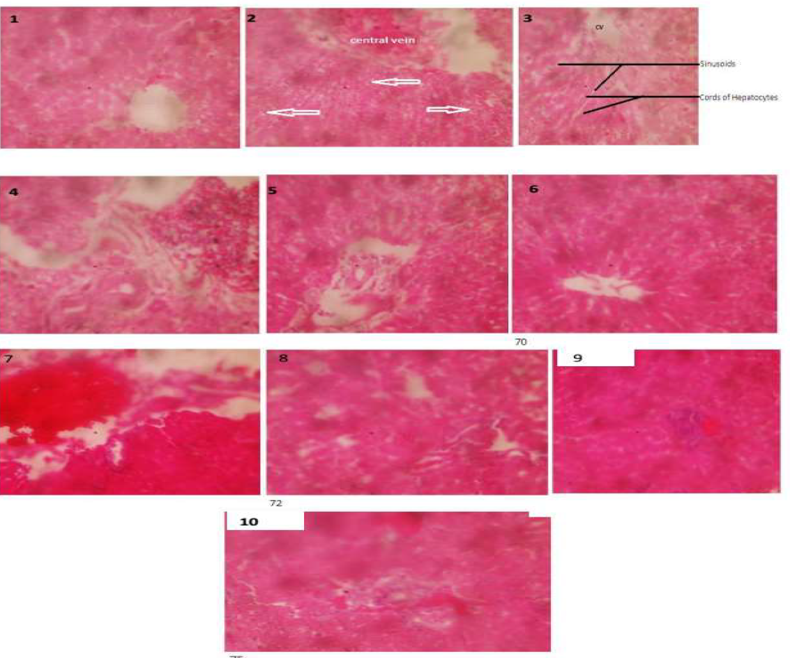
The examination of the photomicrographic section of the liver of the normal control rats (Figure 1) and the groups that received the plant extracts only (Figures 3-6) showed normal liver architecture with patent central vein and hepatocytes. The histological sections of the PbA exposed rats revealed hepatic damage such as congested central portal vein, necrosis and onset of microvessicular steatosis (Figure 2). Concomitant treatment of PbA exposed rats with 200mg/kg J. curcas extract for 28 days induced a moderate restoration of the liver architecture (Figure 8). A similar trend was also observed for the PbA exposed rat group that received 500 mg/kg H. sabdariffa extract (Figure 10).
Photomicrographic liver section from control rat showing normal architecture (Figure 1, x400); PbA treated rats liver section showing congested central vein, necrosis and onset of microvessicular steatosis (white arrow) (Figure 2, x400); 100 mg/kg J. curcas treated rat liver showing normal appearance of hepatocytes and sinusoids (black arrow) (Figure 3 x400); Liver sections of rats treated with 200 mg/kg J. curcas (Figure 4, x400), 250 mg/kg H. sabdariffa (Figure 5, x400), 500 mg/kg H. sabdariffa (Figure 6, x400) all showed normal appearance of hepatocytes. Liver section of rat that received both PbA and 100 mg/kg J. curcas showed minor degree of liver damage (Figure 7, x400). Liver sections of the PbA exposed rats that received 200 mg/kg J. curcas (Figure 8, x400), 250 mg/kg H. sabdariffa (Figure 9, x400) and 500 mg/kg H. sabdariffa (Figure 10, x400) all showed moderately restored hepatic architecture.
Table 1: Changes in Biochemical Parameters of Control and Experimental Groups (N = 6)
| Groups | Serum AST (U/L) | Serum ALT (U/L) | Serum ALP (U/L) | Serum GGT (U/L) | Total Bilirubin (µmol/L) |
| Control | 239.00 ± 10.40b | 70.33 ± 5.40b | 120.00 ± 5.20b | 3.67± 0.67b | 16.70 ± 2.10b |
| PbA | 285.00 ± 12.20a | 82.30± 6.20a | 151.00 ± 10.30a | 8.00 ± 1.33a | 30.00 ± 2.67a |
| JC-100mg | 228.00 ± 13.40ab | 73.60± 3.30b | 110.00 ± 9.80b | 4.00 ± 0.81b | 20.00 ± 1.90b |
| JC-200mg | 238.33 ± 14.60b | 75.00± 4.20b | 123.00 ± 16.70b | 5.40 ± 0.60 | 21.33 ± 2.50b |
| HS-250mg | 221.50 ± 15.10ab | 71.33 ± 3.40b | 128.00 ± 8.10b | 4.00 ± 0.63b | 15.00 ± 1.36b |
| HS-500mg | 238.00 ± 9.00b | 66.67± 2.60b | 126.00 ± 7.90b | 3.25 ± 0.45b | 16.70 ± 1.54b |
| PbA + JC100 | 260.50 ± 12.60ab | 76.00± 3.10ab | 138.00 ± 11.00ab | 7.00 ± 0.90 | 23.00 ± 2.60 |
| PbA + JC 200 | 253.50 ± 18.60ab | 73.90± 2.10b | 130.00 ± 9.60b | 6.00 ± 0.72 | 21.67 ± 2.40b |
| PbA + HS 250 | 237.50 ± 14.40b | 58.20 ± 4.50ab | 123.00 ± 7.20b | 6.33 ± 0.36 | 20.00 ± 2.05b |
| PbA + HS 500 | 249.33 ± 11.50b | 78.00± 3.50ab | 145.00 ± 6.50 | 5.00 ± 0.86b | 23.33 ± 2.73a |
Values are mean ± SE of triplicate determinations. Superscript a and b indicates significant difference from normal and PbA control group respectively (p<0.05) JC- Jatropha curcas HS- Hibiscus sabdariffa AST- Aspartate transaminase, ALT- Alanine transaminase, GGT- Gamma glutamyl transferase, ALP- Alkaline phosphatase
Table 2: Blood MDA, GSH Levels and Antioxidant Enzyme Activities of Control and Experimental Groups (N = 6)
| GROUP | SOD(U/ml) | GSH(µg/ml) | CATALASE (U/ml) | MDA (µmol/ml) |
| Normal Control | 1.16±0.07b | 1.33±0.09 | 5.00±0.23b | 1.20±0.06b |
| Lead acetate (PbA) control | 0.73±0.03a | 1.00±0.03 | 3.58±0.17a | 2.10±0.09a |
| 100mg JC extract | 1.28±0.05b | 1.96±0.12b | 5.33±0.29b | 1.50±0.07 |
| 200mg JC extract | 1.35±0.02b | 2.55±0.26ab | 5.50±0.16b | 1.17±0.04b |
| 250mg HS extract | 1.70±0.04b | 2.11±0.31ab | 5.73±0.21b | 1.32±0.05b |
| 500mg HS extract | 1.95±0.11b | 2.97±0.62ab | 5.82±0.23b | 1.45±0.08b |
| PbA + 100mg JC extract | 0.81±0.07 | 2.27±0.54b | 3.81±0.09 | 1.62±0.07b |
| PbA + 200mg JC extract | 0.93±0.06b | 3.10±0.65b | 4.47±0.11b | 1.65±0.05b |
| PbA + 250mg HS extract | 0.86±0.04 | 3.00±0.08b | 3.65±0.08 | 1.85±0.03 |
| PbA + 500mg HS extract | 0.92±0.05b | 3.55±0.14b | 4.04±0.13 | 1.70±0.05 |
Values are mean ± SE of triplicate determinations. Superscript a and b indicates significant difference from normal and PbA control group respectively (p<0.05) JC- Jatropha curcas HS- Hibiscus sabdariffa SOD- Super oxide dismutase, GSH- Reduced Glutathione

Figures 1-10. The Histological Examination of Livers
Lead toxicity has been associated with the disruption of the normal anatomical organization of hepatic lobules and loss of the distinctive cord-like structure of functional liver cells [5]. The disruption of the liver cell membrane is often accompanied by the leakage of cytosolic content of hepatocytes such as liver enzymes (AST, ALT and ALP) into the blood stream [28]. Thus, the measurements of these enzymes serve as a useful assessment tool for evaluating the function of the liver. These indices were assayed in the present study to evaluate the effect of sub-chronic lead exposure (50 mg/kg PbA for 28 days) on the liver of male Wistar albino rats and the ameliorative potentials of JC and HS aqueous extracts.
The results revealed that lead exposure dose of 50 mg/kg led to significant increase (p<0.05) in the serum AST, ALT and ALP activities in the exposed rats when compared to the control and plant-extract only groups (Groups 3-6). These results are consistent with previous findings on lead exposure [1, 29-30]. Elevated activities of transaminases AST and ALT in serum is usually regarded as indication of degenerative changes in the liver. These changes following lead exposure may be due to oxidative stress mediated alteration of the cell membrane causing the increased release of liver enzymes into the blood stream [31]. The elevated ALP level in the PbA groups also suggest that sub-chronic Pb exposure may have caused damage to the cells of the hepatobiliary tract and could lead to bile duct obstruction and infiltrative disease of the liver [32]. The alteration in ALP activity may also affect various metabolic processes where ALP is involved such as protein, phospholipids and nucleic acid synthesis [33]. Histopathological analysis revealed lesions haemorrhage and inflammation in the PbA control rats further confirming the hepatoxicity of the PbA dosage used in this study.
J. curcas and H. sabdariffa are two plants that have been used in traditional medicine to treat ailments such as anaemia and microbial activities [34-35]. In this study, co-treatment of lead acetate with aqueous extract of J. curcas and H. sabdariffa respectively caused significant decrease in the serum AST, ALT and ALP. The aqueous extracts of JC decreased the concentration of the liver enzymes in a dose dependent manner. In contrast, the higher dose of HS extract (500 mg/kg) resulted in increased AST, ALT and ALP levels. Interestingly, the 250 mg/kg HS extract demonstrated the highest protective effect in terms of reduction of liver enzyme activities when compared to the other treatment groups. This demonstrates the hepatoprotective efficacy of the aqueous extracts of HS since smaller dosage can still effectively attenuate lead induced hepatotoxicity. Other plants that have demonstrated hepatoprotective potentials in various animal models include Bacopa monniera [36], Pomegranate peel and Ginger [37]. The researchers in their respective studies attributed the hepatoprotective properties observed to the presence of potent antioxidant phytochemicals in the plant; their report is consistent with the result in the present study. Both aqueous extracts of J. curcas leaves and H. sabdariffa calyces are rich sources of polyphenols-a potent group of plant antioxidants [38-39]. The flavonoids and phenolic acids present in the plant extracts can prevent free radical accumulation and lipid peroxidation of the liver tissue thereby limiting the extent of its membrane fragility and leakage of liver enzymes into the blood stream.
Elevated serum total bilirubin is as a result of defect in the excretion of bilirubin arising from liver damage and or hemolysis [40]. Result of this study showed an elevated total bilirubin levels in the PbA exposed rats. The observed elevated bilirubin in the PbA control group may be attributed to the combined effects of increased breakdown of red blood cells and liver damage as shown in the histology of the liver tissues of the PbA exposed rats. Treatment with aqueous leaf extract of J. curcas and H. sabdariffa respectively caused a marked decrease in the total bilirubin levels of the treated animals. This suggest that the aqueous leaf extract J. curcas and H. sabdariffa contains phytochemical constituents which has the potential to reduce total bilirubin levels. Shah and Jain [5] have earlier reported that phenolic and flavonoid components present in plant extracts provide strong antioxidant property to plant by acting as ROS scavengers, hydrogen donors, reducing agents, singlet oxygen quenchers and metal chelators. Similarly, in the present study, the protective action of the aqueous extracts of J. curcas leaves and H. sabdariffa calyces against lead-induced hepatotoxicity may be due to the presence of free radical scavenging phytochemicals. Particularly, two major anthocyanin compounds have been identified in the calyx namely delphinidin-3-sambubioside and cyanidin-3-sambubioside [41], they are compounds with high free radical scavenging activities which may account for the high hepatoprotective capacity of the H. sabdariffa extract when compared to the groups that were treated with J. curcas extract.
Antioxidant enzyme activities as well as lipid peroxidation measures serve as reliable means of evaluating the toxic effect of metal contaminants such as lead. This is because oxidative stress is one of the body’s first responses to environmental pollutants [42]. The data obtained in this study showed that there was a significant inhibition of super oxide dismutase (SOD), catalase and reduced glutathione (GSH) in the PbA exposed rats. Moreira et al. [43] had earlier reported significant decreases in the activities of SOD in lead exposed 23-day old rats. A decade later, Khalaf et al. [44] also reported similar trend in the levels of antioxidant enzymes during their study. Catalase and SOD are metalloproteins which accomplish their antioxidant functions by enzymatically detoxifying the peroxides (OH, H2O2) and super oxide anion [45]. The observed decreased activity of SOD and catalase in the PbA exposed rats may be explained by the interaction of between lead and the cofactors copper, zinc and iron. Lead can mimic these divalent ions and bind to the enzymes leading to an inhibition of their activities. Our results show that co-treatment of Pb with J. curcas and H. sabdariffa respectively increased the antioxidant status (SOD, catalase and GSH) in the blood of lead treated rats. The high content of polyphenols and flavonoids in J. curcas and H. sabdariffa may be responsible for its antioxidant activity and these phytochemicals are considered good chelating agents for metal ions.
Microscopical examination of liver sections of control and plant extract-treated rats showed normal histopathological structures, while histological lesions hemorrhage and inflammation were observed in the lead treated rats. These findings were similar to those reported by [29]. They attributed the histological alterations in the liver to lipid peroxidation under the influence of lead exposure. In the current study, co-administration of PbA and J. curcas and H. sabdariffa extracts respectively induced moderate improvement of the histopathological alteration in liver tissue. The improvement observed was in a dose dependent manner with the H. sabdariffa extract demonstrating higher protective effect. Further isolation of bioactive compounds will be useful in large scale implementation of these plant extracts in heavy metal toxicity management.
In the light of the current finding, we suggest that J. curcas and H. sabdariffa extracts are efficacious in promoting an increase in the antioxidant enzyme status. They moderately mitigate hepato-chemical changes induced by lead.
Conflicts of Interest
The authors have none to declare.
Ezejiofor, A.N. et al. “Evaluation of protective effect of aqueous leaf extract of Costus afer on female albino Wistar rats exposed to lead acetate.” EC Pharmacology and Toxicology, vol. 2, 2017, pp. 75–92.
Wani, A.L. et al. “Lead toxicity: A review.” Interdisciplinary Toxicology, vol. 8, no. 2, 2015, pp. 55–64.
Ghosh, D. et al. “Protective effect of aqueous leaf extract of Murraya koenigii against lead-induced oxidative stress in rat liver, heart and kidney: A dose response study.” Asian Journal of Pharmacology and Clinical Research, vol. 5, no. 4, 2012, pp. 54–58.
Singh, N. et al. “Biochemical and molecular bases of lead-induced toxicity in mammalian systems and possible mitigations.” Chemical Research in Toxicology, vol. 31, no. 10, 2018, pp. 1009–1021.
Shah, F. and N. Jain. “An in vitro investigation of ameliorative effect of synthetic and herbal antioxidants on lead-induced toxicity.” Asian Journal of Pharmaceutical and Clinical Research, vol. 9, no. 2, 2016, pp. 364–370.
Mudipalli, A. “Lead hepatotoxicity and potential health effects.” Indian Journal of Medical Research, vol. 126, no. 6, 2007, pp. 518–527.
Bukola, R.O. et al. “Lead-induced damage on hepatocytes and hepatic reticular fibres in rats: Protective role of aqueous extract of Moringa oleifera leaves.” Journal of Biosciences and Medicine, vol. 3, 2015, pp. 27–35.
Shatha, H.K. et al. “The effect of penicillamine in reducing the toxic effects of lead acetate on blood parameters, liver functions and testicular tissue in male rats.” International Journal of Pharmacy and Pharmaceutical Research, vol. 5, 2016, pp. 22–40.
Asuk, A.A. et al. “The biomedical significance of the phytochemical, proximate and mineral compositions of the leaf, stem bark and root of Jatropha curcas.” Asian Pacific Journal of Tropical Biomedicine, vol. 5, no. 8, 2015, pp. 650–657.
Georgiev, V. et al. “Recent advances and uses of grape flavonoids as nutraceuticals.” Nutrients, vol. 6, no. 1, 2014, pp. 391–415.
Syed, D.N. et al. “Pomegranate extracts and cancer prevention: Molecular and cellular activities.” Anticancer Agents in Medicinal Chemistry, vol. 13, no. 8, 2013, pp. 1149–1161.
Chang, H.C. et al. “Hibiscus sabdariffa extract inhibits obesity and fat accumulation and improves liver steatosis in humans.” Food and Function, vol. 5, no. 4, 2014, pp. 734–739.
Lin, H.H. et al. “Chemopreventive properties of Hibiscus sabdariffa on human gastric carcinoma cells through apoptosis and JNK/p38 MAPK activation.” Chemico-Biological Interactions, vol. 165, no. 1, 2007, pp. 59–75.
Zhen, J. et al. “Phytochemistry, antioxidant capacity, total phenolic content and anti-inflammatory activity of Hibiscus sabdariffa leaves.” Food Chemistry, vol. 190, 2016, pp. 673–680.
Prenesti, E. et al. “Antioxidant power quantification of decoction and cold infusions of Hibiscus sabdariffa flowers.” Food Chemistry, vol. 100, no. 2, 2007, pp. 433–438.
Nagappan, R. “Evaluation of aqueous and ethanol extracts of Cassia didymobotrya against immature stages of Culex quinquefasciatus.” Asian Pacific Journal of Tropical Biomedicine, vol. 2, no. 9, 2012, pp. 707–711.
Mishra, S.B. et al. “Evaluation of leaf extract of Jatropha curcas for antihyperglycemic activity in alloxan-induced diabetic rats.” International Journal of Pharmaceutical Sciences, vol. 2, 2010, pp. 482–487.
Dangambo, M.A. et al. “Acute toxicity and hepatocurative effect of aqueous leaf extract of Jatropha curcas in rats.” British Journal of Pharmaceutical Research, vol. 5, no. 1, 2015, pp. 52–57.
Ali, B.H. et al. “Phytochemical, pharmacological and toxicological aspects of Hibiscus sabdariffa: A review.” Phytotherapy Research, vol. 19, no. 5, 2005, pp. 369–375.
Amos, S. et al. “Neuropharmacological effects of Hibiscus sabdariffa aqueous extract.” Pharmaceutical Biology, vol. 41, no. 5, 2003, pp. 325–329.
Onyenekwe, P.C. et al. “Antihypertensive effect of Hibiscus sabdariffa calyx infusion in spontaneously hypertensive rats.” Cell Biochemistry and Function, vol. 17, no. 3, 1999, pp. 199–206.
International Federation of Clinical Chemistry (IFCC). “Approved recommendation on IFCC methods for the measurement of catalytic concentration of enzymes: Part 2. IFCC method for aspartate aminotransferase.” Journal of Clinical Chemistry and Clinical Biochemistry, vol. 24, no. 7, 1985, pp. 481–495.
Misra, H.P. and I. Fridovich. “The role of superoxide anion in the auto-oxidation of epinephrine and a simple assay for superoxide dismutase.” Journal of Biological Chemistry, vol. 247, no. 10, 1972, pp. 3170–3175.
Sedlak, J. and R.H. Lindsay. “Estimation of total, protein-bound and non-protein sulfhydryl groups in tissue with Ellman’s reagent.” Analytical Biochemistry, vol. 25, 1968, pp. 192–205.
Aebi, H. “Catalase in vitro.” Methods in Enzymology, vol. 105, 1984, pp. 121–126.
Ohkawa, H. et al. “Assay for lipid peroxides in animal tissues by thiobarbituric acid reaction.” Analytical Biochemistry, vol. 95, no. 2, 1979, pp. 351–358.
Bancroft, J.D. and M. Gamble. Theory and Practice of Histological Techniques. 5th ed., Churchill Livingstone, 2003.
Sookoian, S. and C.J. Pirola. “Liver enzymes, metabolomics and genome-wide association studies: From systems biology to personalized medicine.” World Journal of Gastroenterology, vol. 21, no. 3, 2015, pp. 711–725.
Samir, D. et al. “Protective effects of Aristolochia longa and Aquilaria malaccensis against lead-induced acute liver injury in rats.” Journal of Acute Disease, vol. 6, no. 5, 2017, pp. 193–199.
El-Latief, H.M.A. “Protective effect of quercetin or zinc against lead toxicity on rat testes.” Global Journal of Pharmacology, vol. 9, no. 4, 2015, pp. 366–376.
Okeke, N. et al. “Biochemical role of taurine in mitigating modifications in male Wistar rats co-exposed to chlorpyrifos and lead.” International Journal of Environment Science and Toxicology, vol. 2, no. 9, 2014, pp. 104–115.
Jarrar, B.M. and N.T. Taib. “Histological and histochemical alterations in the liver induced by chronic lead toxicity.” Saudi Journal of Biological Sciences, vol. 19, no. 2, 2012, pp. 203–210.
Dick, C.F. et al. “Inorganic phosphate as an important regulator of phosphatases.” Enzyme Research, 2011, pp. 1–7.
Nazeema, T.H. and S. Girija. “Comparative phytochemical investigations and in vitro pharmacological studies of two medicinal species of Jatropha from South India.” European Journal of Experimental Biology, vol. 2, no. 2, 2012, pp. 421–426.
Villani, T. et al. “Hibiscus sabdariffa: Phytochemistry, quality control and health properties.” In African Natural Plant Products, Volume II, American Chemical Society, 2013, pp. 209–230.
Velaga, M.K. et al. “Ameliorative effects of Bacopa monniera on lead-induced oxidative stress in different regions of rat brain.” Drug and Chemical Toxicology, vol. 37, no. 3, 2013, pp. 357–364.
Reddy, Y.A. et al. “Ameliorating activity of ginger (Zingiber officinale) extract against lead-induced renal toxicity in male rats.” Journal of Food Science and Technology, vol. 51, no. 5, 2011, pp. 908–914.
Youns, M.A.D. et al. “In vitro evaluation of the antimicrobial and antioxidant activity of Hibiscus sabdariffa.” Journal of Dental and Medical Sciences, vol. 17, no. 4, 2018, pp. 69–74.
Ahaotu, E.O. et al. “Benefits of Jatropha gossipifolia in Nigeria.” International Journal of Agriculture and Biosciences, vol. 2, no. 6, 2013, pp. 349–355.
Hull, T.D. and A. Agarwal. “Bilirubin: A potential biomarker and therapeutic target for diabetic nephropathy.” Diabetes, vol. 63, no. 8, 2014, pp. 2613–2616.
Juliani, H.R. et al. “Chemistry and quality of Hibiscus sabdariffa for developing the natural-product industry in Senegal.” Journal of Food Science, vol. 74, no. 2, 2009, pp. S113–S121.
Romeo, M. et al. “Cadmium and copper display different responses toward oxidative stress in the kidney of the sea bass Dicentrarchus labrax.” Aquatic Toxicology, vol. 48, 2000, pp. 185–194.
Moreira, E.G. et al. “Antioxidant defense in rat brain regions after developmental lead exposure.” Toxicology, vol. 169, 2001, pp. 145–151.
Khalaf, A.A. et al. “The protective effect of green tea extract on lead-induced oxidative and DNA damage in rat brain.” NeuroToxicology, vol. 33, 2012, pp. 280–289.
Jangid, A.P. et al. “Effect of lead on human blood antioxidant enzymes and glutathione.” International Journal of Biochemistry Research and Review, vol. 13, no. 1, 2016, pp. 1–9.