A Prospective Randomized Double Blind Comparative Study of the Effect of Intrathecal Dexmedetomidine and Clonidine as Adjuvants with Ropivacaine in Lower Limb Surgeries
Background and Aim: Sub arachnoid block is most commonly used technique for lower limb surgeries. Various adjuvants are being used with local anesthesia for prolongation of intra operative and post-operative analgesia. Most of the clinical studies about intrathecal alpha-2 agonists are related to clonidine. In view of few evidences of Dexmedetomidine, efficacy as adjuvant to Ropivacaine in Spinal Anesthesia. This study was conducted to explore its usefulness and also to compare this new alpha-2 adrenergic agonist with the previously established and widely used adjuvants like Clonidine on spinal block characteristics and post-operative analgesia. This study meant to compare the effects of Dexmedetomidine 5 mcg versus Clonidine 15 mcg on blockade characteristics and postop analgesia when given as adjuvants to 0.75 % Ropivacaine. The objectives were to compare the onset and duration of sensory and the motor blockade, duration of analgesia, hemodynamic parameters and side effect profile in among two groups. Materials and Methods: 60 adult patients posted for lower limb surgeries were randomly allocated into two groups. Group D received 3 mL 0.75% ropivacaine with 5 mcg of Dexmedetomidine and group C received 3ml of Ropivacaine with 15 mcg of Clonidine. Patients were preloaded with 10mL/kg of Ringer’s lactate. Onset and duration of sensory and motor blockade, peak sensory level, 2 segment regression duration of analgesia and rescue analgesia requirement were noted and compared. Results: Patients in group D had earlier onset of sensory and motor block compared to group C. Mean time of onset of sensory block group D was 3.467±0.507 min and group C was 3.900±0.759 min with a p value of 0.012. Mean time for onset of motor block group D 9.033±1.098 min was group C 10.00±1.66 min with a p value of 0.010. Mean time for two segments regression in group D was 135.5±7.925 min and in group C was 125.567±5.27 min with a p value of 0.000. Duration of motor and sensory block were also significantly prolonged in group D compared to group C. Mean duration of motor block in group D was 339.367±27.031 min and in group C was 306.600±23.940 min with p value of 0.000. Mean duration of sensory block in group D was 421.133±21.968 min and in group C was 360.600±23.940 min with a p value of 0.000. Group D showed a significantly less requirement of analgesia than group C with a p value of 0.000. Hemodynamic were stable and comparable among both groups. Conclusion: We conclude that dexmedetomidine is a better adjuvant than clonidine in Spinal Anesthesia as far as patient comfort, stable cardio-respiratory parameters, intra-operative and post-operative analgesia is concerned. Overall the experience with Dexmedetomidine was quite satisfactory as compared to clonidine.
Keywords
Intrathecal
Ropivacaine
Dexmedetomidine
Clonidine
Lower limb Surgeries
INTRODUCTION
Pain is one of the most dreaded effects of the disease and all medical fraternity should take its relief as one of their main duties. Anesthesiologists have an important role to play in the fascinating field of pain management.
In view of the wider application of regional anesthesia procedure in modern anesthesia practice, there is a need for local anesthetic with desirable properties like longer duration of sensory blockade and lesser duration of motor paralysis. Ropivacaine is newer amide type of local anesthetic having a lower potential for cardiac and central nervous system toxicity [1-4] shows a greater differentiation between sensory and motor blockade [5].
Adjuvant drugs are pharmacological agents possessing little pharmacological effect by themselves, but enhance or potentiate the action of other drugs when given at the same time. Adjuvant drugs modify local anesthetic effects and reduce side effects. Perioperative these drugs effect:
Latency i.e., time of onset of local anesthetic block
Duration of analgesia i.e. duration of sensory and motor block
Quality of analgesia i.e. patient satisfaction, care provider’s impression of pain relief
Various adjuvants that can be added to local anesthetics and administered in central neuraxial blockade are Opioids like Fentanyl [6], Sufentanyl [7], Morphine [8], Alpha2 agonists like Dexmedetomidine [9], Clonidine [10], Benzodiazepines like Midazolam [11] and other adjuvants like Neostigmine [12], Ketamine [13], Epinephrine [14], Magnesium sulphate [15].
Knowledge and use of adjuvant drug therapy has rendered neuraxial analgesia more effective in the management of both acute and chronic pain conditions. Alpha2 agonists have both analgesic and sedative properties when used as adjuvant in regional anaesthesia [16-18].
Dexmedetomidine is a new alpha2-agonist that received FDA approval in 1999 for use as short-term (less than 24 h) sedative, analgesic in the intensive care unit [19]. It causes sedation without causing respiratory depression. It has sedative, analgesic, sympatholytic and anxiolytic effect that blunt many CVS responses in perioperative period. It is an S-enantiomer of Medetomidine used in veterinary medicine. It is thought that intrathecal Dexmedetomidine produces its analgesic effect by inhibiting the release of C fibers transmitters and by hyperpolarization of post-synaptic dorsal horn neurons. The prolongation of motor effect might be caused by direct impairment of excitatory amino acid release from spinal interneurons. Alpha2 agonists produce sedative effect by acting on alpha 2-adrenergic receptors in Locus coeruleus. Alpha2 adrenoreceptors do not have an active role in the respiratory center, therefore, Dexmedetomidine throughout a broad range of plasma concentration, has minimal effects on the respiratory system.
Clonidine is imidazoline derivative with centrally acting alpha 2 adrenergic agonist activity [20]. Inhibition of substance-P release is believed to be involved in the analgesic effect. Clonidine partially inhibits voltage gated sodium and potassium channels and suppresses the generation of action potential in tonic firing spinal dorsal horn neurons. This may contribute to its analgesic effect. It stimulates central alpha2 adrenergic receptors in the brain stem, blocks the release of noradrenaline from nerve terminals leading to decreased peripheral resistance, renal vascular resistance heart rate and blood pressure.
Dexmedetomidine is a highly selective alpha2 agonist with 8 times greater affinity than Clonidine. More clinical experience has been gained with regard to usage of Clonidine compared to Dexmedetomidine by intrathecal route. There are less studies available for these drugs as adjuvants to 0.75%Ropivaciane
In our study doses of study drugs were selected based on previous study with other local anesthetics like Bupivacaine and comparative studies with plain Ropivacaine.
Thus, the present study was undertaken to evaluate clinical effects of adding 5mcg or Clonidine 15mcg to 0.75% Ropivacaine by intrathecal route to study the characteristics of the block, hemodynamic and postoperative pain relief in patients undergoing lower limb surgeries.
MATERIALS AND METHODS
Hospital ethics committee clearance was obtained for this study. Informed consent was taken from all the patients. Patients who denied participation in study were treated by routine protocol of anesthesia with dignity.
Randomization
Randomization was done by using ‘Physical method’, where 60 folded papers were placed in a container, each one labeled either Group D or Group C with each label 30 in number.
Blinding
The syringes containing the study drug were prepared by the third observer anesthesia resident to assure a proper blinding procedure. 60 patients of ASA I and II physical status aged 18-60 years scheduled to undergo elective surgery and satisfying all the inclusion criteria were enrolled in the study.
Inclusion Criteria
Patients of either sex in age group of 18-60 yrs
Patients with Height>140 cms and Weight less than 120 kgs
Patients who are ASA grade I and grade II
Posted for lower limb surgeries
Patients who are willing to give consent for participation in the study
Exclusion Criteria
Patient’s refusal
Patients who are ASA III and above
Patients who are hypersensitive to the local anesthetics
Patients on long term analgesic therapy
Patients with peripheral neuropathy
Patients with local skin infections and spinal deformities
Patients having coagulation abnormalities.
A sample size of 30 patients each randomly allocated into two groups using power analysis based on previous studies [21-22].
Study Procedure
A pre-anesthetic evaluation comprising of history of previous medical and surgical illnesses, previous anesthesia exposures, drug allergies; clinical examination; airway examination and baseline investigation of complete blood picture, blood sugar, serum creatinine, chest radiograph and 12 lead ECG will be done. Patients were explained about the procedure of spinal anesthesia.
Informed/written consent was obtained from each patient before starting the study. Every patient was supplied with patient information sheet before taking the consent
A specially designed proforma was used to collect the data including patient’s particulars, patient’s written informed consent, indication for surgery, the anesthetic details, intra-operative monitoring, post-operative follow up, etc
Anesthetic Procedure
Intraoperatively, the following protocol was followed:
Intravenous line was secured with 18/20 Gauge cannula. All standard monitors (NIBP, ECG, Pulse oximeter) were attached before giving spinal anesthesia
Subarachnoid block under strict aseptic conditions was performed in sitting position at L3-L4 or L4-L5 interspines space with 27 Gauge Whitacre spinal needle and drug was administered after confirming the free flow of cerebrospinal fluid
Study groups received spinal anesthesia with:
Group D: 3 ml of 0.75% Ropivacaine + Dexmedetomidine (5 µg) (total vol. – 3.5 ml)
Group C: 3 ml of 0.75% Ropivacaine + Clonidine (15 µg) (total vol.- 3.5ml)
The following parameters were observed:
Sensory Blockade
It was subjectively studied by cold swab. The patients were tested every 30 seconds at one fixed dermatome level T10 level. Then highest dermatomal level was noted. Time required for regression of level to S1 is total duration of sensory block.
Motor Blockade
It was assessed by straight leg raising while lying supine and was graded according to modified Bromage scale Table 1.
The time taken for the onset of motor blockade and the duration of motor blockade were noted. The time from injection of drug to attain bromage 3 was considered as onset of blockade and time to regain bromage score of 0 was considered as duration of motor blockade.
Hemodynamic
Heart rate and blood pressure were monitored immediately after injection and then after every 5 minutes for first 30 minutes and then every 10 minutes thereafter throughout the surgery and every hour till the complete recovery from block. The anesthesia record was maintained and changes in heart rate, blood pressure were noted. Any decrease in heart rate below 60 beats/min was considered as bradycardia and was treated with 0.3 mg of Atropine. Similarly any fall ofsystolic blood pressure of more than 30 % of baseline was considered as Hypotension was treated with 6 mg of ephedrine and crystalloids and oxygen as needed.
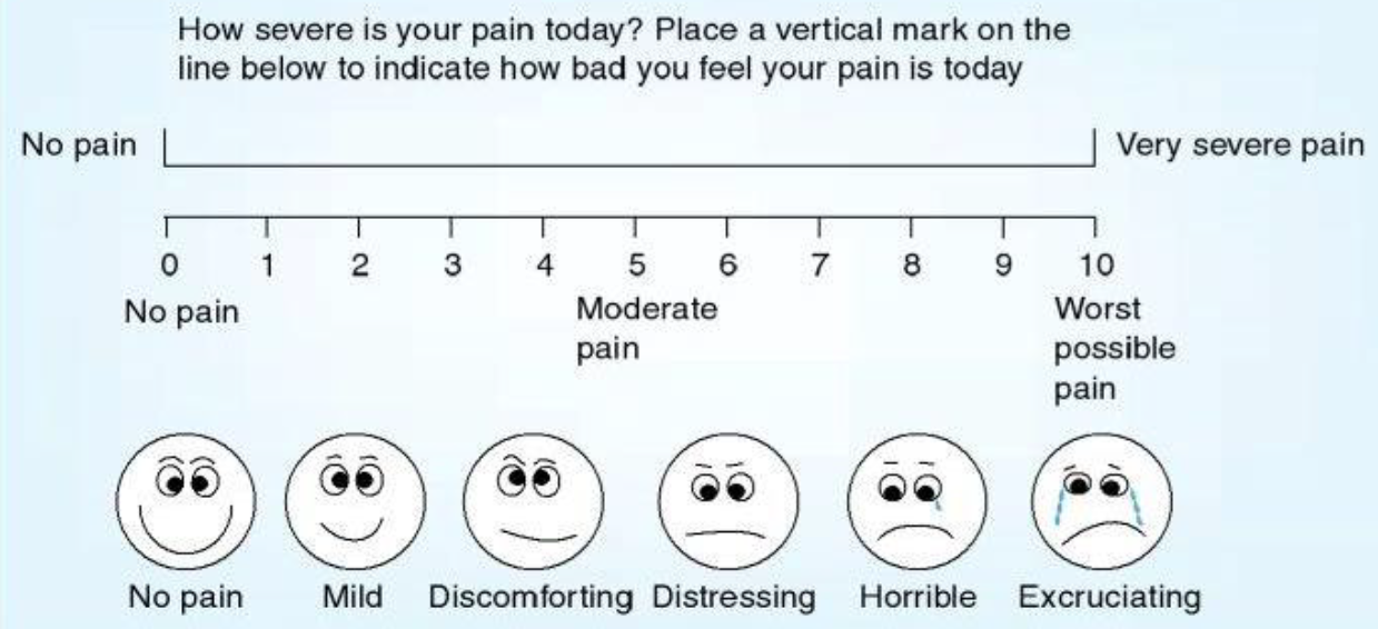
Figure 1: Visual Analogue Score
Table 1: Modified Bromage Scale
Grade
Criteria
Degree Of Block
1
Free movement of legs and feet
None
2
Just able to flex knee with free movement of feet
Partial 33%
3
Unable to flex knee but with free movement of feet
Partial 66%
4
Unable to move legs and feet
Complete paralysis
Sedation
Sedation was graded according to Modified Ramsey Sedation Scale:
Patient is Anxious, Agitated, Restless or both
Patient is Cooperative, Oriented, and Tranquil
Patient responds to commands only
Patients exhibit brisk response to light glabellar tap or loud auditory stimulus
Patients exhibits a sluggish response to light glabellar tap or loud auditory stimulus
Patients exhibit no Response
Pain
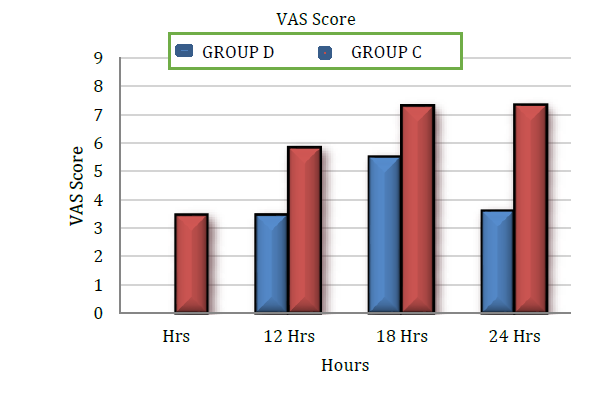
Assessed by visual analogue scale at 6th hour, 12th hour, 18th hour, 24th hour. Injection tramadol 2mg/kg iv (maximum of 100mg) was given as a rescue analgesic when VAS score was greater than or equal to 4.
Side Effects
Intra-operative side effects like sedation, nausea, vomiting, shivering, backache, bradycardia, and hypotension requiring active treatment were noted. Following variables were observed:
Sensory blockade – onset and duration
Motor blockade – onset and duration
hemodynamic parameters
Sedation
Maximum level of sensory block and time to reach maximum level
Time to reach two segment regression
Duration of Analgesia
Side effects
Rescue analgesic requirement
All the parameters were recorded as per the proforma and statistically analyzed.
Statistical Methods: Method of Statistical Analysis: The following methods of statistical analysis were used in this study. Data were expressed as Mean+Standard deviation, percentages (%) and numbers (n). The statistical analysis was performed by a statistician using Windostat version 9.2.
Proportions were compared using chi-square test of significance
The student t test was used to determine whether there was a statistical difference between two treatment groups in the parameters measured
One-way analysis of variance (Anova)
Differences in parameters such as heart rate, systolic blood pressure, diastolic blood pressure, VAS score, rescue analgesics and adverse effects over time were analyzed using ANOVA (analysis of variance)
In the entire above test p value<0.05 was accepted as indicating statistical significance.
RESULTS
Sixty patients posted for lower limb surgeries were randomly divided into two groups:
Group D: received 0.75%Ropivacaine with 5mcg Dexmedetomidine
Group C: received 0.75% Ropivacaine with 15mcg of Clonidine
Observations and results were obtained from two groups by statistical analysis and calculation the p-value.
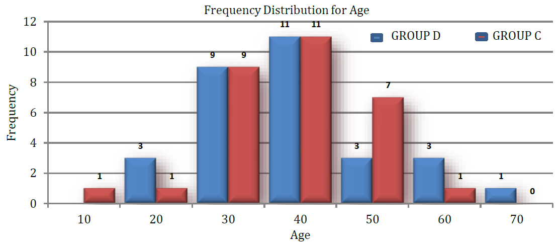
According to Figure 1 there is no significant difference between mean age (years) in group D and group C as p-value is>0.05.
Table 2: Comparison of Mean Age in Group D and Group C
Group
Number of Patients
Age (in Years)
p-Value
Mean
SD
Group D
30
38.967
11.556
0.879
Group C
30
38.533
10.352
Figure 1: Bar Diagram Showing Age Distribution
X axis – Age in years, Y axis – number of patients
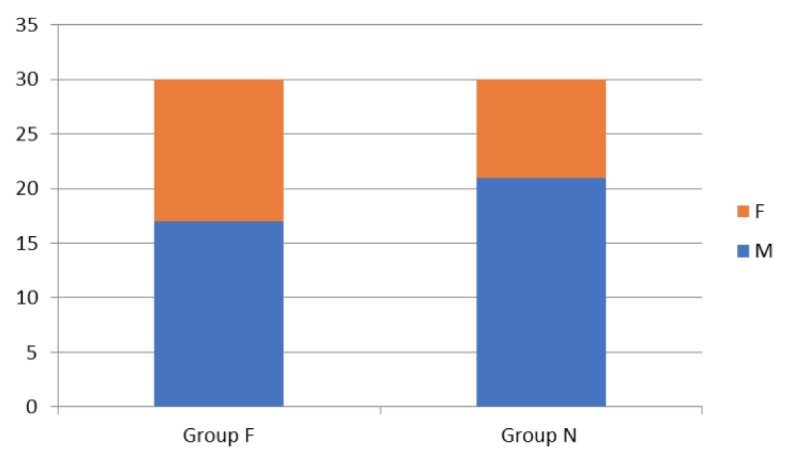
According to Figure 2 there is no significant difference between proportion of gender in group D and group C as p-value is>0.05.
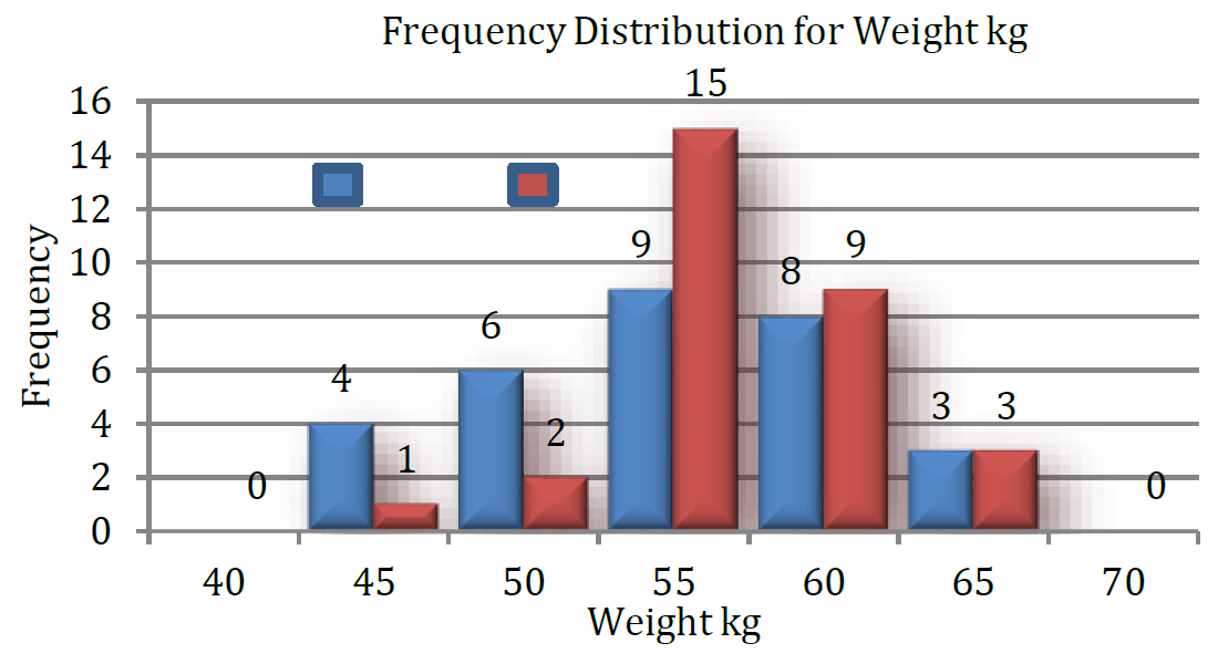
According to Figure 3 there is no significant difference between the mean weight (kgs.) in group D and group C, as the p-value is>0.05.
According to Figure 4 there is no significant difference in the mean weight of the group D and group C, as the p-value is>0.05.
According to Figure 5 there is no significant difference in the mean weight of the group D and group C, as the p-value is>0.05.
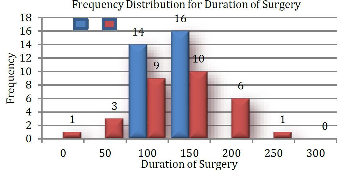
According to Figure 6 in our study mean duration of surgery in group D was 125.90+23.458 and group C was133.667±51.238 and p-value was 0.453, thus both groups were comparable with regards to duration of surgery.
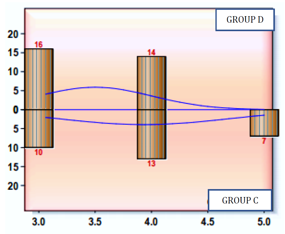
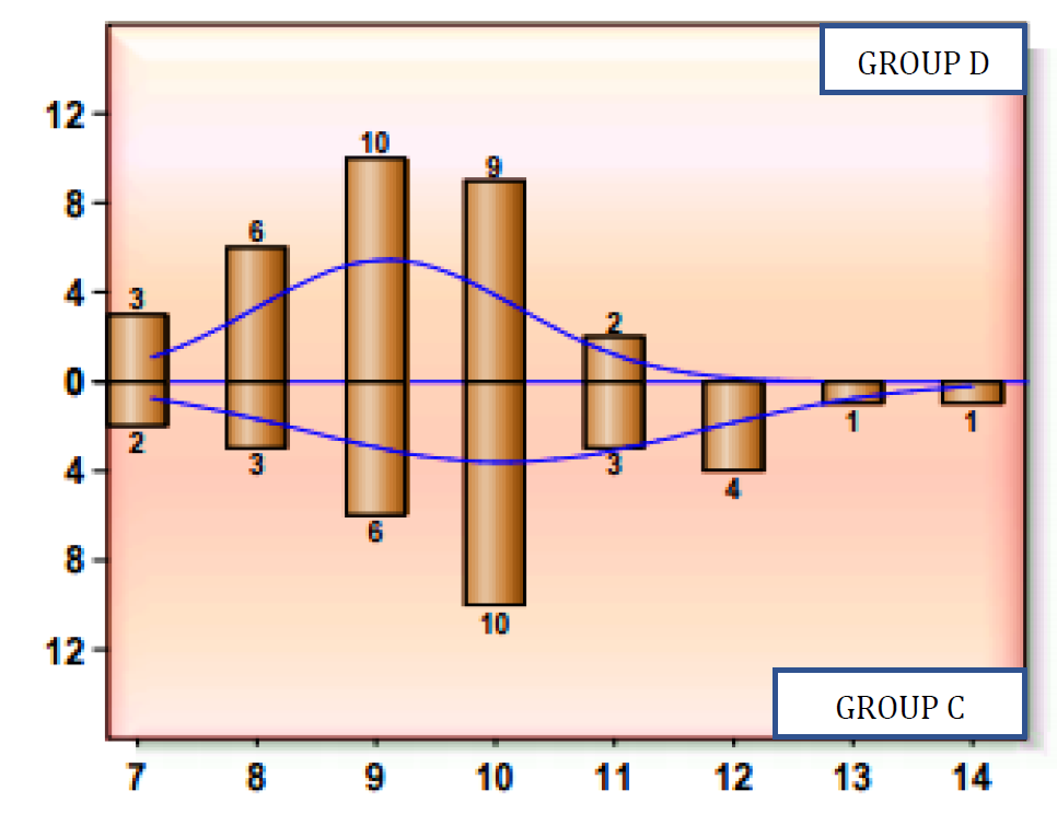
According to Figure 7 in present study, mean time of onset of sensory block in Group D was 3.467±0.507 min and with Group C was 3.900±0.759 with a (p = 0.012). Mean time of onset of motor block in Group D was 9.033±1.098 and in Group C was 10.00±1.66 min with (p = 0.010). This shows that the onset of sensory and motor block was faster in group D compared to group C and was statistically significant.
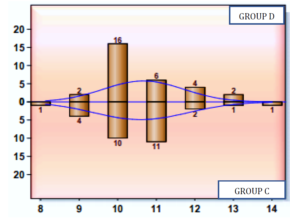
According to Figure 8 mean time required to reach the maximum level of sensory block in Group D was 10.600±1.037 min and in Group C was 11.413±1.15min. (p = 0.005) which was statistically significant, thus the mean time required to achieve a maximum sensory level was lower with group D compared to group C.
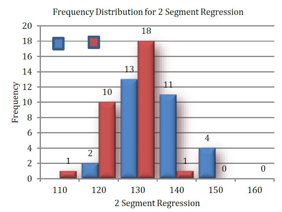
In our study the mean Time required for two segment regression of sensory level in group D was 135.5±7.925 min while in group C this time was 125.567±5.27 min. The p-value was 0.00 which is statistically significant (Table 1).
Graph shows that the time required for two segment regression of sensory level in group was greater in group D (Dexmedetomidine and Ropivacaine) (Table 2).
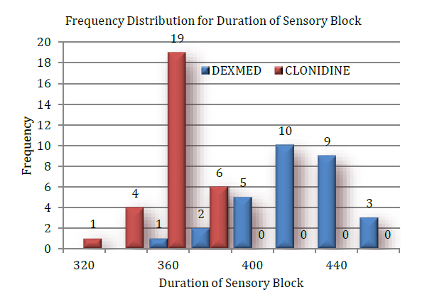
In our study, duration of sensory block in Group D was 421.133±21.968 min and in Group C was 360.600 ±23.940 min with p value of 0. 000.Thus duration of sensory block is statistically significant (p = 0.000) in Group D as compared to Group C with Dexmedetomidine group having longer total sensory blockade compared to Clonidine.
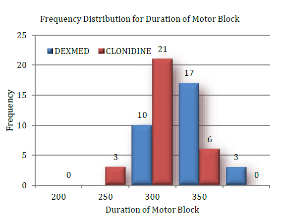
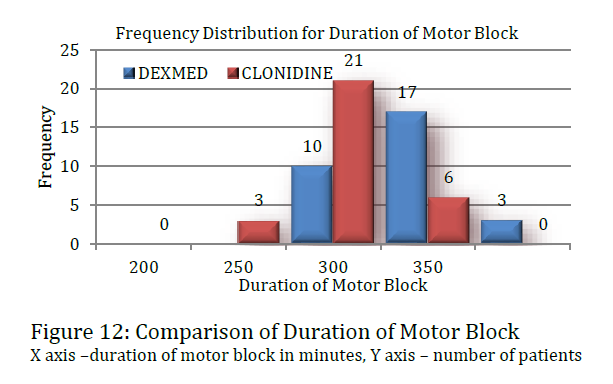
Duration of motor block in Group D was 339.367±27.031 min and in Group C was 339.367±27.031 min with p value of 0.000. Thus, duration of motor block was statistically significant (p = 0.000) in Group D as compared to Group C with Dexmedetomidine group having longer duration of total motor blockade.
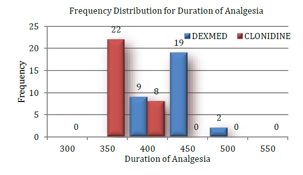
Total duration of Post - operative analgesia in Group D was 434.700±27.507 min and in Group C was 365.767±12.716 min with p value of 0.000. Thus, duration of Post - operative analgesia was statistically significant with Dexmedetomidinehaving longer total duration of Post - operative analgesia Table 3.
According to Figure 9 Mean sedation Group D was 1.900±0.607 and in Group C 2.167±0.699 was with p value of 0.109. Thus, the level of sedation was comparable among both groups.
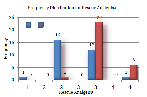
According to Figure 10 The mean requirement of rescue analgesics in group D was 2.433±0.626 and in group C was 3.167±0.461 and the P value was 0.000 which shows that there was a significantly lower requirement of analgesics in group D compared to group C.
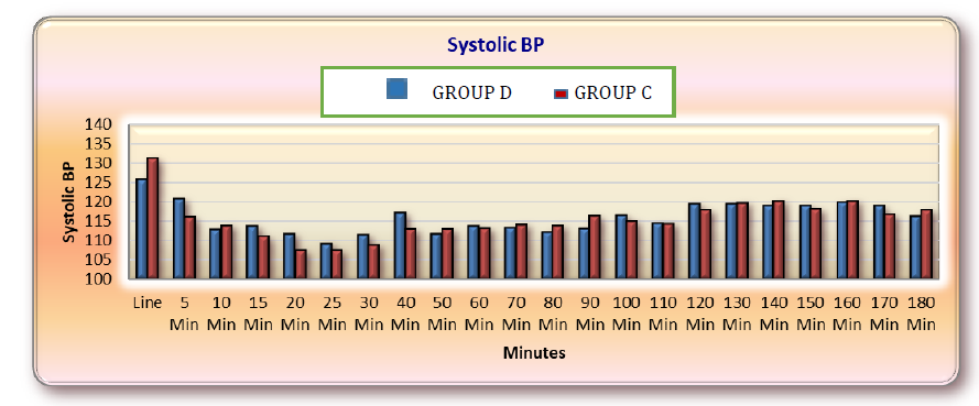
According to Figure 11 In our study, a fall in Systolic Blood Pressure of more than 30% was taken as hypotension and was treated with Inj. ephedrine 6mg iv.
In studied Groups, the mean baseline systolic blood pressure, in Group D was 125.767 mm of Hg and in Group C was 131.300 mm of Hg. (p = 0.067). Both groups were comparable. The mean systolic blood pressure was measured at 0, 5min, 10min, 15min, 20min, 25min, 30min, 40min, 50 min, 60 min, 70 min, 80 min, 90min, 100 min, 110 min, 120 min, 130 min, 140 min, 150 min, 160 min, 170 min, 180 min period. In our study the systolic blood pressure was comparable in both groups.
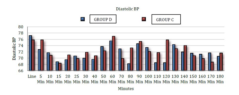
According to Figure 12 In studied Groups, the mean baseline diastolic blood pressure, in Group D was mm 77.333±11.595 of Hg and in Group C was 78.467±9.961 mm of Hg. (p = 0.686). Thus, both groups were comparable in baseline pressures.
The mean diastolic blood pressure was measured at 0, 5min, 10min, 15min, 20min, 25min, 30min, 40min, 50min, 60min, 70min, 80min, 90min, 100min, 110min, 120min, 130min, 140min, 150min, 160min, 170min, 180min period. In our study the diastolic blood pressure was comparable in both groups.
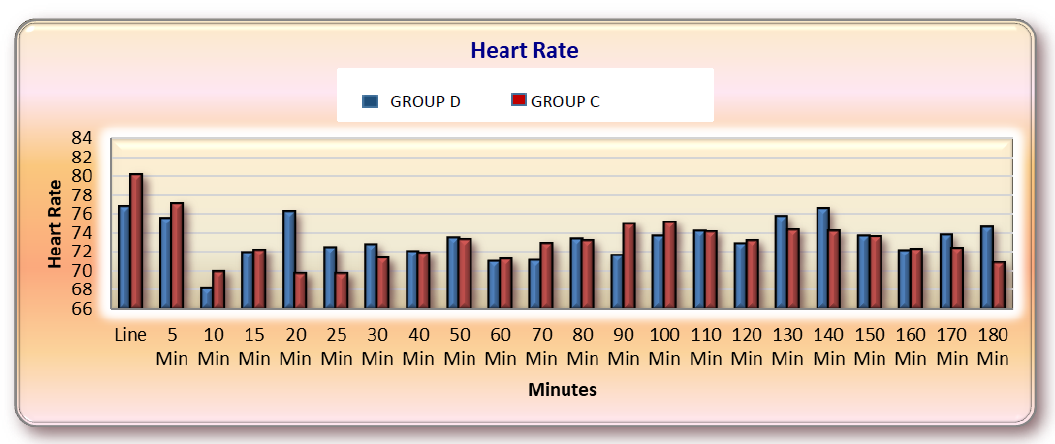
According to Figure 13 In our study heart rate less than 60 beats per min was taken as bradycardia and was treated with Inj. Atropine. In studied Groups, the mean baseline Heart rate, in Group D is 77.500±7.375. And in Group C was79.700±6.182 with a p-value of 0.216. Baseline heart rates were comparable. The mean heart rates were measured at 0, 5min, 10min, 15min, 20min, 25min, 30min, 40min, 50min, 60min, 70min, 80min, 90min, 100min, 110min, 120min, 130min, 140min, 150min, 160min, 170min, 180min. In our study the Heart rate was comparable in both groups with no significant difference.
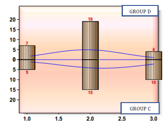
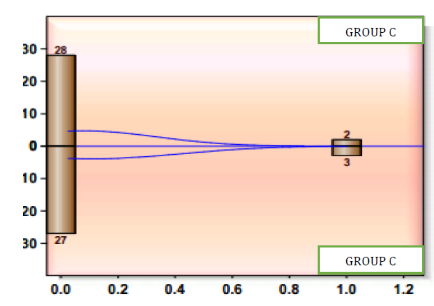
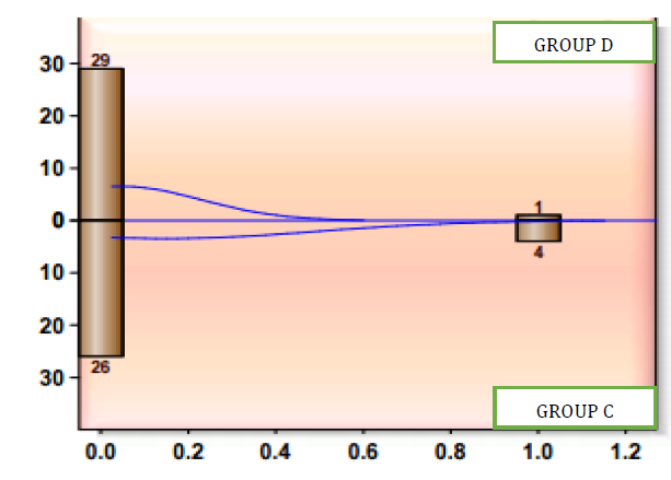
There incidence of hypotension in group D was 6.66%, the incidence of hypotension in group C was 10% which was promptly treated with Inj. ephedrine 6mg iv stat. Similarly, incidence of bradycardia in group D was 3.0 %, while the incidence of bradycardia in group C was 13.0% which was treated with Inj. Atropine 0.3 mg iv bolus. Incidence of bradycardia and hypotension were comparable among both groups. The Incidence of nausea/vomiting and shivering in group D and C was 0% Table 4.
According to Figure 14 Graph shows that the incidence of bradycardia was higher in group C but that was statistically insignificant with p-Value 0.167.
So, from our study we concluded that incidence of hypotension and bradycardia was comparable among both groups. There was no incidence of nausea/vomiting and shivering in both groups.
According to Figure 15 Mean VAS scores in group D were lower than in group C. Mean value in group D was 3.142±2.051 and in group C was 5.983±1.805 with a p value of 0.000.
Table 3: Gender Wise Distribution of Patients in Group D and Group C
Gender
Group
p-Value
D
C
Male
17
21
0.873
Female
13
9
Total
30
30
Figure 2: Bar Diagram Showing Frequency Distribution of Gender
Table 4: Comparison Mean Weight (Kilograms) In Group D and Group C
Group
Number of Patients
Weight (Kgs)
p-Value
Mean
Sd
Group D
30
55.267
5.711
0.239
Group C
30
56.833
4.403
Figure 3: Bar Diagram Showing Weight Distribution
There is no significant difference between the mean weight (kgs.) in group D and group C, as the p value is>0.05
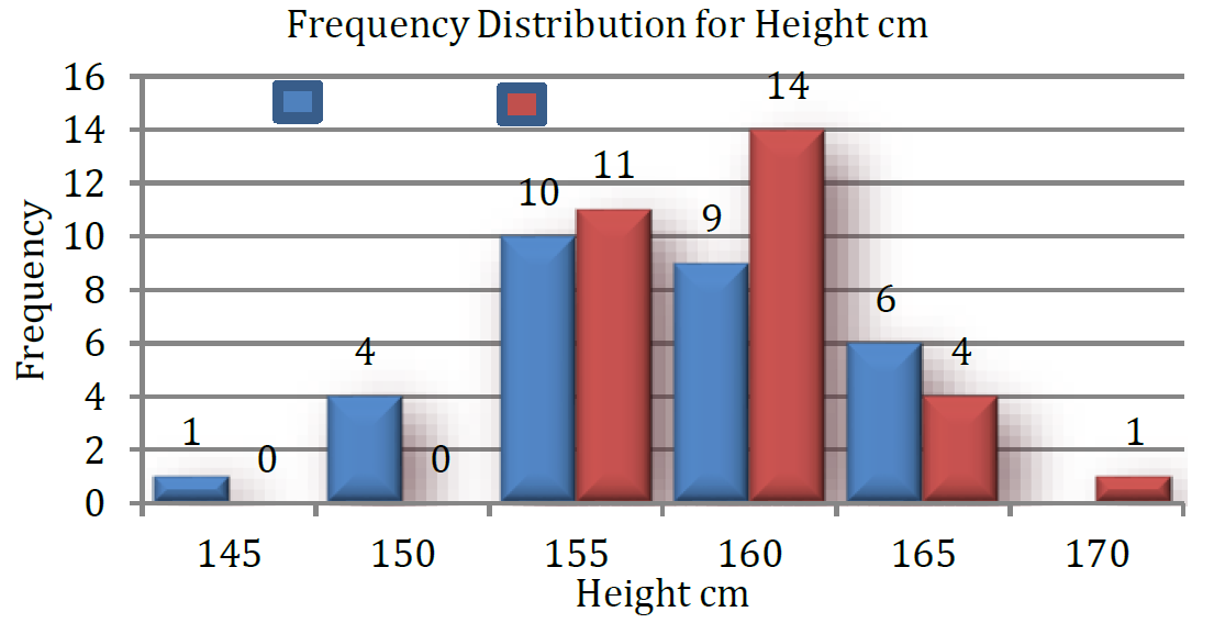
Table 5: Comparison of Mean Height (Cm) In Group D and Group C
Group
Number Of Patients
Height (Cm)
p-Value
Mean
Sd
Group D
30
157.433
5.257
0.191
Group C
30
159.033
4.038
Figure 4: Bar Diagram Showing Height Distribution
There is no significant difference in the mean weight of the group D and group C, as the p value is>0.05
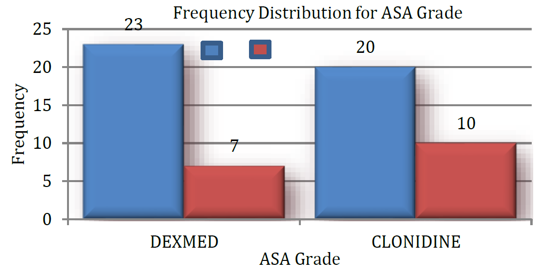
Figure 5: Bar Diagram Showing ASA Grade
There is no significant difference in the mean weight of the group D and group C, as the p value is>0.05
Figure 6: Bar Diagram Showing Duration of Surgery
(X axis –duration of surgery, in minutes, Y axis – number of patients)
In our study mean duration of surgery in group D was 125.90+23.458 and group C was133.667±51.238 and P value was 0.453, thus both groups were comparable with regards to duration of surgery.
Table 6: Distribution of Patients with Respect To ASA Grade
ASA Grade
Group
p-Value
D
C
I
23
20
0.399
II
7
10
Total
30
30
Table 7: Comparison of Duration of Surgery between Group D and Group C
Group
Number of Patients
Duration of Surgery
p-Value
Mean
Sd
Group D
30
125.90
23.458
0.453
Group C
30
133.667
51.238
Comparison of Study Parameters
In present study, mean time of onset of sensory block in Group D was 3.467±0.507 min and with Group C was 3.900±0.759 with a (p = 0.012). Mean time of onset of motor block in Group D was 9.033±1.098 and in Group C was 10.00±1.66 min with (p = 0.010). This shows that the onset of sensory and motor block was faster in group D compared to group C and was statistically significant.
Mean time required to reach the maximum level of sensory block in Group D was 10.600±1.037 min and in Group C was 11.413±1.15min. (p = 0.005) which was statistically significant, thus the mean time required to achieve a maximum sensory level was lower with group D compared to group C.
In our study the mean Time required for two segment regression of sensory level in group D was 135.5±7.925 min while in group C this time was 125.567±5.27 min. The p-value was 0.00 which is statistically significant.
Figure 7: Comparison of Onset of Sensory Block
(X axis –onset of sensory block in minutes, Y axis – number of patients)
Figure 8: Comparison of Onset of Motor Block
(X axis –onset of sensory block in minutes, Y axis – number of patients)
Figure 9: Comparison of Time to Achieve Peak Sensory Level
X axis –time to achieve peak level in minutes, Y axis – number of patients
Figure 10: Comparison of 2 Segment Regression
(X axis –time for 2 segment regression in minutes, Y axis – number of patients) Graph shows that the time required for two segment regression of sensory level in group was greater in group D (Dexmedetomidine and Ropivacaine)
Figure 11: Comparison of Duration of Sensor Block
(X axis –duration of sensory block in minutes, Y axis – number of patients
Figure 12: Comparison of Duration of Motor Block
X axis –duration of motor block in minutes, Y axis – number of patients
Table 8: Onset of Sensory and Motor Blockade
Parameter
Group D
Group C
p-Value
Sensory (Min)
Blockade
Mean
Sd
Mean
Sd
3.467
0.507
3.900
0.759
0.012
Motor (Min)Blockade
9.033
1.098
10.00
1.66
0.010
Table 9: Comparison of Time to Achieve Peak Sensory Level
Parameter
Group D
Group C
p-Value
Time To Achieve Peak Sensory Level
Mean
Sd
Mean
Sd
10.600
1.037
11.413
1.15
0.005
Table 10: Comparison of 2-Segment Regression Time
Parameter
Group D
Group C
p-Value
2-Segment Regression
Mean
Sd
Mean
Sd
135.5
7.925
125.567
5.27
0.000
Table 11: Duration of Sensory, Motor Blockade and Duration of Analgesia
Parameter
Group D
Group C
p-Value
Duration of Sensory block
Mean
Sd
Mean
Sd
421.133
21.968
360.600
23.940
0.000
Duration of Motor Block
339.367
27.031
306.600
23.940
0.000
Duration of Analgesia
434.700
27.507
365.767
12.716
0.000
Table 12: Comparison of Level of Sedation
Parameter
Group D
Group C
p-Value
Sedation
Mean
Sd
Mean
Sd
1.900
0.607
2.167
0.699
0.109
Table 13: Requirement of Rescue Analgesics
Parameter
Group D
Group C
p-Value
Mean
Sd
Mean
Sd
Rescue Analgesics
2.433
0.626
3.167
0.461
0.000
Figure 12: Comparison of Duration of Motor Block
X axis –duration of motor block in minutes, Y axis – number of patients
Figure 13: Comparison of Duration of Analgesia
(X axis –duration of analgesia in minutes, Y axis – number of patients)
Figure 14: Comparison of Level of Sedation
Figure 15: Comparison of Rescue Analgesic Requirement
Table 13: Requirement of Rescue Analgesics
Parameters
Group D
Group C
p-value
Mean
Sd
Mean
Sd
Rescue analgesics
2.433
0.626
3.167
0.461
0.000
Table 14: Comparison of Systolic Blood Pressure
Sbp
Group D
Group C
p-Value
Mean
+Sd
Mean
+Sd
Baseline
125.767
+14.330
131.300
+7.580
0.067
5 Min
120.667
+11.351
116.100
+10.873
0.117
10 Min
112.733
+13.172
113.733
+10.389
0.745
15 Min
113.800
+ 9.863
111.033
+7.717
0.231
20 Min
111.767
+ 8.245
107.400
+7.740
0.039
25 Min
109.233
+ 7.664
107.533
+6.202
0.349
30 Min
111.333
+ 6.900
108.833
+8.192
0.206
40 Min
117.133
+ 7.267
112.933
+7.652
0.033
50 Min
111.667
+ 4.880
113.00
+7.439
0.382
60 Min
113.600
+ 5.864
113.200
+7.439
0.818
70 Min
113.167
+ 5.106
114.100
+5.833
0.512
80 Min
112.033
+ 9.550
113.767
+10.441
0.505
90 Min
113.100
+ 6.900
116.433
+10.510
0.152
100 Min
116.533
+11.175
114.300
+7.429
0.545
110 Min
114.400
+ 7.655
114.300
+7.429
0.959
120 Min
119.500
+ 6.394
117.833
+6.524
0.322
130 Min
119.500
+ 6.421
119.800
+5.088
0.842
140 Min
118.900
+ 5.241
120.200
+4.859
0.323
150 Min
118.967
+ 6.825
118.100
+6.288
0.611
160 Min
119.767
+ 5.283
120.100
+7.218
0.839
170 Min
119.000
+ 4.749
116.733
+5.632
0.097
180 Min
116.167
+10.171
117.967
+4.979
0.388
Pooled
115.852
+9.154
115.424
+9.076
0.335
Table 15: Comparison of Diastolic Blood Pressure
Dbp
Group D
Group C
p-value
Mean+Sd
Mean + Sd
Baseline
77.133+9.895
75.733+9.777
0.584
5 min
72.700+10.242
75.733±8.792
0.223
10 min
71.700±7.979
70.867±5.097
0.632
15 min
68.833±6.696
68.367±6.441
0.784
20 min
69.467±7.642
71.000±5.408
0.373
25 min
70.667±5.554
69.833±4.654
0.531
30 min
69.967±5.592
71.833±6.193
0.225
40 min
69.567±5.569
70.733±4.927
0.394
50 min
73.667±5.542
72.267±6.186
0.360
60 min
75.433±8.924
76.900±8.138
0.509
70 min
72.900±9.960
69.867±8.046
0.200
80 min
68.200±11.109
73.200±7.712
0.047
90 min
74.533±7.519
75.267±8.263
0.720
100 min
73.333±8.527
72.033±10.033
0.591
110 min
68.533±6.580
71.733±8.403
0.106
120 min
68.533±6.580
75.733±7.216
0.000
130 min
74.267±6.085
72.967±5.461
0.387
140 min
71.933±4.025
74.000±4.077
0.053
150 min
71.533±3.866
70.733±3.413
0.399
160 min
71.200±3.398
69.800±3.576
0.125
170 min
71.633±3.285
68.733±5.051
0.011
180 min
70.567±3.775
71.600±6.049
0.431
Pooled
71.650+7.397
72.224+7.074
0.133
Figure 16: Comparison of Systolic Blood Pressure
Table 16: Comparison of Heart Rate
Sr. No
HR
Group I
Group II
p-Value
Mean ± SD
Mean ± SD
1.
Baseline
77.500±7.375
79.700±6.182
0.216
2.
5 Min
75.567±9.971
77.167±6.721
0.469
3.
10 Min
68.133±9.291
70.000±10.062
0.458
4.
15 Min
71.967±10.601
72.267±9.078
0.907
5.
20 Min
76.267±6.721
69.800±7.609
0.001
6.
25 Min
72.467±7.065
69.733±6.470
0.124
7.
30 Min
72.833±5.670
71.433±6.632
0.383
8.
40 Min
72.033±6.911
71.933±3.393
0.944
9.
50 Min
73.500±8.046
73.400±5.556
0.956
10.
60 Min
71.033±5.149
71.333±6.305
0.841
11.
70 Min
71.167±7.202
72.933±4.955
0.273
12.
80 Min
73.400±5.661
73.233±6.611
0.917
13.
90 Min
71.700±5.724
75.000±5.777
0.030
14.
100 Min
73.700±5.161
75.233±4.493
0.225
15.
110 Min
74.200±4.701
74.200±5.635
1.000
16.
120 Min
72.933±4.185
73.233±3.997
0.777
17
130 Min
75.800±3.388
74.400±4.312
0.167
18.
140 Min
76.567±2.700
74.233±3.126
0.003
19.
150 Min
73.700±4.450
73.667±3.889
0.975
20.
160 Min
72.133±3.340
72.300±4.786
0.876
21.
170 Min
73.833±2.902
72.300±2.583
0.059
22.
180 Min
74.633±5.129
70.867±4.224
0.003
23.
Pooled
73.380±6.543
73.138±6.206
0.469
Figure 17: Comparison of Diastolic Blood Pressure
Figure 18: Comparison of Heart Rate
Table 17: Comparison of Side Effects
Parameter
Group D
Group C
p-Value
Mean ± SD
Mean ± SD
Hypotension
0.067±0.254
0.100±0.305
0.647
Bradycardia
0.033±0.183
0.133±0.346
0.167
Table 18: Frequency of Side Effect
Parameter
Group D (n = 30)
Group C (n = 30)
Hypotension
2
3
6.66%
10.00%
Bradycardia
1
4
3.33%
13.330%
Nausea/Vomiting
0
0
00%
00%
Shivering
0
0
00%
00%
Graph shows that the time required for two segment regression of sensory level in group was greater in group D (Dexmedetomidine and Ropivacaine).
In our study, duration of sensory block in Group D was 421.133±21.968 min and in Group C was 360.600±23.940 min with p value of 0. 000.Thus duration of sensory block is statistically significant (p = 0.000) in Group D as compared to Group C with Dexmedetomidine group having longer total sensory blockade compared to Clonidine.
Duration of motor block in Group D was 339.367±27.031 min and in Group C was 339.367±27.031 min with p value of 0.000. Thus, duration of motor block was statistically significant (p = 0.000) in Group D as compared to Group C with Dexmedetomidine group having longer duration of total motor blockade.
Total duration of Post - operative analgesia in Group D was 434.700±27.507 min and in Group C was 365.767±12.716 min with p value of 0.000. Thus, duration of Post-operative analgesia was statistically Mean sedation Group D was 1.900±0.607 and in Group C 2.167±0.699 was with p value of 0.109. Thus, the level of sedation was comparable among both groups.
Figure 19: Comparison of Hypotension
Figure 20: Comparison of Bradycardia
Table 19: Comparison of Vas Score
Hours
Group D
Group C
p-Value
Mean + Sd
Mean + Sd
6th Hour
0.000 + 0.000
3.467 + 0.507
0.000
12th Hour
3.467 + 0.507
5.833 + 0.985
0.000
18 Th Hour
5.500 + 0.508
7.300 + 0.952
0.000
24 Th Hour
3.600 + 0.674
7.333 + 0.994
0.000
Pooled
3.142 + 2.051
5.983 + 1.805
0.000
Figure 21: Comparison of Vas Score
significant with Dexmedetomidine having longer total duration of Post - operative analgesia.
The mean requirement of rescue analgesics in group D was 2.433±0.626 and in group C was 3.167±0.461 and the p-value was 0.000 which shows that there was a significantly lower requirement of analgesics in group D compared to group C.
In our study, a fall in Systolic Blood Pressure of more than 30% was taken as hypotension and was treated with Inj. ephedrine 6mg iv.
In studied Groups, the mean baseline systolic blood pressure, in Group D was 125.767 mm of Hg and in Group C was 131.300 mm of Hg. (p = 0.067). Both groups were comparable. The mean systolic blood pressure was measured at 0, 5min, 10min, 15min, 20min, 25min, 30min, 40min, 50min, 60min, 70min, 80min, 90min, 100min, 110min, 120min, 130min, 140min, 150min, 160min, 170min, 180min period. In our study the systolic blood pressure was comparable in both groups.
In studied Groups, the mean baseline diastolic blood pressure, in Group D was mm 77.333±11.595 of Hg and in Group C was 78.467±9.961 mm of Hg. (p = 0.686). Thus, both groups were comparable in baseline pressures.
The mean diastolic blood pressure was measured at 0, 5min, 10min, 15min, 20min, 25min, 30min, 40min, 50min, 60min, 70min, 80min, 90min, 100min, 110min, 120min, 130min, 140min, 150min, 160min, 170min, 180min period. In our study the diastolic blood pressure was comparable in both groups.
In our study heart rate less than 60 beats per min was taken as bradycardia and was treated with Inj. Atropine. In studied Groups, the mean baseline Heart rate, in Group D is 77.500±7.375. And in Group C was79.700±6.182 with a p value of 0.216. Baseline heart rates were comparable. The mean heart rates were measured at 0, 5min, 10min, 15min, 20min, 25min, 30min, 40min, 50min, 60min, 70min, 80min, 90min, 100min, 110min, 120min, 130min, 140min, 150min, 160min, 170min, 180min. In our study the Heart rate was comparable in both groups with no significant difference.
There incidence of hypotension in group D was 6.66%, the incidence of hypotension in group C was 10% which was promptly treated with Inj. ephedrine 6mg iv stat. Similarly, incidence of bradycardia in group D was 3.0 %, while the incidence of bradycardia in group C was 13.0% which was treated with Inj. Atropine 0.3 mg iv bolus. Incidence of bradycardia and hypotension were comparable among both groups. The Incidence of nausea/vomiting and shivering in group D and C was 0%.
Graph shows that the incidence of bradycardia was higher in group C but that was statistically insignificant with p-Value 0.167.
So, from our study we concluded that incidence of hypotension and bradycardia was comparable among both groups. There was no incidence of nausea/vomiting and shivering in both groups.
Mean VAS scores in group D were lower than in group C. Mean value in group D was 3.142±2.051 and in group C was 5.983±1.805 with a p value of 0.000.
DISCUSSION
The use of Alfa 2 agonists intrathecally are being extensively evaluated as an alternative to opioids due to opioid related side effect profile like respiratory depression, nausea, urinary retention, pruritis.
The pharmacologic properties of α-2 agonists have been extensively studied and have been employed clinically to achieve the desired effects in regional anesthesia like sedation, analgesia, anxiolysis, hypnosis and sympatholysis. Clonidine has been used successfully over the last decade for the said purpose and the introduction of dexmedetomidine has further widened the scope of α-2 agonists in regional anesthesia. The faster onset of action of local anesthetics, rapid establishment of both sensory and motor blockade, prolonged duration of analgesia into the post-operative period, dose-sparing action of local anesthetics and stable cardiovascular parameters makes these agents a very effective adjuvant in regional anesthesia.
We have undertaken a study to evaluate the efficacy of Inj. Dexmedetomidine (5mcg) with 0.75% Ropivacaine by intrathecal route compared to Inj. Clonidine (15mcg) with 0.75% Ropivacaine by intrathecal route in patients undergoing Lower limb surgeries.
The demographic profile of our patients was comparable with respect to mean age, body weight, height, sex distribution, and duration of surgeries.
Onset of Sensory and Motor Blockade
Onset of sensory block is the time lapse between drug administration and loss of cold sensation at T10 dermatome tested by cold swab. Onset of motor block is the time lapse between drug administration and attainment of Bromage 3.
In our study the mean time of onset of sensory block in group D is (3.467±0.507) and group C is (3.900±0.759) with a p value of 0.012.
Mean time for the onset of motor block in group D is 9.033±1.098, group C 10.00±1.66 with a p value of 0.010, thus indicating a faster onset of sensory and motor block in group D compared to group C.
[24] Conducted a double-blind randomized control study of intrathecal ropivacaine 7.5mg with dexmedetomidine 5mcg versus ropivacaine 7.5mg with clonidine 15 mcg in patients undergoing TURP. The study showed an earlier onset of sensory and motor blockade.
In another study conducted by [25] a Comparative evaluation of dexmedetomidine and fentanyl for epidural analgesia in lower limb orthopedic surgeries, Onset of sensory analgesia at T10 was earlier in dexmedetomidine group (7.12±2.44min) compared to fentanyl group (9.14±2.94min).
Peak sensory level and two segment regression:
In our study the mean peak sensory level achieved is comparable in both groups but the mean time to reach the peak sensory level is significantly shorter in group D (10.600±1.037) than in group C (11.413±1.150) with a p value of 0. 005. The mean two segment regression time is also prolonged in group D.
[26] Conducted a randomized, prospective, double-blind study of intrathecal hyperbaric ropivacaine with Dexmedetomidine 5 mcg and hyperbaric ropivacaine with clonidine 50 mcg in lower abdominal surgeries. They concluded that time to two segment regression was significantly greater with dexmedetomidine group (148.16+14.42 min) compared to clonidine group (102.16 +14.69 min) with a p value of<0.001.
Duration of Sensory and Motor Blockade:
Mean duration of sensory block in group D is (421.133±21.968) and in group C is (360.600±23.940) with a p value of 0.000. Mean duration of motor block in group D is (339.367±27.031) and group C is (306.600±23.940) with a p value of 0.000. Thus, the duration of both sensory and motor block is significantly prolonged in group D compared to group C.
Similar prolongations were observed in a study conducted by [27]. Who compared dexmedetomidine, clonidine, fentanyl as adjuvants to hyperbaric bupivacaine. They concluded that intrathecal dexmedetomidine as adjuvant was associated with a prolonged motor and sensory blockade with a better postoperative analgesia and reduced requirement of rescue analgesia in first 24 hrs. Compared to clonidine and fentanyl groups.
In another study conducted by [21] they observed that the total duration of motor blockade was prolonged in dexmedetomidine group as compared to fentanyl group (421±21 min vs. 149.3±18.2 min, p-value˂0.0001).
[28], conducted a randomized, prospective study between intrathecal isobaric ropivacaine 0.75% plus dexmedetomidine and isobaric ropivacaine 0.75% plus fentanyl for lower limb surgeries. They concluded that dexmedetomidine at dose of 5 mcg prolonged the duration of motor blockade (136.733 min) as compared to fentanyl (94.866 min) with p value of 0.00.
Duration of Analgesia and Rescue Analgesia
In our study mean duration of analgesia in group D is 434.700±27.507 and group C is 365.767±12.716 with a p value of 0.000. The mean VAS scores were lower in group D compared to group C. The mean requirement for rescue analgesia in group D is (2.433±0.626) and in group C is (3.167±0.461) with a p value of 0.000. Thus, the duration of analgesia is statistically significantly prolonged in group D with a significant lower requirement of rescue analgesia.
[29] Studied the effect of intrathecal ropivacaine and dexmedetomidine and demonstrated that dexmedetomidine significantly prolongs the sensory motor blockade and provides a better analgesia with reduced requirement of analgesics.
[30] Conducted a prospective study of ropivacaine with dexmedetomidine versus clonidine by epidural route and concluded that dexmedetomidine is a better neuraxial adjuvant compared to clonidine in providing early blockade and prolonged postoperative analgesia with stable cardiorespiratory parameters.
[31] Conducted a randomized double-blind study on the effects of adding intrathecal dexmedetomidine to hyperbaric bupivacaine for saddle spinal block in adults undergoing Peri-anal surgeries. They concluded that the duration of analgesia was significantly prolonged and analgesic consumption was significantly reduced in group with dexmedetomidine (501.35+63.90) with a p value of 0.001.
Sedation Score:
Sedation in our study was assessed by Ramsay sedation scale. The mean Ramsay sedation (RSS) score of Group D was comparable to Group C with p value of 0.109 showing no statistical significance between the two groups.
[27], who compared dexmedetomidine, clonidine, fentanyl as adjuvants to hyperbaric bupivacaine showed no significant difference in the sedation scores.
Hemodynamic Parameters and Side Effects:
Average mean systolic blood pressure in group D (dexmedetomidine and ropivacaine) was115.852+9.154 and group C (clonidine and ropivacaine) was 115.424+9.076 with a p value of 0.335. Average mean diastolic blood pressure in group D (dexmedetomidine and ropivacaine) was 71.650+7.397 and group C was 72.224+7.074 with a p value of 0.133. Average mean heart rate in group D (dexmedetomidine and ropivacaine) 73.380+6.543and group C was 73.138+ 6.206 with a p-value of 0.469.
In our study hemodynamic parameters and side effects are comparable in both the groups.
[21] Conducted a study of Dexmedetomidine as an intrathecal adjuvant for postoperative analgesia and demonstrated a no significant change in haemodynamic parameters.
[27] Who compared dexmedetomidine, clonidine, fentanyl as adjuvants to hyperbaric bupivacaine showed no significant changes in hemodynamic parameters and side effects both were comparable intraoperatively and postoperatively.
[32] Conducted study on Effect of low-dose dexmedetomidine or clonidine on the characteristics of bupivacaine spinal blockade divided 60 patients into 3 groups. Group B received 12 mg of hyperbaric bupivacaine, group D received 12 mg of bupivacaine supplemented with 3 µg of dexmedetomidine and group C received 12 mg of bupivacaine supplemented with 30 µg of clonidine. He observed that the mean arterial pressure, heart rate and level of sedation were similar in the three groups intra-operatively and post-operatively.
[24] Conducted a double-blind randomized control study of intrathecal ropivacaine 7.5mg with dexmedetomidine 5mcg versus ropivacaine 7.5mg with clonidine 15 mcg by intrathecal route in patients undergoing TURP.
CONCLUSION
From the present study, we draw the following conclusion:
Dexmedetomidine as an adjuvant with ropivacaine intrathecally as compared to clonidine
Shortens the onset of sensory and motor block
Prolongs the duration of sensory and motor blockade
Provides effective postoperative analgesia and prolongs the duration for first rescue analgesia
Provides desirable sedation intraoperatively
Does not result in any major adverse effects
Thus, intrathecal Dexmedetomidine in a dose of 5mcg as an adjuvant to ropivacaine increases postoperative analgesia without any significant adverse effects.
Recommendations
Adjuvants used in neuraxial blockade with local anaesthetics prolong the duration of analgesia into postoperative period. Complications associated with neuraxial blockade can be avoided if the adjuvant added to intrathecal local anaesthetic provides adequate analgesia for prolonged periods.
Our experience with dexmedetomidine as an adjuvant to ropivacaine have been very satisfactory in terms of blockade characteristics, post-operative analgesia and smooth and stable maintenance of hemodynamic with no significant side effects as compared to clonidine. So, we recommend the use of dexmedetomidine in a dose of 5 mcg as an adjuvant to ropivacaine for prolonged duration surgeries and those with significant post-operative pain. Further ropivacaine has an added advantage of less cardio toxicity and better differential sensory and motor blockade.
We recommend further studies to evaluate dexmedetomidine as a smaller number of studies are available.
REFERENCE
Leone S et al. “Pharmacology, toxicology and clinical use of new long acting local anaesthetics, ropivacaine and levobupivacaine.” Acta Biomed, vol. 79, no. 2, 2008, pp. 92–105.
Simpson D, et al. “Ropivacaine.” Drugs, vol. 65, no. 18, 2005, pp. 2675–2717.
Wille M. “Intrathecal use of ropivacaine: a review.” Acta Anaesth Belg, vol. 55, 2004, pp. 251–259.
Graf B. et al. “Differences in cardiotoxicity of bupivacaine and ropivacaine are the result of physicochemical and stereoselective properties.” Anaesthesiology, vol. 96, no. 6, 2002, pp. 1427–1434.
McNamee D. et al. “Spinal anaesthesia: comparison of plain ropivacaine 5 mg/ml with bupivacaine 5 mg/ml for major orthopaedic surgery.” British Journal of Anaesthesia, vol. 89, no. 5, 2002, pp. 702–706.
Biswas B. et al. “Intrathecal fentanyl with hyperbaric bupivacaine improves analgesia during caesarean delivery and in early post-operative period.” Indian Journal of Anaesthesia, vol. 46, no. 6, 2002, pp. 469–472.
Dahlgren G. et al. “Intrathecal sufentanil, fentanyl, or placebo added to bupivacaine for caesarean section.” Anesthesia & Analgesia, vol. 85, no. 6, 1997, pp. 1288–1293.
Culebras X, et al. “Advantages of intrathecal nalbuphine, compared with intrathecal morphine, after cesarean delivery: an evaluation of postoperative analgesia and adverse effects.” Anaesthesia & Analgesia, vol. 91, no. 3, 2000, pp. 601–605.
Al-Ghanem S. et al. “Effect of adding dexmedetomidine versus fentanyl to intrathecal bupivacaine on spinal block characteristics in gynecological procedures: a double blind controlled study.” American Journal of Applied Sciences, vol. 6, no. 5, 2009, pp. 882–887.
Sethi B.S. et al. “Efficacy of analgesic effects of low dose intrathecal clonidine as adjuvant to bupivacaine.” Indian Journal of Anaesthesia, vol. 51, 2007, pp. 415–419.
Valentine J. et al. “The effect of intrathecal midazolam on post-operative pain.” European Journal of Anaesthesiology, vol. 13, no. 6, 1996, pp. 589–593.
Jain A. et al. “Analgesic efficacy of low dose intrathecal neostigmine in combination with fentanyl and bupivacaine for total knee replacement surgery.” Journal of Anaesthesiology Clinical Pharmacology, vol. 28, no. 4, 2012, pp. 486–490.
Kathirvel S. et al. “Effects of intrathecal ketamine added to bupivacaine for spinal anaesthesia.” Anaesthesia, vol. 55, no. 9, 2000, pp. 899–904.
Abouleish E. et al. “A clinical and laboratory study to compare the addition of 0.2 mg of morphine, 0.2 mg of epinephrine, or their combination to hyperbaric bupivacaine for spinal anaesthesia in caesarean section.” Anesthesia & Analgesia, vol. 77, no. 3, 1993, pp. 457–462.
Khalili G. et al. “Effects of adjunct intrathecal magnesium sulphate to bupivacaine for spinal anaesthesia: a randomized, double-blind trial in patients undergoing lower extremity surgery.” Journal of Anesthesia, vol. 25, no. 6, 2011, pp. 892–897.
Kamibayashi T, and Maze M. “Clinical uses of alpha-2 adrenergic agonists.” Anaesthesiology, vol. 93, 2000, pp. 1345–1349.
Scafati A. “Analgesia and alpha agonists 2.” Medens Review, vol. 4, 2004, pp. 4–7.
Gabriel J, and Gordin V. “Alpha 2 agonists in regional anaesthesia and analgesia.” Current Opinion in Anaesthesiology, vol. 14, 2001, pp. 751–753.
Gertler R. et al. “Dexmedetomidine: a novel sedative analgesic agent.” Baylor University Medical Centre Proceedings, vol. 14, no. 1, 2001, pp. 13–21.
Dobrydnjov I. et al. “Clonidine combined with small-dose bupivacaine during spinal anesthesia for inguinal herniorrhaphy: a randomized double-blinded study.” Anesthesia & Analgesia, vol. 96, 2003, pp. 1496–1503.
Gupta R. et al. “Dexmedetomidine as an intrathecal adjuvant for postoperative analgesia.” Indian Journal of Anaesthesia, vol. 55, no. 4, 2011, pp. 347–352.
Gupta R. et al. “A comparative study of intrathecal dexmedetomidine and fentanyl as adjuvants to bupivacaine.” Journal of Anaesthesiology Clinical Pharmacology, vol. 27, no. 3, 2011, pp. 339–344.
Barash, P.G. Textbook of Clinical Anaesthesia. 7th ed., chapter “Acute Pain Management, Visual Analogue Scale,” 2013, pp. 56–1619.
Dhasmana S. et al. “Dexmedetomidine versus clonidine as an adjunct to intrathecal small dose ropivacaine in patients undergoing transurethral resection of prostate.” Ain-Shams Journal of Anaesthesiology, vol. 7, no. 4, 2014, pp. 534–540.
Bajwa S. et al. “Comparative evaluation of dexmedetomidine and fentanyl for epidural analgesia in lower limb orthopedic surgeries.” Saudi Journal of Anaesthesia, vol. 5, no. 4, 2011, pp. 365–370.
Singh N. et al. “A comparative study of analgesic effects of intrathecal hyperbaric ropivacaine with dexmedetomidine and hyperbaric ropivacaine with clonidine in lower abdominal surgery.” Journal of Medical Society, vol. 29, no. 3, 2015, pp. 164–168.
Mahendru V. et al. “A comparison of intrathecal dexmedetomidine, clonidine, and fentanyl as adjuvants to hyperbaric bupivacaine for lower limb surgery: a double blind controlled study.” Journal of Anaesthesiology Clinical Pharmacology, vol. 29, no. 4, 2013, pp. 496–502.
Ravipati P. et al. “A comparative study between intrathecal isobaric ropivacaine 0.75% plus dexmedetomidine and isobaric ropivacaine 0.75% plus fentanyl for lower limb surgeries.” Anaesthesia: Essays and Researches, vol. 11, no. 3, 2017, pp. 621–626.
Parmar N. et al. “Effect of intrathecal ropivacaine with dexmedetomidine for operative and post-operative analgesia: a prospective randomized study.” Journal of Evolution of Medical and Dental Sciences, vol. 3, no. 11, 2014, pp. 2917–2925.
SaravanaBabu M. et al. “A comparative study in the post-operative spine surgeries: epidural ropivacaine with dexmedetomidine and ropivacaine with clonidine for post-operative analgesia.” Indian Journal of Anaesthesia, vol. 57, no. 4, 2013, pp. 371–376.
Gautam B. et al. “Effects of adding intrathecal dexmedetomidine to hyperbaric bupivacaine for saddle spinal block in adults undergoing perianal surgeries.” Journal of Nepal Health Research Council, vol. 16, no. 1, 2018, pp. 43–47.
Kanazi G.E. et al. “Effect of low dose dexmedetomidine or clonidine on the characteristics of bupivacaine spinal block.” Acta Anaesthesiologica Scandinavica, vol. 50, 2006, pp. 222–227.
Recommended Articles
Research Article
Evidence Guided, Experience Based and Patient Compatible 3-Step Protocol for Managing Moderate to Severe Category of COVID19 Infections
Farah Husain,
...
Kirti N Saxena
Published: 05/01/2021
Download PDF
Research Article
A Descriptive Cross-Sectional Study to Evaluate the Knowledge about Symptoms and Risk Factors for Heart Attack (Myocardial Infarction) Among the General Population of New Delhi
Aastha Sehgal,
...
Avneet Kaur
Published: 06/05/2023
Download PDF
Research Article
Knowledge About Risk and Preventive Factors for Osteoarthritis Knee among Ortho OPD Patients in Regional Hospital, Keylong in District Lahaul and Spiti of Himachal Pradesh
Ajay Thakur,
Jaspal Chaudhary
Published: 20/06/2022
Download PDF
Research Article
A Randomized Controlled Trial Comparing the Effect of Oral Preemptive Pregabalin Compared to IV Lignocaine in Attenuating Hemodynamic Response to Laryngoscopy
P. Aashik,
...
B.N. Reddy
Published: 17/05/2023
Download PDF
PO Box 101, Nakuru
Kenya.
Email: office@iarconsortium.org
Editorial Office:
J.L Bhavan, Near Radison Blu Hotel,
Jalukbari, Guwahati-India
Varsha, T., Bhupathy, S. & Sekhar, C. C. (2021). A Prospective Randomized Double Blind Comparative Study of the Effect of Intrathecal Dexmedetomidine and Clonidine as Adjuvants with Ropivacaine in Lower Limb Surgeries. IAR Journal of Anaesthesiology and Critical Care, 2(1), 1-14.
MLA
Varsha, Tipparaju, S. Bhupathy and C.N. C. Sekhar. "A Prospective Randomized Double Blind Comparative Study of the Effect of Intrathecal Dexmedetomidine and Clonidine as Adjuvants with Ropivacaine in Lower Limb Surgeries." IAR Journal of Anaesthesiology and Critical Care 2.1 (2021): 1-14.
Chicago
Varsha, Tipparaju, S. Bhupathy and C.N. C. Sekhar. "A Prospective Randomized Double Blind Comparative Study of the Effect of Intrathecal Dexmedetomidine and Clonidine as Adjuvants with Ropivacaine in Lower Limb Surgeries." IAR Journal of Anaesthesiology and Critical Care 2, no. 1 (2021): 1-14.
Harvard
Varsha, T., Bhupathy, S. and Sekhar, C. C. (2021) 'A Prospective Randomized Double Blind Comparative Study of the Effect of Intrathecal Dexmedetomidine and Clonidine as Adjuvants with Ropivacaine in Lower Limb Surgeries' IAR Journal of Anaesthesiology and Critical Care 2(1), pp. 1-14.
Vancouver
Varsha T, Bhupathy S, Sekhar CC. A Prospective Randomized Double Blind Comparative Study of the Effect of Intrathecal Dexmedetomidine and Clonidine as Adjuvants with Ropivacaine in Lower Limb Surgeries. IAR Journal of Anaesthesiology and Critical Care. 2021 Jan;2(1):1-14.


 Download PDF
 Download XML