+91 6002993949
submission@iarconsortium.org
Open Access
ISSN (Print) : 2788-8843
ISSN (Online) : 2788-8851
Peripheral smear study is used to diagnosis the disease condition in blood components, for the peripheral smear stain the most commonly used staining is Romanowsky-type stain they are neutral stain and it is most widely used for the examination of blood from the blood films. Leishman stain is the type of Romanowsky stain, Leishman stain is the mixture of Methylene blue and Eosin dye, it stains the blood cells in violet color, Leishman stain is habitually used stain in hematology. Field’s stain is named after physician John Willian Field, he developed it in 1941. Field’s stain is the combination of Methylene blue, Azure I, Eosin Y dye and the fixative is methanol. Field’s stain is rapid stain used primarily on thin films for malarial parasites, Field’s stain is routinely used in hematology and cytopathologyp. Field’s satin is extremely fast stain. In the present study was comparing the Leishman stain and Field’s stain for this study 60 patient sample is used. Two peripheral smears were prepared from each sample and totally 120 smears, each patients samples were stained in parallel with Leishman stain and Field’s stain. Staining time taken for Leishman stain is 15 minutes and the staining time taken for Field’s stain is 1 minute and the slides were observed under microscope 4, 10, 40 and 100× with oil immersion. The percentage were analyzed statistically. This study shows that Field’ stain better alternative for Leishman stain for routine peripheral smear study and this Field’ stain has advantages in the staining time, clarity view of blood cells and easy to identify the different leukocytes with crisp and clear view of granule of cell and the cytoplasm. Field’ s stain is the better alternative for Leishman stain.
Peripheral smear study is used to analyze and diagnose the disease condition in blood components, blood is a specialized body fluid and it consist of a fluid various composition of erythrocyte, leucocyte and thrombocyte. Peripheral smear is two type they are thick layer blood smear, thin layer blood smear [1-5].
Romanowsky stain refers to a class of histological stains used in the field of pathology to prepare and visualize cells and cellular components. These stains are named after their developer, Dr. Romanowsky. The staining technique is named after the Russian physician Dmitri Leonidovich Romanowsky (1861-1921), who was one of the first to recognize its potential for use as a blood stain. These stains are commonly used for staining blood smears to examine and differentiate various blood cells, such as red blood cells, white blood cells and platelets. They can also be used for staining other types of cells, such as bone marrow cells and microorganisms. Romanowsky stain which is combination of an acidic stain and a basic stain so that it is said as neutral staining and this stain are used in hematology and cytopathology in department for study about the cells. Romanowsky staining methods are used to stain the blood, bone marrow film, fine needle aspirated sample and cerebrospinal fluid sample from lumbar puncture. When applied to a blood smear, the Romanowsky stain imparts different colors to various cellular structures. The basic components of the stain interact with different cellular components, resulting in distinct colorations. For example, the eosin component stains acidic structures, such as eosinophilic granules and the cytoplasm of eosinophils, red or pink [6]. On the other hand, the methylene blue component stains basic structures, such as the nucleus of cells, blue or purple. The Romanowsky stain is particularly useful in the examination of blood smears for the diagnosis of various blood disorders, including leukemia, anemia and infections. By evaluating the size, shape and staining characteristics of different blood cells, it can identify abnormalities and make accurate diagnosis. These stains are important tools in the field of pathology and hematology as they provide valuable information about cellular morphology and can helps in diagnosis of various disease conditions related to blood cells and other cell types.
Field’s Stain
Field's stain is a histological stain used in the field of pathology. It is primarily used for staining blood smears to aid in the identification and differentiation of various blood cell. Field stain is named after physician John William Field, who developed it in 1941. It is a combination of two stains; Field's stain A and Field's stain B. Field's stain A - It is a mixture of methylene blue and azure dye, Field’s stain B - It is a mixture of eosin Y dye [7]. Field's stain A selectively stains acidophilic structures, such as red blood cells, eosinophils and some granules within basophils. On the other hand, Field stain B stains basophilic structures, such as nuclei and granules within neutrophils and basophils. The combination of Field's stain A and B allows for differential staining method for examination of various blood cell components, aiding in identification and classification of different types of blood cells [8-12].
Field’s stain is quick method for staining thick film for visualization of malarial parasite. Field's stain comes under the classification of Romanowsky-type stain, which is used in histology that produce a characteristic pattern of staining in cells and cell structures. Field's stain has used to identification and characterization of blood cell abnormalities, aiding in the diagnosis of various hematological disorders [13-16].
Leishman Stain
Leishman stain is a vital stain commonly used in the field of microscopy for staining blood cells, particularly for the examination of blood smears. It was named after the British pathologist William Boog Leishman, who introduced the stain in 1901. Leishman stain is a Romanowsky-type stain, which means it is a combination of different dyes that react with cellular components to produce distinct staining patterns. The original formulation of Leishman stain contained a mixture of methylene blue and eosin dyes [17].
Leishman stain improved upon these methods by providing better differentiation between different types of blood cells, such as red blood cells, white blood cells and their various subtypes. Over time, modifications and variations of the original Leishman stain formula have been developed to enhance its effectiveness and specificity. These modifications include altering the pH, adjusting dye concentrations and incorporating additional chemicals to improve staining quality and cell differentiation [18].
Objective
The Field’s stain and Leishman stain are compared based on the appearance of blood cells.
The stains are compared based on the staining time of Field’s stain and Leishman stain.
The sources of literature referenced from the various electronic database such as Google scholar, Science Direct, PubMed, ResearchGate, library books search, used for our study, the experiment of the study in the site of Central laboratory in Department of pathology of ACS Medical College and Hospital, in the period of six months [19].
Leishman stain most commonly used as a staining method for peripheral smear study. In this study Field’s stain is compared with Leishman stain in accordance with manual differential count, erythrocyte appearance and platelet appearance in peripheral smear study [20]. A totally 120 smear preparation were made, (2 smear for each sample) 60 smears were stained with Leishman stain, 60 smear were stained with Field’s stain. Thin blood films stained with both Leishman stain and Field’s stain will be evaluated based on their appearance of blood cells.
Material Required
About 2 mL of Ethylene Diamine Tetra Acetic Acid (EDTA) blood sample
Microscopical slides
Micropipette
Microscope
Reagent-Leishman stain and buffer solution
Reagent-Field’s stain- A, Field’s stain -B and methanol
Blood sample is collected to study morphology of blood cells by vein puncture in the antecubital vein 0.2 mL of venous blood drawn and collected in Ethylene Diamine Tetra Acetic Acid (EDTA) vacutainer. The sample is analysed in the department of Haematology [21].
Peripheral Smear Blood Preparation
One hundred twenty slides were are taken to prepare a set two slides of smear from each blood sample to compare the both staining. Slide is labelled with glass pencil by the name and unique number of the particular patient from whom the blood sample is taken .Place a drop blood in 3-4 mm diameter near the near the end of the slide, take another slide use as a spreader, rest the spreader slide at 25 degree angles on the slide, the drop should flow to the edges of the spreader slide approximately 1-2 mm in depth, keep the spreader at 25 degree angles with light but firm pressure against the horizontal slide. The smear should cover ½-¾ of the slide and finish with a feathered edge obtain the tongue shaped smear over the slide, allow the slide to air dry [22-26].
Staining Procedure
Leishman Stain: The air-dried slide is taken and placed in staining rack, a 1mL of Leishman stain solution wait for 3 minutes, all twice the amount of buffer solution wait for 10 minutes and wash in running tape water. Observe under 4×, 10×, 40× and oil immersion (100×) [27].
Field’s Stain: The air-dried slide is taken and placed in staining rack, fix the slide with methanol spray allow it for air dry, add 1 mL of Field’s stain B solution and wait for 5 seconds, wash in tap water, add 1 mL of Field’s stain A solution and wait for 40 seconds and wash in tap water, observe under 4×, 10×, 40× and oil immersion (100×) [28-30].
Observation
Observation of blood cell under microscope, the appearance of erythrocyte, leucocyte and thrombocyte [31-34]. Field’s stain appearance of erythrocyte in pinkish purple and in Leishman stain appearance of erythrocyte in pink, Field’s stain appearance of thrombocyte in pale bluish and in Leishman stain appearance of thrombocyte in violet, Field’s stain appearance of neutrophil nucleus in purple and the cytoplasmic granules in pale iliac and in Leishman stain appearance neutrophil nucleus in dark purple and the cytoplasmic granules in pale, Field’s stain appearance of eosinophil nucleus in purple and the cytoplasmic granules in orange and in Leishman stain appearance eosinophil nucleus in blue and the cytoplasmic granules in red to orange, Field’s stain appearance of basophil nucleus in purple and cytoplasmic granules in dark blue and Leishman stain appearance basophil nucleus in purple and the cytoplasmic granules in dark purple, Field’s stain appearance of lymphocyte nucleus in dark blue and cytoplasm in pale blue and Leishman stain appearance basophil nucleus in dark purple and the cytoplasm sky blue, Field’s stain appearance of monocyte in smokey purple and in Leishman stain appearance in grey blue, Field’s stain appearance of parasite in dark purple and Leishman stain appearance of parasite in dark blue (photographic Figure (1a-r) are shown below) [35].
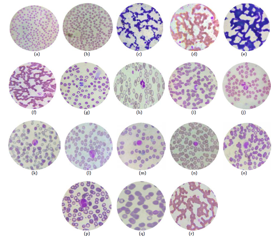
Figure 1(a-r): (a) RBC stained with Field’s stain, 100× (b) RBC stained with Leishman stain,100× (c) Platelet stained with Field’s stain,100× (d) Platelet stained with Leishman stain,100× (e) Cluster of Platelet stained with Field’s stain,100× (f) Cluster of Platelet stained with Leishman stain,100× (g) Segmented Neutrophil stained with Field’s stain,100x (h) Segmented Neutrophil stained with Leishman stain,100× (i) Band shaped Neutrophil stained with Field stain,100× (j) Band shaped Neutrophil stained with Leishman stain,100× (k) Eosinophil stained with Field’s stain,100× (l) Eosinophil stained with Leishman stain,100× (m) Lymphocyte stained with Fields’s stain,100× (n) Lymphocyte stained with Leishman stain,100× (o) Monocyte stained with Field’s stain,100× (p) Monocyte stained with Leishman stain,100× (q) MALARIAL PARASITE: (Trophozoite Stage) stained with Field’s stain,100× (r) MALARIAL PARASITE: (Trophozoite Stage) Stained with Leishman stain, 100×
A total of 60 participants we included in the study according to the inclusion criteria. A smear which is prepared were stained according to Leishman and Field’s staining procedure totally 120 peripheral blood smears were made (2 smear for each sample) 60 smears were stained with Leishman stain, 60 smear were stained Field’s stain. Differential count was made by the manual cell counter, which the count of each leucocyte cell by the both was staining. The appearance of erythrocyte and thrombocyte have been given by grading method (0-Better and 1-Excellent) which is observed peripheral smear of Leishman stain and Field’s stain [36,37].
The observation of both staining methods evaluated by the appearance of RBC, Platelet by scoring method and the for WBC cell is calculated by the manual differential count and the mean value is taken for each cell. Based on the score of the erythrocyte and thrombocyte cells; In Field’s stain- RBC, Platelet excellent percentage is 93.3, better percentage is 6.6. In Leishman stain RBC, Platelet excellent percentage is 71.6, better percentage is 28.3. Mean value of Field’s stain DC of Neutrophil segment is 58.7, Neutrophil band is 4.6, Neutrophil totally count is 63.1, Eosinophil is 5.4, Monocyte is 8.016, Lymphocyte is 23.6. Mean value of Leishman stain DC of Neutrophil segment is 61.3, Neutrophil band is 2.35, Neutrophil totally count is 63.6, Eosinophil is 4.91, Monocyte is 7.85, Lymphocyte is 23.4. Based on this percentage, Field satin is the better alternative for Leishman stain [38-40].
Statistical Analysis
Grade are based on the appearance of erythrocyte and thrombocyte; differential count of Leucocyte is counted by differential counting chamber and from that mean value are calculated.
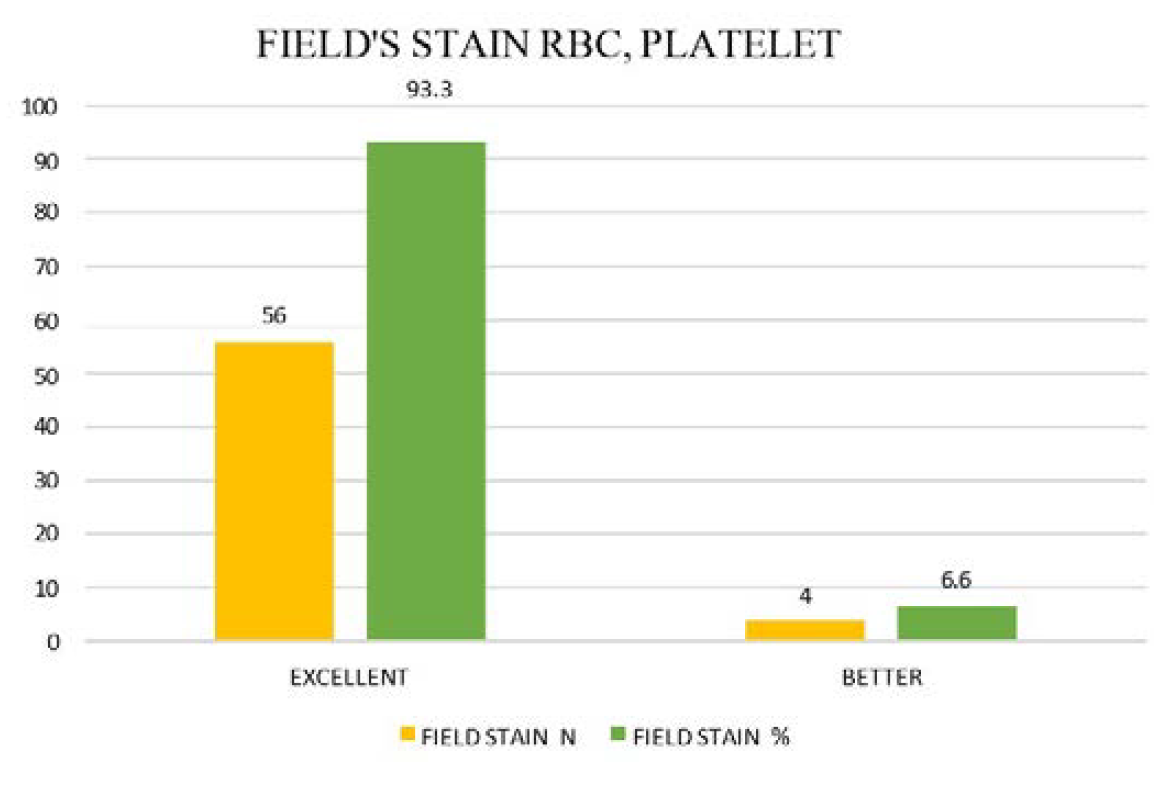
Figure 2: Graphical Representation 1
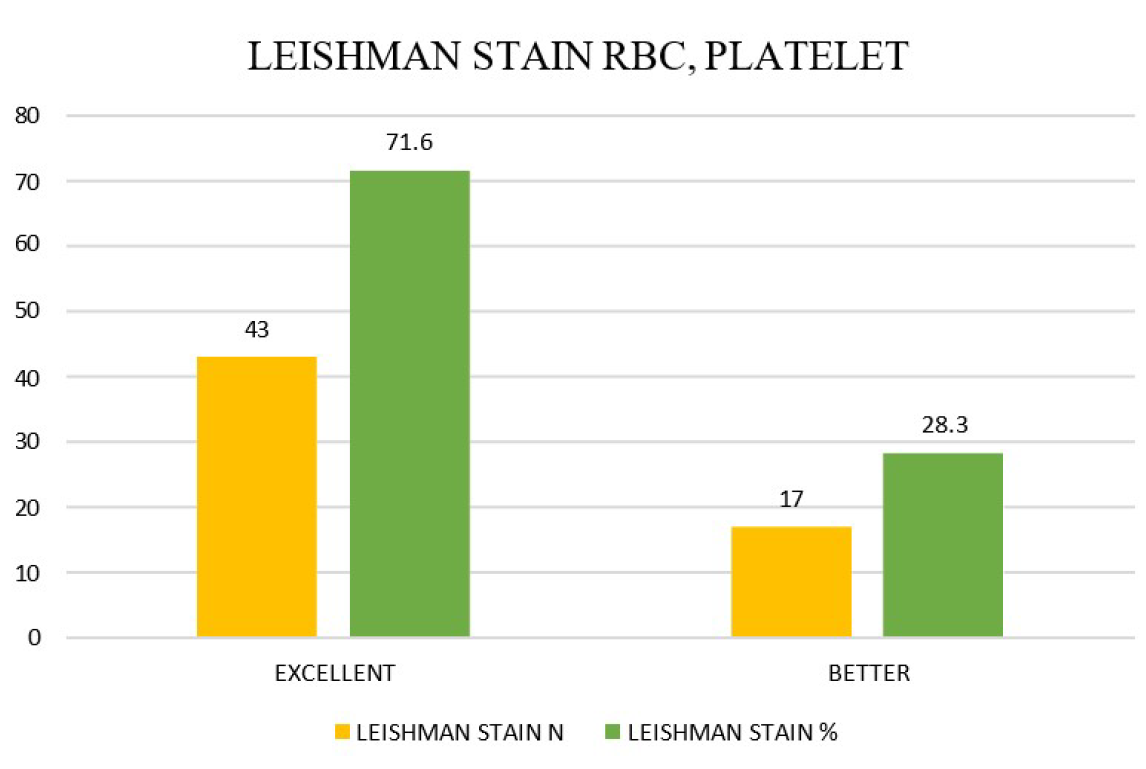
Figure 3: Graphical Representation 2
Table 1: Quality Grading of RBC and Platelet Staining by Field’s and Leishman Methods
| RBC And Platelet | Field’s Stain | RBC, Platelet | Leishman Stain | ||
N | % | N | % | ||
Excellent | 56 | 93.3 | Excellent | 43 | 71.6 |
Better | 4 | 6.6 | Better | 17 | 28.3 |
Total | 60 | 100 | Total | 60 | 100 |
Table 2: Mean and Standard Deviation of Differential Leukocyte Count by Field’s and Leishman Stains
| Staining | Field’s Stain | Leishman Stain | |||
Parameter | Mean | SD | MEAN | SD | |
Neutrophil | SEG | 58.7 | 6.14 | 61.3 | 6.55 |
Band | 4.6 | 0.942 | 2.35 | 1.527 | |
Total | 63.1 | 6.28 | 63.6 | 6.76 | |
Eosinophil | 5.4 | 2.62 | 4.91 | 2.22 | |
Basophil | 0 | 0 | 0 | 0 | |
Monocyte | 8.016 | 1.75 | 7.85 | 1.592 | |
Lymphocyte | 23.6 | 5.37 | 23.4 | 5.97 | |
For the appearance of erythrocyte and thrombocyte grade are given:
Excellent Better
1 0
The RBC, Platelet grade of the Field’s stain and Leishman stain (Table 1).
Graphical representation 1,2 (Figure 2 and 3).
It is also calculated mean from the differential count of Leucocyte (Table 2).
A comparative study between Field’s stain and Leishman stain in routine peripheral smear study. This study the main parameter is RBC, Platelet appearance by both the staining the overall excellent percentage of field’s stain is 93.3% and Leishman stain is 71.6%, the better percentage of Field’s stain is 6.6% and Leishman stain is 28.3%. Based on this percentage field satin is the better alternative for Leishman stain. Field’s stain has a better clarity view of all cell than the Leishman stain view and the appearance granules are very crisp and clear and it is easy to recognize and differentiate the leucocyte, clear appearance erythrocyte and thrombocyte.
Field’s stain time taken is less than Leishman stain, Field’s stain time taken for staining procedure is 3 minutes but for Leishman stain time taken for staining is 15 minutes.
In field’s stain it has good clarity view of cytoplasmic granules of WBC cells than the Leishman stain’s view.
Field’s stain has the advantage of being used in high volume hospital/diagnosis center for rapid staining and shorter than the Turn Around Time (TAT).
By all this note we conclude that field’s stain is better than the Leishman stain for routine peripheral smear study. Field’s stain is better alternative for Leishman stain.
Acknowledgment
I am heartly grateful and very thankyou to our educational institute Dr. M.G.R. Educational and Research Institute, ACS Medical College and Hospital.
Adewoyin, A.S. and B. Nwogoh. “Peripheral Blood Film – A Review.” 2014.
Mayada, S.H. Blood Smear Preparation Practical Biology. 2014.
“Blood Sampling and Blood Film Preparation and Examination.” March 2006.
International Committee for Standardization in Haematology. “ICSH Reference Methods for Staining of Blood and Bone Marrow Films by Azure B and Eosin Y (Romanowsky Stain).” British Journal of Haematology, 1984.
Greer, J.P. et al. Wintrobe’s Clinical Hematology. 11th ed., Lippincott, 2003.
Marshall, P.N. et al. “A Rapid Thin-Layer Chromatographic System for Romanowsky Blood Stains.” Stain Technology, 1974.
Houwen, B. “Blood Film Preparation and Staining Procedures.” Laboratory Hematology, 2000.
Gulati, G. et al. “Criteria of Blood Smear.” 2013.
Jaber, M.A. Blood Film. Donna Virginia Ward, 2020.
Al-Aswad, S.I.H. and A. Al-Hindi. “Blood Smear Preparation and Staining.” 2016.
Sood, R. Medical Laboratory Technology: Method and Interpretation. 2014.
Munster, M. “The Role of the Peripheral Blood Smear in the Modern Haematology Laboratory.” 2013.
Crocker, J. and D. Burnett. General Stains: The Science of Laboratory Diagnosis. 2005.
Riley, R.S. et al. “How to Prepare and Interpret Peripheral Blood Smears.” 2013.
Peterson, L.C. et al. “Review of the Peripheral Blood Smear: When and Why.” Laboratory Hematology, 2001.
Javidian, P. et al. “Review of the Peripheral Film.” Pathologist, 1993.
Perkins, S. Diagnosis of Anaemia. 2013.
Beutler, E. et al. “Examination of the Blood.” 2001.
Wickramasinghe, S.N. and W.N. Erber, editors. Blood and Bone Marrow Pathology. 2011.
Basu, S. Blood Cell and Bone Marrow Morphology. 2005.
Constantino, B.T. and B. Cogionis. “Nucleated RBCs—Significance in the Peripheral Blood Film.” 2000.
Bainton, D.F. “Morphology of Neutrophils, Eosinophils and Basophils.” 2006.
Bain, B.J. Blood Cell Morphology in Health and Disease. 2012.
Chunge, C.N. et al. “A Rapid Staining Technique for Leishmania Parasites in Splenic Aspirate Smears.” 1989.
Cole, E.R. and A.K. Sewell. “A Simplified Direct Preparation of Field’s Stain.” 1956.
Cole, E.R. and A.K. Sewell. “The Preparation and Stability of Field’s Stain.” 1953.
Singh, T. and K.U. Chaturvedi. Practical Pathology. 2015.
Chakraborthy, P. and G. Chakraborthy. Practical Pathology. 2002.
Sanyal, S. and A. Bhattacharya. A Practical Manual. 2017.
Vissa, S. “Peripheral Smear – WBC Histopathology.” Histopathology Guru.
Mundhra, D. et al. “Analyzing Microscopic Images of Peripheral Blood Smear Using Deep Learning.” 2017.
Wolman, M. “Field’s Stain.” 1943.
Niranjan, M.B. et al. “Field’s Stain: A Quick Alternative to MGG (May Grunwald Giemsa).” 2004.
Manmadhan, A.A. “A Comparison between Conventional Leishman Stain and a Modified Blood Stain for the Evaluation of Hematologic Elements.” National Journal of Laboratory Medicine.
Mathur, A. et al. “Scalable System for Classification of White Blood Cells from Leishman-Stained Blood Stain Image.” 2013.
De Burgh, P.M. “Notes on Field’s Stain.” 1946.
Wilson, C.I. et al. “The Peripheral Blood Smear in Patient with Sickle Cell Trait: A Morphological Observation.” August 2000.
Jain, M. and T.K. Dolai. ABC of Peripheral Smear. 2020.
Field, J.W. and A. Sandosham. “The Romanowsky Stain-Aqueous or Methanolic.” 1964.
Barrelet, A. et al. “Staining Technique: Romanowsky Stain-Overview.” 2001.
