+91 6002993949
submission@iarconsortium.org
Open Access
ISSN (Print) : 2789-6099
ISSN (Online) : 2789-6102
Intraperitoneal adhesions are abnormal adhesions between tissues and organs, usually between the omentum, bowel loops and abdominal wall. One of the pathogenesis is tissue hypoxia as a result of impaired blood supply to mesothelial cells and the inflammatory process. Nitric Oxide (NO) is a pleitropic mediator of hypoxia, especially iNOS. Intraperitoneal adhesion was proposed to develop in hypoxic condition through the increasing of iNOS, which contributed directly and indirectly through reduce peristaltic in promoting intraperitoneal adhesion.
Intraperitoneal adhesions are abnormal adhesions between tissues and organs, usually between the omentum, bowel loops and abdominal wall. This adhesion can be a thin layer of connective tissue, or a thick fibrous tissue containing blood vessels and nerve tissue, as well as direct contact between two organ surfaces [1]. The pathogenesis of adhesion involves three important trauma-induced processes: trauma or direct tissue damage induces inhibition of the fibrinolytic and extracellular matrix degradation systems, an inflammatory response with the production of cytokines, particularly TGF- and interleukins and tissue hypoxia as a result of impaired blood supply to mesothelial cells and the inflammatory process, which are responsible for collagen formation and angiogenesis [2].
Hypoxia
Hypoxia is a condition in which the oxygen concentration in cells is very low. Hypoxia is caused by cell injury whereas cell injury is caused by several factors; ischemia, anemia, decreased oxygen perfusion caused by blood problems and others [3]. In inflammatory conditions, there is an increase in growth factors, chemokines, cytokines and ROS balance which results in gene mutations. There will be various pro-inflammatory conditions that will form a lot of free radicals, causing a state of hypoxia [4]. In hypoxic conditions, ROS levels will increase. Several ROS such as O2, H2O2, OH- produced during normal aerobic metabolism are known to act as signaling molecules that induce various biological processes, such as stimulation of protein phosphorylation, Ca2+ -signaling, phospholipid hydrolysis and activation of transcription factors. In hypoxic conditions there is also an increase in levels of Reactive Oxygen Species (ROS) [5].
Hypoxia induces inflammation through the release of inflammatory mediators by hypoxic parenchymal and endothelial cells. Neutrophils as one of the effectors of acute inflammation work by generating free radicals. The hypoxia-induced transcription factor complex, Hypoxia-Inducible Factor (HIF)-1 is regulated by hypoxia as well as by various inflammatory mediators [6]. After extravasation from these vessels, cell activity is further enhanced by stimulation of HIF-1 by proinflammatory cytokines such as IL-1, IL-6, TNF-alpha and locally expressed tissue factor. TNF- induced HIF-1α stimulation requires NF-kB at the level of HIF-1α protein stabilization without affecting its mRNA level [7].
Under normal physiological conditions, eukaryotic cells are under aerobic conditions and have an innate defense system against ROS that contributes to the maintenance of a balance between prooxidant (free radical species) and antioxidant capabilities. When excessive ROS production or free radical eradication mechanisms are disrupted, oxidative stress occurs, in which ROS accumulate and damage important biomolecules such as nucleic acids, proteins and lipids, as well as cells in the body [8]. Under hypoxic conditions, the mitochondrial respiratory chain also produces nitric oxide (NO) which can generate reactive nitrogen species (RNS; ONOO−, Nitrite (NO2-), Nitrate (NO3-), which in turn produces other reactive species such as the reactive aldehyde -malondialdehyde and 4-hydroxynonenal which can attack lipid membranes to initiate a free radical chain reaction known as lipid peroxidation. At the molecular level, hypoxia decreases tPA and increases PAI expression in human peritoneal fibroblasts in vitro and in peritoneal tissue in vivo, thereby reducing plasmin, inhibiting fibrin lysis and increasing adhesion formation [9].
Free radicals are compounds or molecules that contain one or more unpaired electrons in their outer orbit. The presence of unpaired electrons causes the compound to be very reactive looking for a partner, by attacking and binding the surrounding molecular electrons. In living cells, free radicals are formed in the plasma membrane, mitochondria, peroxisomes, endoplasmic reticulum and cytosol through enzymatic reactions that take place in metabolic processes. The body has a protective mechanism that is formed to neutralize free radicals, including the enzymes Superoxide Dismutase (SOD), Catalase (CAT) and glutathione peroxidase [10].
Proteins, unsaturated fatty acids and lipoproteins, as well as elements of DNA including carbohydrates are the main targets for free radicals, but the most vulnerable are unsaturated fatty acids. Free radical attack on the surrounding molecules will cause a chain reaction, which then produces new radicals. The impact of free radical reactivity varies, ranging from cell or tissue damage, autoimmune diseases, degenerative diseases to cancer. High levels of free radicals in the body can be indicated by high levels of malondialdehyde in plasma and low activity of antioxidant enzymes [11].
Free radicals are generated from endogenous and exogenous sources. Immune cell activation, inflammation, ischemia, infection, cancer, excessive exercise, mental stress and aging are all responsible for the production of endogenous free radicals. Exogenous free radical production can occur as a result of exposure to environmental pollutants, heavy metals (Cd, Hg, Pb, Fe and as), certain drugs (cyclosporine, tacrolimus, gentamicin and bleomycin), chemical solvents, cooking (smoked meat, olive oil, etc.) used and fat), cigarette smoke, alcohol and radiation. When these exogenous compounds penetrate the body, they are degraded or metabolized and free radicals are generated as by-products [12].
Hypoxia Induced Intraperitoneal Adhesion
In intraperitoneal adhesion, tissue injury from suirgery cause devascularization, which induced tissue hypoxia. Later on, oxidative stress happens, increasing HIF-1alpha and NF-kB. By these two mediators, TGF beta, IRF-1, TNF-alpha, IFN-1gamma, hydrogen peroxide and VEGF were increased. The pathway that author concerned about is the TNF that increase heat shock protein 90, later increasing iNOS, NO, OONO-, then protein nitration. This pathway also contributed to lower peristaltic condition due to vasodilatation effect dominated in high NO level. Finally, the effect accumulated to the development of intraperitoneal adhesion [13] (Figure 1).
Perhaps, the best-known free radical that acts as a signaling molecule is Nitric Oxide (NO). It is an important cell-to-cell messenger required for proper modulation of blood flow, is involved in thrombosis and is essential for normal neuronal activity. NO is also involved in nonspecific host defense, required for the elimination of intracellular pathogens and tumor cells. Another physiological activity of free radicals is the induction of mitogenic responses. In summary, free radicals, when maintained at low or moderate levels, are essential for human health [14].
Nitric Oxide (NO)
Nitric oxide is a compound that is toxic and short-lived, in the form of gas molecules produced by inducible NO synthase (iNOS) by converting the amino acid L-arginine into NO and citrulline. NO can easily diffuse freely across cell membranes to adjacent cells, then react with ferrous sulfur of some macromolecules and inhibit the occurrence of ribonucleotide reductase. In the synthesis of DNA ribonuclease is converted into DNA, DNA synthesis is inhibited and cell proliferation is stopped and is a mechanism of phagocytes to inhibit inflammation [15]. NOS in humans (and mice) has three forms, namely:
Nitric Oxide (nNOS or NOS¯1) neurons found in nerve cells act as neuromodulators or neuromediators
Endothelial Nitric Oxide (eNOS or NOS¯3) which is found in endothelial cells of blood vessels functions to maintain low blood vessel pressure and prevents the adhesion of leukocytes and platelets to the blood vessel walls
Inducible Nitric Oxide Synthase (iNOS or NOS¯2) found in macrophages. Stimulation of macrophages by IFN-け, TNF-, IL-8 and Lipopolysaccharide (LPS) will stimulate gene transcription that causes an increase in NOS levels. NO secretion will increase following the increase in NOS. The levels of nNOS and eNOS in the body are relatively stable, while the levels of iNOS can be affected by the presence of inflammation
Nitric oxide (NO) is a pleitropic mediator of inflammation, synthesized from L-Arginine by the enzyme Nitric Oxide synthase (NOS). NO is a free radical in the form of diatomic gas that functions as an endogenous vasodilator produced by the endothelium. Three types of NOS have been identified: neuronal NOS (NOS-1), macrophage or induced NOS (NOS-II) or endothelial NOS (NOS-III). NOS-III is a major factor in the process of controlling smooth muscle tone [16].
Nitric oxide has been shown to reduce leukocyte recruitment to tissues, inhibit platelet aggregation and increase tissue perfusion through regulation of vascular tone. Studies show that NO precursors reduce adhesion formation and this effect is thought to be from the cofactors NO, iNOS and eNOS. NO has an important modulating role in inflammatory processes and intestinal motility. The role of iNOS in the regulation of leukocyte recruitment has been studied in inflammatory models [17].
After its formation, NO decomposes into other nitrogen oxides, namely nitrite (NO2) and nitrate (NO3). NO also reacts with superoxide anions to produce peroxynitrite (ONOO¯), which is a potent cytotoxic molecule and damages epithelium, increasing the recruitment of inflammatory cells. High NO concentrations have an adverse effect on the immune system and initiate inflammation. The presence of inflammatory stimuli will produce a thousand times more NO. Excess amount of NO will be converted into peroxynitrite form (ONOO¯) which has a cytotoxic effect [9].
Adhesion formation begins with the formation of a fibrin matrix, which is then gradually regulated and replaced by one containing fibroblasts, macrophages and giant cells. In studies of inhibition of iNOS, especially after the third day of injury, fewer clinical adhesion scores and less fibrosis were shown to be histopathologically dominant. Shi et al. studied the role of iNOS in wound healing and stated that iNOS knockout synthesizes less collagen and contracts collagen matrix [18].
One of the factors that control the physiology of gastrointestinal peristalsis is nitric oxide or NO which is produced by nitric oxide synthase. NO causes smooth muscle relaxation and vasodilation by activating guanylate cyclase. Guanylate cyclase causes an increase in intracellular cGMP levels which inhibits the entry of calcium into cells and decreases calcium levels in cells resulting in smooth muscle relaxation. NO causes smooth muscle relaxation and reduces the function of the peristalsis activator which is regulated by the cajal interstitial cell network [19]. Nitrogen oxide is thought to act as a neurotransmitter in the central nervous system, but the mechanism is still unclear. It is thought to play a role in efferent motor neuron activity and potentiation of long-term memory function. The NOS enzyme found in the posterior hypothalamus is thought to be associated with the excretion of oxytocin and vasopressin, while in the anterior hypothalamus it is associated with the excretion of corticotropin, growth hormone and thyroid stimulating hormone. In the intestine, NOS enzymes are found in Auerbach's plexus NANC neurons and myenteric plexus that play a role in the peristalsis process [20]. Nitric oxide (NO) is believed to be the predominant inhibitor of nonadrenergic neurotransmitters in the gastrointestinal tract, especially in the form of NO synthetase in enteric neurons. Mice undergoing laparotomy, intestinal manipulation, given NO synthetase inhibitors, significantly increased gastrointestinal transit [17]. In addition, it has been demonstrated that leukocyte-induced nitric oxide synthetase (iNOS) and cyclooxygenase 2 (COX-2) play an important role in the pathogenesis of postoperative ileus [21]. Thus, increasing of NO cause decreased peristaltic and later on contributed to the intraperitoneal adhesion development.
Adhesion fibroblasts produce less NO than normal fibroblasts. Adhesion fibroblasts manifest increased expression of adhesion factors such as increased production of adhesion transcription factors and their proteins (HIF-1α, NF-κB), increased production of cytokines (TGF-β1 and TGF-β, IL, TNF-α, increased VEGF), increased glycoproteins (-smooth muscle actin), increased ECM components (type I and type III collagen and fibronectin) and reduced proteolytic enzyme production (decreased tPA/PAI-1 ratio and matrix metalloproteinases). The main pathway for NO removal is through a controlled near-diffusion controlled interaction with O2- which produces ONOO−. Peroxynitrite, like HOCl, is a strong oxidant that can react with tyrosine residues to form stable nitrotyrosine adducts. Hypochlorous acid also induces the production of TGF-1 and type 1 collagen, known as fibroblast adhesion [22].
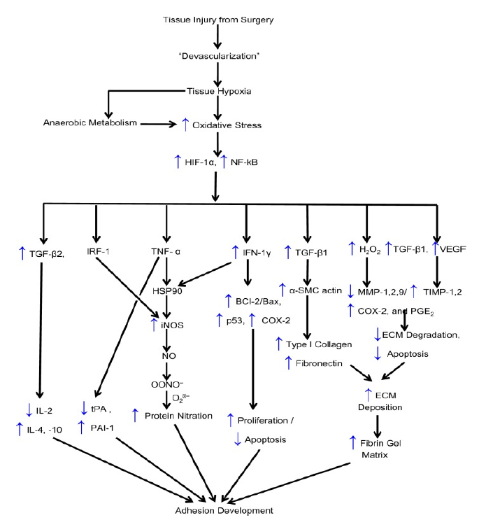
Figure 1: Mechanism of Hypoxia in Intraperitoneal Adhesion [13]
BCl-2: B Cell CLL/lymphoma 2, BAX: BCl2-Associated X, COX-2: Cyclooxygenase 2, ECM: Extracellular Matrix, HO: Hydroxyl Radical, H2O2: Hydrogen Peroxide, HIF: Hypoxia-Induced Factor, HSP90: Heat Shock Protein 90, IFN-γ: Interferon-Gamma, IL: Interleukin, iNOS: Inducible Nitrous Oxide Synthase, IRF-1: Interferon Regulatory Factor-1, MMP: Matrix Metallo Proteinases, NADP: Nicotine Adenine Dinucleotide Phosphate, NF-kB: Nuclear Factor-kB, NO: Nitric Oxide, NOS: Nitric Oxide Synthase, O2•−: Superoxide, ONOO−, Peroxynitrite, P53: Tumor Protein 53, PAI-1: Plasminogen Activator Inhibitor, PGE2: Prostaglandin E2, TGF-β1: Transforming Growth-Beta1, TIMP: Tissue Inhibitor of Matrix Metallo Proteinases, TNF-α: Tumor Necrosis Factor, TPA: Tissue Plasminogen Activator, VEGF: Vascular Endothelial Growth Factor
Intraperitoneal adhesion was proposed to develop in hypoxic condition through the increasing of iNOS, which contributed directly and indirectly through reduce peristaltic in promoting intraperitoneal adhesion.
Brüggmann, D. et al. “Intra-abdominal adhesions: definition, origin, significance in surgical practice and treatment options.” Deutsches Ärzteblatt International, vol. 107, no. 44, November 2010, pp. 769–775.
Fatehi Hassanabad, A., et al. “Post-operative adhesions: a comprehensive review of mechanisms.” Biomedicines, vol. 9, no. 8, July 2021.
Della Rocca, Y. et al. “Hypoxia: molecular pathophysiological mechanisms in human diseases.” Journal of Physiology and Biochemistry, July 2022.
Mittal, M., et al. “Reactive oxygen species in inflammation and tissue injury.” Antioxidants & Redox Signaling, vol. 20, no. 7, March 2014, pp. 1126–1167.
Tafani, M. et al. “The interplay of reactive oxygen species, hypoxia, inflammation and sirtuins in cancer initiation and progression.” Oxidative Medicine and Cellular Longevity, 2016.
Eltzschig, H.K. and P. Carmeliet. “Hypoxia and inflammation.” The New England Journal of Medicine, vol. 364, no. 7, February 2011, pp. 656–665.
Imtiyaz, H.Z. and M.C. Simon. “Hypoxia-inducible factors as essential regulators of inflammation.” Current Topics in Microbiology and Immunology, vol. 345, 2010, pp. 105–120.
Nita, M. and A. Grzybowski. “The role of reactive oxygen species and oxidative stress in the pathomechanism of age-related ocular diseases and other pathologies of the anterior and posterior eye segments in adults.” Oxidative Medicine and Cellular Longevity, 2016.
Saed, G.M. and M.P. Diamond. “Modulation of the expression of tissue plasminogen activator and its inhibitor by hypoxia in human peritoneal and adhesion fibroblasts.” Fertility and Sterility, vol. 79, no. 1, January 2003, pp. 164–168.
Phaniendra, A. et al. “Free radicals: properties, sources, targets and their implication in various diseases.” Indian Journal of Clinical Biochemistry, vol. 30, no. 1, January 2015, pp. 11–26.
Lobo, V. et al. “Free radicals, antioxidants and functional foods: impact on human health.” Pharmacognosy Reviews, vol. 4, no. 8, July 2010, pp. 118–126.
Pham-Huy, L.A., et al. “Free radicals, antioxidants in disease and health.” International Journal of Biomedical Science, vol. 4, no. 2, June 2008, pp. 89–96.
Thakur, M. et al. “Is there a genetic predisposition to postoperative adhesion development?” Reproductive Sciences, vol. 28, no. 8, August 2021, pp. 2076–2086.
Pizzino, G. et al. “Oxidative stress: harms and benefits for human health.” Oxidative Medicine and Cellular Longevity, 2017.
Bruckdorfer, R. “The basics about nitric oxide.” Molecular Aspects of Medicine, vol. 26, nos. 1–2, February–April 2005, pp. 3–31.
Hussain, S.P. et al. “Nitric oxide, a mediator of inflammation, suppresses tumorigenesis.” Cancer Research, vol. 64, no. 19, October 2004, pp. 6849–6853.
Hickey, M.J. “Role of inducible nitric oxide synthase in the regulation of leucocyte recruitment.” Clinical Science, vol. 100, no. 1, January 2001, pp. 1–12.
Pata, O. et al. “The effect of inducible nitric oxide synthase on postoperative adhesion formation in rats.” European Journal of Obstetrics, Gynecology and Reproductive Biology, vol. 117, no. 1, November 2004, pp. 64–69.
Förstermann, U. and W.C. Sessa. “Nitric oxide synthases: regulation and function.” European Heart Journal, vol. 33, no. 7, 2012, pp. 829–837.
Peng, X. et al. “Distribution of nitric oxide synthase in stomach myenteric plexus of rats.” World Journal of Gastroenterology, vol. 7, no. 6, December 2001, pp. 852–854.
Türler, A. et al. “Leukocyte-derived inducible nitric oxide synthase mediates murine postoperative ileus.” Annals of Surgery, vol. 244, no. 2, August 2006, pp. 220–229.
Awonuga, A.O. et al. “Advances in the pathogenesis of adhesion development: the role of oxidative stress.” Reproductive Sciences, vol. 21, no. 7, July 2014, pp. 823–836.
