
+91 6002993949
submission@iarconsortium.org
Open Access
ISSN (Print) : 2789-6056
ISSN (Online) : 2789-6064
The triazole-scaffold plays a crucial role as a specific and selective detection of Fe3+ ion in biological and environmental systems. These probes provide an efficient molecular platform for sensing of Fe3+ ion at ultra-low concentrations and bio-imaging applications. The N-rich triazole ring facilitate strong metal-ligand complexation and induces photo-induced electron transfer (PET)-based fluorescence quenching upon Fe3+ binding. The spectroscopic analyses (UV-vis absorption, fluorescence emission) revealed the 1:1 stoichiometry ratio and complex formation followed by distinct “turn-Off” and “turn-On” response, as well as bathochromic and hypsochromic shifts indicative of charge transfer effect. These probes exhibit ultralow detection limits, high selectivity, excellent cell permeability and good biocompatibility enabling real-time Fe3+ visualization. Overall, this review highlights that trizaole-scaffold-based sensor provide a versatile, cost-effective and reliable platform for environmental and biomedical applications.
The monitoring of metal ions plays a crucial role in fields including environmental chemistry, bio-chemistry, as well as biomedical science [1]. In biological systems, metal ions serve as Lewis’s acids and redox mediators, cellular signalling, oxygen transport, regulating electron transport, gene expression, neural function and photosynthesis mechanisms [2-6]. Vital metal ions like Na+, K+, Zn2+, Ca2+, Mg2+, Fe2+ and Fe 3+are required to maintain homeostasisand proper biological function. However, anthropogenic activities including industrial effluent discharge, mineral operations and coal combustion introduce toxic and non-essential ions (e.g. Cu2+, Ni2+, Hg2+, Cd2+, Co2+, Pb2+, As3+, Cr3+/Cr6+) into the environment, posing severe ecological and health hazards. These ions accumulate in food chains and ecosystems, resulting in severe environmental and health hazards [7-11]. Most of these toxic ions are classified as soft acids or thiophilic elements, which exhibit strong affinities towards biological ligands, thereby exacerbating their toxic effects [12-15].
Among heterocyclic compounds, triazole derivatives are especially significant for sensing applications. Triazole,an aromatic heterocycle containing three N-atoms, was first reported by Bladdin in 1885. It occurs in two structural isomeric forms, namely 1,2,3-triazole and 1,2,4-triazole, which differ in the arrangement of nitrogen atoms within the ring. Triazoles occur naturally in microorganisms, fungi and marine organisms and several triazole-based natural products have been reported with potent biological activity. These compounds are crystalline, aromatic and soluble in water and alcohol and they have been widely used in pharmaceuticals, displaying activities such as anticonvulsant, antimalarial, anticancer [16,17], antioxidant [18] antifungal, antibacterial and antidiabetic [19].
Triazoles severe as remarkably adaptable ligands because of their N-rich structures, which efficiently coordinates with transition metal ions such asCu2+, Ni2+, Hg2+, Cd2+, Co2+, Pb2+, Pd2+, As3+, Rh3+, Pt4+, Ir4+, Cr3+/Cr 6+ and Mo6+[20]. The coordination often involves ligand-to-metal charge transfer (LMCT), intramolecular charge transfer (ICT) or photoinduced electron transfer (PET) pathways, allowingtriazole derivatives to act as potent fluorometric and colorimetric chemosensors. Their sensing efficiency is further influenced by protonation/ deprotonation dynamics, host-guest interactions and solvent polarity providing versatility for the detection of diverse analytes [21-27].
Iron (Fe) serves as a vital transition metal in living systems, performing key roles in oxygen transport via hemoglobin and myoglobin, collagen synthesis, neurotransmitter regulation and enzymatic catalysis [28]. However, an imbalance in Fe³⁺ levels can result in serious pathological conditions. Excessive iron catalyzes Fenton-type reactions, producing reactive oxygen species (ROS) that induce oxidative damage to nucleic acids, proteins and lipids, thereby contributing to disorders such as Parkinson’s disease, Alzheimer’s disease, hepatic fibrosis, diabetes and cancer. On the other hand, iron deficiency results in anemia, impaired organ function and developmental disorders. Hence, selective and sensitive detection of Fe³⁺ ions is highly important for environmental monitoring and biomedical diagnostics. Iron is essential for many physiological functions and its nutritional deficiency is known to cause disability and death, affecting billions of people worldwide. Low intake of iron in diet causes anaemia while the concomitant accumulation of Fe (III) in ferritin proteins can be harmful to humans, potentially leading to severe conditions such as respiratory distress syndrome, cardiac arrest, cancer and irregularity in biosynthetic activity etc. According to WHO recommendations, the daily iron intake for adult males and females is approximately 9.1 mg/day and 19.6 mg/day, respectively. Iron is also an important component of water and soil needed for proper growth of plants and aquatic animals. Detection of different oxidation states, [Fe (II) and Fe (III)], is required due to their distinct properties with the development of selective, cost effective and easy to use probes for their detection and to clearly distinguish between the two oxidation states. While many probes can detect both Fe (II) and Fe (III) simultaneously, fewer can distinguish between the two.
In this context, triazole Schiff base derivatives have gained attention as efficient Fe³⁺ ion sensors. The combination of the imine (–C=N–) linkage and triazole nitrogen atoms provides multiple coordination sites for Fe³⁺, leading to strong binding interactions accompanied by distinct optical responses. Such systems often display “turn-On” or “turn-Off” fluorescence in response to Fe3+ ions, maintaining high specificity and in the presence of competing metal ions. Moreover, triazole-scaffold are easily synthesized, cost-efficient and multifunctional, making them suitable for Fe3+ion detection in both biological and environmental samples.
Chemosensing Performance of Triazole-Based Probes
Neliswa Mama and Aidan Battison developed a triazolyl-coumarin based fluorescent chemosensor (Probe 1 = S1 & S2) (Figure 1), highlighting the triazole moiety as a promising framework for trace level Fe3+ detection.
Figure 1: Methoxy based triazole-based Schiff base as sensor for Fe3+ ions
The probe exhibited excellent selectivity and sensitivity for Fe³⁺ ions in both aqueous and mixed media, displaying a pronounced “turn-off” fluorescence response via a photoinduced electron transfer (PET) mechanism. Binding studies confirmed a 1:1 stoichiometry between the triazole-based Schiff base sensor and Fe3+, with a detection limit of 1.4 µM. The Stern–Volmer constants (Kₛᵥ) were measured as 1.32 × 10⁴ M-1 in water and 1.28 × 10⁴ M-1 in acetonitrile, reflecting strong fluorescence quenching efficiency upon Fe3+ coordination. Additionally, the sensor exhibited a fluorescence quantum yield (Φ) of 0.27 in ethanol, indicating favourable photophysical properties. Overall, these findings establish the triazole coumarin scaffold as an effective and reliable platform for selective detection of Fe3+ ions at trace levels [29].
Joseph et al. reported a triazole- and amide-functionalized pillar [5] arene derivative (Probe 2 = APA) was synthesized as a versatile supramolecular fluorescent chemosensor. The triazole moieties served as effective coordination sites for Fe3+, enabling selective fluorescence quenching among eleven biologically relevant metal ions (Figure 2).
Figure 2: Schematic representation of triazolyl silane Schiff for Fe3+ ions identification
Upon titration with Fe3+, APA exhibited significant fluorescence quenching (~265-fold) with no notable response toward other ions such as Na+, K+, Ca2+, Mg2+, Mn2+, Fe2+, Co2+, Ni2+, Cu2+ and Zn2+, confirming its high selectivity. UV–visible and 1H NMR titrations further verified Fe3+ complexation through triazole and amide coordination sites, with a detection limit of 689 ppm. Interestingly, the in situ Fe3+ complex of APA (FeAPA) demonstrated dual “turn-on” sensing behavior toward fluoride and cysteine via Fe3+ displacement, restoring the fluorescence of free APA. Fe3+-APA exhibited ~12-fold fluorescence enhancement for F-(limit 434 ppm) and ~120-fold for cysteine (limit 1740 ppm), with negligible interference from other anions or amino acids. The triazole groups in APA played a crucial role in Fe3+ binding and reversible de-chelation, offering a dynamic platform for both ion and biomolecule recognition. Thus, the triazole-functionalized pillar [5] arene represents an efficient, water-soluble and reversible fluorescent sensor for selective Fe3+ ion detection and displacement-driven sensing applications [30].
Singh et al. identified a Schiff base derived bis (1,2,3-triazolyl-γ-propyltriethoxysilanes) (Probe 3) have been prepared by utilizing and efficient and biocompatible Cu (I) catalysed click chemistry approach and investigated their sensing property by using UV-vis absorption study. The result revealed that two absorption peaks appeared at 278 and 364 nm. However, upon addition of Fe3+ ions to the solution, the absorption band exhibited a hyperchromic shift at 278 nm and a hypochromic shift at 364 nm (from 364 to 358 nm), along with the appearance of an additional band at 274 nm. A Benesi-Hildebrand plot was used to determine the association constant, limit of detection and the 1:1 stoichiometry of the Probe– Fe3+ complex. The association constant and limit of detection were calculated to be 0.5006 × 10⁵ M-1 and 0.4462 µM, respectively (Figure 3) [31].

Figure 3: Schematic representation of silica nanoparticles containing triazole derivatives as sensor for Fe3+ ions
Zhang et al. have prepared two novel triazole-based fluorescent probes, (Probe 4a = NC-TAZ) and (Probe 4b = CC-TAZ), as efficient dual-mode (fluorimetric/ colorimetric) sensors for the specific detection of Fe3+ ions in aqueous media. Both probes exhibited a distinct fluorescence quenching and a color transformation from colorless to yellow upon Fe3+ interaction, enabling sensitive and visual detection. The fluorescence intensity of NC-TAZ and CC-TAZ decreased by approximately 84% and 79%, respectively, upon interaction with Fe3+ (1×10⁻⁵ M), while negligible responses were observed with other metal ions such as Cu2+, Mn2+ and Cr3+, confirming high selectivity. The calculated limits of detection (LOD) were 8.2×10⁻⁷ M for NC-TAZ and 9.3× 10⁻⁷ M for CC-TAZ significantly lower than the U.S. EPA maximum contaminant level for Fe3+ in drinking water (5.54 × 10⁻⁶ M), highlighting their excellent sensitivity. The Stern-Volmer quenching constants (Ksv) were determined to be 1.99×10⁹ M⁻¹ (NC-TAZ) and 1.44×10⁹ M⁻¹ (CC-TAZ), with binding constants (n) of 1.7 and 1.5, respectively. Spectroscopic analyses (UV–Vis, IR and mass spectrometry) confirmed Fe3+ coordination through the triazole nitrogen and amine functionalities. Both probes maintained stable sensing performance across various pH levels and real water samples, demonstrating the strong potential of triazole-based materials as selective, sensitive and practical fluorescent sensors for Fe3+ ion detection (Figure 4) [32].

Figure 4: Turn-off fluorescent triazole-based probes NC-TAZ and CC-TAZ for selective detection of Fe3+ ions
Zhang et al. have prepared three newly designed intermolecular charge-transfer (CT) materials, namely Probe 5a (T1), Probe 5b (T2), andProbe5c (T3),were explored as efficient fluorescent sensors for Fe3+ ion detection. These D-A structured molecules exhibited strong intramolecular charge transfer, confirmed by DFT calculations showing HOMO-LUMO localization on the donor and acceptor moieties, respectively. The photophysical studies revealed absorption around 350 nm corresponding to the charge transfer transition and emission peaks at 460, 444 and 478 nm for T1, T2 and T3, respectively. Photoluminescence studies showed a decrease in fluorescence intensity accompanied by slight red-shifts upon incremental addition of Fe3+, attributed to the formation of non-radiative complexes between Fe3+ and the electron-withdrawing triazole units, which modulate the intramolecular charge transfer (ICT) of the CT materials. The detection limits for (Probe 5a, 5b and 5c) were determined to be 1.7 × 10⁻⁷, 1.2× 10⁻⁷ and 3.6× 10⁻⁷ M, respectively, the Stern-Volmer constant were determined to be 1.3 × 10⁴ M-1, 2.3 × 10⁴ M-1 and 1.8 × 10⁶ M-1respectively, demonstrating high sensitivity and selectivity even in the presence of competing metal ions. Moreover, successful application in real water samples confirmed their excellent practicality. The quenching mechanism was ascribed to Fe3+ coordination with triazole acceptor sites, which hindered electron transfer within the CT framework, leading to fluorescence suppression (Figure 5) [33].
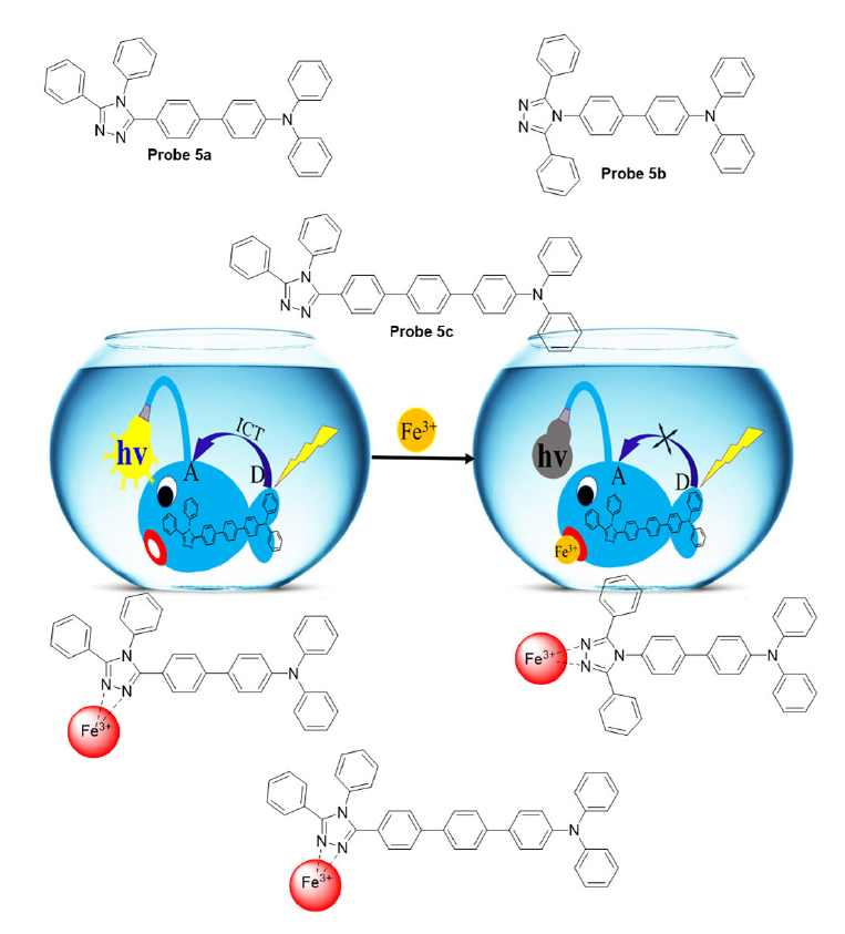
Figure 5: Schematic representation of intermolecular charge-transfer (CT) materials-based sensor (Probe 5a, 5b and 5c) for Fe (III) detection
A novel triazole-based chemosensor (Probe 6) was designed and synthesized by K. Shah et al. through a copper(I)-catalyzed alkyne–azide click reaction and comprehensively characterized using EI-MS, ¹H NMR and FT-IR spectroscopy. The sensing ability of Probe 6 toward Fe3+ ions was systematically evaluated using UV-Vis spectroscopy in a water-acetone medium against a broad spectrum of metal ions, including NH4+, Na+, K+, Li+, Ag+, Cu+, Ca2+, Ni2+, Co2+, Cd2+, Mg2+, Cu2+, Zn2+, Pb2+, Hg2+, In3+, La3+ and Y3+. The sensor exhibited outstanding selectivity toward Fe3+ ions, showing a pronounced enhancement in absorption intensity at 262 nm upon complexation, while other metal ions produced negligible effects. Job’s plot analysis confirmed a 1:1 stoichiometric ratio for the Probe-Fe3+ complex. The probe demonstrated a low detection limit of 3.21 μM and was successfully applied for Fe3+ detection in human blood plasma and laboratory tap water, with the complex exhibiting stability under basic conditions (pH 8–12), highlighting its practical applicability as a sensitive and selective tool for monitoring iron ions in biological and environmental samples (Figure 6) [34].
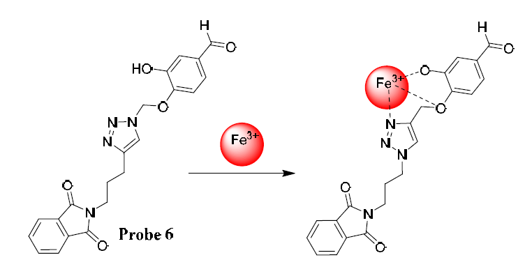
Figure 6: Schematic structure of Probe 6 selectivity sensing towards Fe3+ ions
Gahlyan et al. reported the synhtesis of a novel rhodamine-based triazole fluorescent chemosensor (Probe 7), synthesized via a copper (I)-catalyzed click chemistry approach, which functions as a sensor for Fe3+ ions. Focusing on Fe3+ detection, the probe exhibits a fluorescence turn-on effect and a color change from colorless to pink, enabling naked-eye recognition. Spectroscopic analyses, including UV–Vis, fluorescence, FT-IR and ¹H NMR studies, revealed that the probe forms a 1:2 stoichiometric complex with Fe3+ ions, with a binding constant of 2.93 × 10⁷ M2 and a detection limit of 0.33 μM, exhibiting excellent selectivity even in the presence of other competing metal ions. The opening of the rhodamine spirolactam ring upon Fe3+ coordination underlies the fluorescence enhancement, while theoretical calculations support the proposed binding mode. This probe shows potential applicability for sensitive and selective detection of Fe3+ in both environmental and biological systems [35].
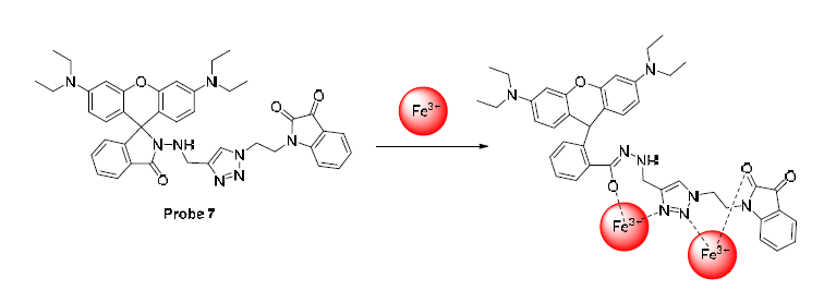
Figure 7: Schematic structure of rhodamine-based triazole sensor for Fe3+ and probable complex between probe and iron ions
Kamal et al. synthesized a selective electrochemical sensor based on a newly synthesized ferrocene-appended quinoline-triazole (Probe 8) derivative for the detection of Fe3+ ions. Cyclic voltammetry of the probe showed a reversible voltammogram corresponding to the ferrocene/ ferrocenium redox couple, which exhibited an anodic shift upon Fe³⁺ addition, indicating the formation of a Probe–Fe3+ complex. Differential pulse voltammetry (DPV) confirmed this interaction with a distinct anodic shift and a color change from yellow to olive, while competitive experiments demonstrated high selectivity of the probe towards Fe3+ over other metal ions such as Na+, Ca2+, Cu2+, Zn2+, Co2+, Ni2+, Hg2+, Cd2+, Pb2+ and Fe2+. Spectroscopic studies using fluorescence and UV–vis techniques supported a 1:1 stoichiometry and strong binding affinity, with a stability constant (Ka) of 1.82 × 10³ M-1 and a negative free energy change (-18.6 kJ mol-1), verifying the formation of the complex. Voltammetric sensors functioned over a concentration range of 5.0 × 10⁻⁶ to 1.0 × 10⁻⁴ M, with a detection limit of 2.33 × 10⁻⁷ M, while potentiometric solid-contact electrodes displayed a Nernstian slope of 18.7 mV per decade and a detection limit of 2.94 × 10⁻⁸ M. The sensors demonstrated high precision, reproducibility and selectivity, making them effective tools for quantifying Fe3+ ions in medicinal, agricultural and environmental samples (Figure 8) [36].
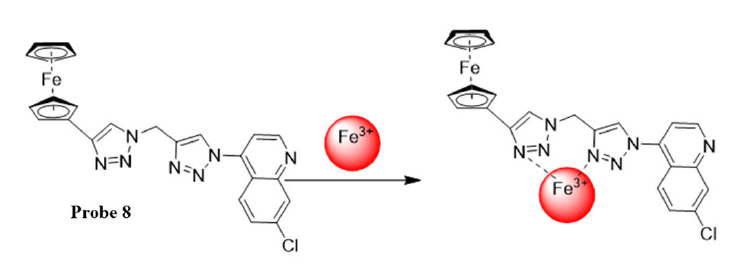
Figure 8: Ferrocene-appended quinoline-triazole based sensor used as sensor for Fe3+ ion
Wang et al. synthesized two new triazole-containing 8-(alkoxy) quinoline derivatives (Probes 9a & 9b) were efficiently synthesized via ‘click’ chemistry and evaluated as fluorescent sensors for Fe3+ ions. The sharp UV-Visible spectra of Probes 9a & 9bappeared at 240 nm and 300 nm because of π–π*transition. Both probes showed intense fluorescence emission with maxima near 400 nm in various organic solvents, attributed to the suppression of the excited-state intramolecular proton transfer (ESIPT) process. In aqueous media, however, their fluorescence was significantly quenched and exhibited a red-shift, likely due to hydrogen bond–induced intermolecular excited-state proton transfer. Similarly, the fluorescence of both probes was quenched under acidic conditions (pH < 4) and in the presence of Fe3+. Job’s plot analysis confirmed a 1:1 binding stoichiometry between the probes and Fe3+. Notably, these sensors exhibited high sensitivity and selectivity for Fe3+ over other metal ions, including Cr3+ and Cu2+, which is attributed to the metal-to-ligand charge transfer (MLCT) from Fe3+ to the quinoline ligands. The limit of detection for Probes 9a & 9bwere obtained to be 2.8 µM and 0.4 µM, respectively. The reversibility studies revealed that upon the addition of EDTA, the quenched emission intensity was restored, confirming the reversible binding nature of the probes towards (Figure 9) [37].
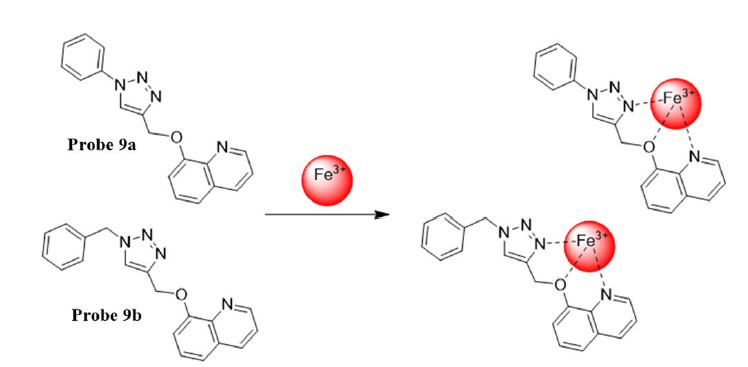
Figure 9: 8-(alkoxy) quinoline derivatives as sensor for Fe3+ ions
Joseph et al. reported the synthesis of a quinoline-functionalized pillar [5] arene (Probe 10) and its application for the selective detection of Fe3+ ions in aqueous media. The probe exhibited pronounced fluorescence quenching exclusively upon interaction with Fe3+ among eleven biologically relevant metal ions, achieving a quenching efficiency of approximately 31-fold, while other competing ions induced minimal or negligible effects, confirming its excellent selectivity toward Fe3+. The formation of a 1:2 Probe–Fe³⁺ complex was confirmed by MALDI-TOF mass spectrometry, UV–Vis spectroscopy and 1H NMR titration studies, which showed notable broadening and slight chemical shifts of the triazole and quinoline proton signals, indicative of strong complexation. Competitive titration experiments further demonstrated that the probe could selectively recognize Fe3+ even in the presence of other metal ions. The detection limit of the probe for Fe3+ ions was determined to be 157 ppm, whereas the Fe–probe complex exhibited detection limits of 289 ppm, 3.57 × 10⁻³ ppm (Figure 10) [38].
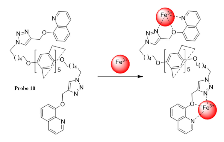
Figure10: Schematic structure of quinoline-functionalized pillar [5] arene bis traizole sensor for iron ion
Puthiyedath and Bahulayan developed a triazole-coumarin derivative (Probe 11, Figure 11) as a selective fluorescent chemosensor for Fe3+ ions, demonstrating a turn-off response based on a photoinduced electron transfer (PET) mechanism. The fluorescence of the probe is substantially quenched in the presence of Fe3+, while other metal ions show minimal interference. UV–vis titration revealed a progressive enhancement in absorption intensity with increasing Fe3+concentration and fluorescence titration showed a corresponding decrease in emission at 473 nm, with a linear response suitable for quantitative analysis. The limit of detection for Fe3+ was found to be 2.61 μM from fluorescence measurements and the association constant was determined to be 6.97 × 10⁸ M-1, indicating strong binding affinity. Fluorescence lifetime studies showed a decrease from 2.04 ns to 1.75 ns upon Fe3+addition, confirming the quenching effect, while Job’s plot indicated a 1:1 stoichiometric complexation between the probe and Fe3+. Density functional theory (DFT) calculations indicated that the HOMO energy level of the Probe– Fe3+ complex lies between that of the coumarin acceptor, thereby facilitating photoinduced electron transfer (PET)-mediated fluorescence quenching. FT-IR analysis further confirmed the coordination of Fe3+ with the ester carbonyl group of TC1, shifting the carbonyl stretching frequency from 1740 cm-1 to 1721 cm-1. Real sample analysis in tap water and simulated wastewater demonstrated the practical applicability of the probe for sensitive and selective detection of Fe3+ions [39].
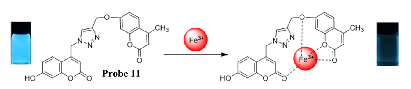
Figure 11: Schematic structure of Coumarin-based triazole sensor for Fe3+ ion detection
Geng et al. synthesized a novel amide derivative of [1,2,4] triazolo [1,5-a] pyrimidine (Probe 12) and developed it as a highly selective and sensitive fluorescence probe for Fe3+ ions. The probe exhibits rapid fluorescence quenching within 5 seconds upon binding with Fe3+ and demonstrates a low limit of detection of 0.82 μM, maintaining strong selectivity even in the presence of common interfering metal ions and strong chelating agents such as EDTA. Fluorescence titration revealed a linear decrease in emission at 450 nm with increasing Fe3+ concentration, while reversibility studies confirmed the recovery of fluorescence upon addition of OH⁻, indicating that probe can be reused multiple times. Job’s plot and high-resolution mass spectrometry confirmed a 1:3 stoichiometric complex formation between probe and Fe3+. The probe shows excellent photophysical properties, stability across a wide pH range and good cell permeability with low cytotoxicity, enabling effective bio-imaging of Fe3+ in living HeLa cells. Furthermore, a probe-12-based fluorescent test paper was developed, allowing rapid and convenient identification of Fe3+, highlighting the practical applicability of this probe for both environmental and biological monitoring of Fe3+ ions (Figure 12) [40].
Figure 12: Schematic representation of triazole based probe 12 depicting sensing towards metal ions and cell imaging behaviour
A novel ruthenium (II) complex, [(Ru(bpy)2(dabpy))] [PF6]2, was developed as a highly sensitive and selective luminescent probe for visualizing nitric oxide (NO) generation in living cells. Upon reacting with NO under aerobic aqueous conditions, the complex rapidly forms its triazole derivative (Probe 13 = [Ru(bpy)2(T-bpy)]2+), as depicted in Figure 13a, accompanied by a remarkable enhancement in luminescence quantum yield (from 0.13 % to 2.2 %). This conversion showed bright intracellular fluorescence, rendering its effectiveness for bio-imaging utilization. The native Ru (II) complex readily crosses the cell membranes, enabling efficient uptake in animal and plant cells, in contrast, the triazole derivative remains membrane-impermeable, ensuring that fluorescence originates exclusively from intracellular NO-activation. Luminescence imaging revealed bright red emission in macrophage and gardenia cells, verifying NO mapping and its concentration near the nuclear region. Thus, the in-situ formation of the triazole derivative within cells provides a robust, selective and water-soluble probe for real-time monitoring of NO production, exhibiting superior photo-stability, a large Stokes shift and wide pH tolerance compared to conventional organic NO sensors (Figure 13 b, c, & d) [41].

Figure 13: Schematic representation illustrating (a) the synthetic route of the ruthenium-based triazole derivative; (b) bright-field (left) and luminescence (right) images of mouse macrophage cells treated with [Ru(bpy)2(dabpy)]2+ in the absence and presence of NOC 13; (c) bright-field and luminescence images of gardenia cells incubated with [Ru(bpy)2(T-bpy)][PF6]2 for 5 h; and (d) bright-field and luminescence images of gardenia cells treated with [Ru(bpy)2(dabpy)]2+ at different incubation times
Tricarbocyanine N-triazoles based (Probe 14 = CIR) were designed as a new class of near-infrared (NIR) dyes to overcome the brightness and photo-stability limitations of conventional tricarbocyanines for long-term in vivo imaging, as depicted in (Figure 14a).
Among them, CIR38M emerged as a highly stable (Figure 14a), non-transferable and biocompatible fluorophore, suitable for real-time tracking of therapeutic T cells with superior performance over commercial NIR dyes such as DiR, IR800CW and ICG. A brief 2 min incubation with 10 µM CIR38M resulted in intense, near-saturated fluorescence in CD4+ T cells. Upon stimulation, CIR38M-labelled T cells retained strong emission with only minor fluorescence loss after 3 days, while CFSE exhibited significant decline, indicating suitability for multi-cycle proliferation tracking. Co-culture of CD45.2+ CIR38M-labelled T cells with unlabelled CD45.1+ T cells showed no dye transfer, confirming its excellent intracellular retention through covalent interaction with intracellular proteins. CIR38M showed no cytotoxicity or cytokine alteration (TNF-α, GM-CSF) under stimulatory conditions. Fluorescence microscopy revealed much brighter cytosolic staining than IR800CW-SE, with preferential mitochondrial accumulation (Figure14b). Flow cytometry confirmed >12-foldhigher fluorescence intensity relative to IR800CW-SE and no extracellular leakage. In vivo, CIR38M detected as few as 4 × 10³ T cells, about three-fold more sensitive than DiR (Figure 14c). Following antigen challenge, CIR38M-labelled CD4⁺ T cells accumulated specifically in draining lymph nodes, with >2-fold higher fluorescence versus control, maintaining 40% signal at day 4 and 15% at day 7. No dye leakage, immune interference or systemic toxicity was observed. Human CD4⁺ T cells treated with 10 µM CIR38M for 2 min also displayed >10× brighter fluorescence than ICG, with stable emission for ≥3 days and no cytotoxic or cytokine effects. Overall, CIR38M demonstrates superior photo-stability, intracellular retention and NIR brightness, establishing it as a next-generation triazole-based NIR probe for longitudinal, non-invasive T-cell imaging in both murine and human systems [42].

Figure 14: (a) Chemical synthesis of CIR-traizole-based fluorophores, their absorbance, fluorescence and time-course analysis; (b) fluorophore cell imaging of CIR38M labelled T cells; (c) determination of limit of detection in suspension of CIR38M labelled.
A novel triazole-based fluorogenic (Probe 15a & 15b = coumOCT (9 & 10)), was synthesized by coupling a cyclooctyne unit with a coumarin fluorophore for strain-promoted azide-alkyne cycloaddition (SPAAC)-based imaging of azido-glycoconjugates in living HeLa cells. The probe exhibited good cell permeability and emitted fluorescence only upon triazole formation, thereby minimizing background emission and allowing real-time intracellular monitoring of glycan trafficking. Spectral data recorded in PBS buffer (10% DMSO, pH 7.4) revealed that compound 1 displayed weak emission at 405 nm with a low quantum yield (Φf= 0.011) upon excitation at 330 nm, while the corresponding triazoles (Probe 15a = (9)& 15b = (10)), as depicted in (Figure 15a), showed strong emissions at 435 nm with Φf = 0.23 and 0.21, respectively. The SPAAC coupling between compound 1 and Man Naz happened correctly, with more than 90% substrate conversion in 40 min and fluorescence saturation within one hour, (Figure 15c). In live-cell imaging, HeLa cells treated with Ac4ManNAz and exposed to coumOCT for 30 min at 37 °C exhibited strong fluorescent labelling, whereas control cells displayed negligible emission (Figure 15b). Confocal microscopy revealed blue emission overlapping with Golgi apparatus staining, confirming selective and specific labelling of azido-sialic acid glycol-conjugates (Figure 15 b). Under no-wash conditions, time-dependent imaging displayed a progressive emission intensity enhancement, reaching maximum intensity after ~ 1.5 h (Figure 15c). Furthermore, cells labelled with Ac₄GlcNAz and Ac₄GalNAz also showed distinct fluorescence localization patterns (Figure 15d). Importantly, NMR analyses confirmed that coumOCT and its triazole adducts were inert towards thiol-yne or thiol-ene reactions, thereby eliminating non-specific binding. Collectively, these results establish coumOCT as a bright, selective and biocompatible triazole-based turn-on probe for real-time intracellular imaging of azido-labeled glycol-conjugates with excellent photo-stability and negligible background fluorescence [43].
Figure 15: (a) Synthesis of the SPAAC-based fluorogenic probe 1 and its triazole products 15a and 15b; (b) Confocal microscopy showing fluorescence and intracellular localization of probe-labeled HeLa cells; (c) Time-lapse imaging of intracellular sialylated glycoconjugates via SPAAC in live cells; (d) Comparative fluorescence imaging of live cells treated with different azido-sugars
The triazole-scaffold-based fluorescent sensor proves to be a highly efficient and selective system for the detection of Fe3+ ions in trace amounts. The presence of the triazole ring, with its nitrogen-rich coordination sites, facilitates strong metal-ligand interaction, leading to a distinct fluorescence “turn-off” or “turn-on” response upon Fe3+ binding. Spectroscopic and analytical studies determine the binding stoichiometry, high binding affinity and ultralow detection limit in the µM to nM range, displaying the probe’s remarkable sensitivity. Furthermore, the probe displayed remarkable sensitivity and selectivity even in presence of various competing metal ions, confirming its practical applicability in real water and biological samples. The distinct fluorescence change, fast response and easy synthesis make the triazole-based probe an efficient and user-friendly analytical device. Apart from the sensing properties of traizole for investigation of toxic metal ions, it displaying excellent biocompatibility and cellular permeability, enabling its application in bio-imaging to investigate transport and distribution processes in living systems. This review highlights not only sensing properties towards Fe3+ ions but also displayed robust and adaptable molecular platform for preparation of advanced fluorophore. The exceptional, selectivity, specificity and sensitivity of triazole-based sensors depict its wide applicability, from environmental metal detection to bio-imaging, thereby making a substantial contribution to chemical sensing, biological function detection and ecological detection.
Cao, D. et al. "Coumarin-based small-molecule fluorescent chemosensors." Chemical Reviews, vol. 119, no. 18, 2019, pp. 10403–10519.
Hu, H. et al. "Optical sensing at the nanobiointerface of metal ion–optically-active nanocrystals." Nanoscale, vol. 10, no. 11, 2018, pp. 5035–5046.
Juvekar, V. et al. "Recent progress in the two-photon fluorescent probes for metal ions." Coordination Chemistry Reviews, vol. 427, 2021, p. 213574.
Saleem, M. et al. "Microwave assisted synthesis of a novel optical chemosensor for selective Fe3+ detection." Journal of Luminescence, vol. 162, June 2015, pp. 14–24.
Wu, D. et al. "Recent progress in the development of organic dye based near-infrared fluorescence probes for metal ions." Coordination Chemistry Reviews, vol. 354, 2018, pp. 74–97.
Wang, Y. et al. "Fluorescent probe for mercury ion imaging analysis: strategies and applications." Chemical Engineering Journal, vol. 406, 2021, p. 127166.
Liu, Y. and Z. Zhong. "Extraction of heavy metals, dichromate anions and rare metals by new calixarene-chitosan polymers." Journal of Inorganic and Organometallic Polymers and Materials, vol. 28, 2018, pp. 962–967.
Liu, Y. and Z. Zhong. "Synthesis and characterization of new calixarene-chitosan polymers." Journal of Macromolecular Science, Part A: Pure and Applied Chemistry, vol. 54, no. 10, 2017, pp. 678–683.
Kumar, R. et al. "Bis-triazolylated-1,4-dihydropyridine—highly selective hydrophilic fluorescent probe for detection of Fe3+." Dyes and Pigments, vol. 147, December 2017, pp. 420–428.
Maurya, N. and A.K. Singh. "Indirect approach for CN− detection via Cu2+ induced turn-off sensor: using novel AIEE fluorophore with logic gate and antimicrobial application." Dyes and Pigments, vol. 147, 2017, pp. 484–490.
Bhardwaj, S. et al. "Chromone based fluorescent organic nanoparticles for high-precision in-situ sensing of Cu2+ and CN− ions in 100% aqueous solutions." Sensors and Actuators B: Chemical, vol. 260, 2018, pp. 753–762.
Zhao, M. et al. "Development of a Si-rhodamine-based NIR fluorescence probe for highly specific and quick response of Hg2+ and its applications to biological imaging." Microchemical Journal, vol. 171, 2021, p. 106855.
Bhalla, V. et al. "Fe3+-ensemble of triazole appended pentacenequinone derivative for ‘turn-on’ detection of fluoride ions." Talanta, vol. 105, February 2013, pp. 152–157.
Roy, A. et al. "Dual chemosensors for metal ions: a comprehensive review." Trends in Analytical Chemistry, vol. 138, 2021, p. 116204.
Chen, S.Y. et al. "Small molecular fluorescent probes for the detection of lead, cadmium and mercury ions." Coordination Chemistry Reviews, vol. 429, 2021, p. 213691.
Mohammad, Y. et al. "Synthesis and biological evaluation of novel 3-O-tethered triazoles of diosgenin as potent antiproliferative agents." Steroids, vol. 118, 2017, pp. 1–8.
Huang, M. et al. "Synthesis and biological evaluation of salinomycin triazole analogues as anticancer agents." European Journal of Medicinal Chemistry, vol. 127, 2017, pp. 900–908.
Karrouchi, K. et al. "Synthesis, antioxidant and analgesic activities of Schiff bases of 4-amino-1,2,4-triazole derivatives containing a pyrazole moiety." Annales Pharmaceutiques Françaises, vol. 74, 2016, pp. 431–438.
Wang, G. et al. "Synthesis, in vitro evaluation and molecular docking studies of novel triazine-triazole derivatives as potential α-glucosidase inhibitors." European Journal of Medicinal Chemistry, vol. 125, 2017, pp. 423–429.
Fukuhara, G. "Analytical supramolecular chemistry: colorimetric and fluorimetric chemosensors." Journal of Photochemistry and Photobiology C: Photochemistry Reviews, vol. 42, 2020, p. 100340.
Liu, R.Q. et al. "A triazole-based fluorescence probe for detecting Hg2+ ions and its biological application." Luminescence, vol. 35, no. 1, 2020, pp. 129–137.
Bhalla, V. et al. "Fe3+-ensemble of triazole appended pentacenequinone derivative for ‘turn-on’ detection of fluoride ions." Talanta, vol. 105, February 2013, pp. 152–157.
Sun, M.Y. and D.M. Chen. "A porous Zn(II)-based metal–organic framework for highly selective and sensitive Fe3+ ion detection in water." Polyhedron, vol. 147, June 2018, pp. 80–85.
Geng, Y. et al. "A novel [1,2,4]triazolo[1,5-a]pyrimidine derivative as a fluorescence probe for specific detection of Fe3+ ions and application in cell imaging." Analytica Chimica Acta, vol. 1187, December 2021, p. 339168.
Aktara, M.N. et al. "A sensorial colorimetric detection method for Hg2+ and Cu2+ ions using single probe sensor based on 5-methyl-1,3,4-thiadiazole-2-thiol stabilized gold nanoparticles and its application in real water sample analysis." Microchemical Journal, vol. 147, 2019, pp. 1163–1172.
Kang, H. et al. "A highly selective fluorescence switch for Cu2+ and Fe3+ based on a new diarylethene with a triazole-linked rhodamine 6G unit." Tetrahedron, vol. 74, no. 33, August 2018, pp. 4390–4399.
Wu, C. et al. "Coumarin-based Hg2+ fluorescent probe: synthesis and turn-on fluorescence detection in neat aqueous solution." Sensors and Actuators B: Chemical, vol. 243, 2017, pp. 678–683.
Joshi, S. et al. "Experimental and theoretical study: determination of dipole moment of synthesized coumarin–triazole derivatives and application as turn-off fluorescence sensor: high sensitivity for iron(III)." Sensors and Actuators B: Chemical, vol. 220, 2015, pp. 1266–1278.
Mama, N. and A. Battison. "Synthesis and application of a fluorescent ‘turn-off’ triazolyl-coumarin-based fluorescent chemosensor for the sensing of Fe3+ ions in aqueous solutions." Arkivoc, no. 5, 2020, pp. 59–84.
Joseph, R. "Selective detection of Fe3+, F− and cysteine by a novel triazole-linked decaamine derivative of pillar[5]arene and its metal ion complex in water." ACS Omega, vol. 5, no. 11, March 2020, pp. 6215–6220.
Singh, G. et al. "Schiff base derived bis-organosilanes: immobilization on silica nanosphere and Cu2+ and Fe3+ dual ion sensing." Inorganica Chimica Acta, vol. 514, 2021, p. 120028.
Zhang, Y. et al. "Highly sensitive triazole-based fluorimetric/colorimetric dual-channel Fe3+ probe." Asian Journal of Organic Chemistry, vol. 9, no. 7, July 2020, pp. 1081–1086.
Zhang, Z. et al. "Novel Fe3+ fluorescence probe based on the charge-transfer molecules." Sensors and Actuators B: Chemical, vol. 255, February 2018, pp. 1878–1883.
Shah, K. et al. "A new highly selective chemosensor for the detection of iron ion in aqueous medium based on click generated triazole." Sensors and Actuators B: Chemical, vol. 249, 2017, pp. 515–522.
Gahlyan, P. et al. "Isatin-triazole-functionalized rhodamine: a dual sensor for Cu2+ and Fe3+ ions and its application to cell imaging." ChemistrySelect, vol. 4, no. 25, July 2019, pp. 7532–7540.
Kamal, A. et al. "Selective sensing ability of ferrocene appended quinoline-triazole derivative toward Fe3+ ions." Sensors and Actuators B: Chemical, vol. 221, 2015, pp. 370–378.
Wang, Z. et al. "Synthetically simple, click-generated quinoline-based Fe3+ sensors." Methods and Applications in Fluorescence, vol. 5, no. 2, 2017, p. 024015.
Joseph, R. et al. "Quinoline appended pillar[5]arene (QPA) as Fe3+ sensor and complex of Fe3+ (FeQPA) as a selective sensor for F−, arginine and lysine in the aqueous medium." Spectrochimica Acta Part A: Molecular and Biomolecular Spectroscopy, vol. 224, 2020, p. 117390.
Puthiyedath, T. and D. Bahulayan. "A click derived triazole–coumarin derivative as fluorescence on-off PET based sensor for Ca2+ and Fe3+ ions." Sensors and Actuators B: Chemical, vol. 272, 2018, pp. 110–117.
Geng, Y. et al. "A novel [1,2,4]triazolo[1,5-a]pyrimidine derivative as a fluorescence probe for specific detection of Fe3+ ions and application in cell imaging." Analytica Chimica Acta, vol. 1187, December 2021, p. 339168.
Zhang, R. et al. "Development of a ruthenium(II) complex based luminescent probe for imaging nitric oxide production in living cells." Chemistry—A European Journal, vol. 16, no. 23, June 2017, pp. 6884–6891.
Mellanby, R.J. et al. "Tricarbocyanine N-triazoles: the scaffold-of-choice for long-term near-infrared imaging of immune cells in vivo." Chemical Science, vol. 9, no. 36, 2018, pp. 7261–7270.
Shie, J.J. et al. "A cell-permeable and triazole-forming fluorescent probe for glycoconjugate imaging in live cells." Chemical Communications, vol. 53, no. 9, 2017, pp. 1490–1493.
