+91 6002993949
submission@iarconsortium.org
Open Access
ISSN (Print) : 2788-9556
ISSN (Online) : 2788-9564
In addition to polymerization of Heamoglobin S in red blood cells under deoxygenated conditions, recent studies have demonstrated that an imbalance in the oxidant – antioxidant system is also involved in the pathophysiology of sickle cell disease. This oxidative stress plays a very critical role in the symptoms and complications of sickle cell disease and conventional therapies directed to restore this balance have proved beneficial. Extracts of the leaves of Terminalia catappa L. which have been shown by recent researchers to have antisickling activity were investigated for its ability to restore the oxidant – antioxidant balance in this study. The ability of the n-Hexane and ethanol extracts of T. catappa leaves to restore nitric oxide availability under physiological conditions was assessed using the Griess reaction assay and results show that only higher concentrations (≥600 µg/ml) of the ethanol extracts exhibited nitric oxide generating activity. Standard Fe2+ Chelation Assay was used to evaluate the ability of the extracts to reduce Iron overload under physiological conditions and only the n-Hexane extract was able to chelate free iron.
Sickle cell disease is a devastating chronic genetic disorder caused by inheritance of sickle heamoglobin gene which is reported to results to various clinical manifestations such as vaso-occlusive crisis, heamolysis, anaemia, risk of infection and other complications [1]. This genetic defect is said to be due to a substitution of Glutamic acid, a hydrophilic amino acid with valine, which is hydrophobic, at the sixth position of the beta-globin chain [2]. Heamoglobin S (HbS), the heamoglobin that is produced owing to this abnormality, is known to be poorly soluble and polymerizes when deoxygenated [3]. On the other hand Oxidative stress is known to plays a very critical role in the pathophysiology of SCD and its complications [4-5]. Generally, Oxidative stress is defined as an imbalance between oxidants/free radical and antioxidants [6]. Several molecular mechanisms have been proposed to contribute towards an increased oxidative burden in sickle cell patients. However for this research, the excessive level of Fe2+ radicals due to free heamoglobin and the impaired bioavailability of endogenous nitric oxide in SCD were assessed.
Nitric Oxide (NO) imbalance in SCD is known to be responsible for the pain crisis in a sickle cell carrier. NO released from the endothelium is a potent endogenous vasodilator that acts by reducing intracellular calcium concentration in smooth muscle through a cascade of events therefore producing relaxation and dilation of the vascular smooth muscles and increases blood flow through the blood vessels [7-9]. In addition, nitric oxide also suppresses platelet aggregation and promotes expression of cell adhesion molecules on endothelial cells [10-13] .
Recent studies have demonstrated that patients with sickle cell disease suffer from impaired bioavailability of the endogenous vaso-dilator, nitric oxide and this could be due to reduced plasma L-arginine level, scavenging of nitric oxide by cell-free plasma heamoglobin and by reactive oxygen species [14-18].
There are several methods which are being used widely in biological system for assay of NO; however, the Griess reaction assay as demonstrated by Johann Peter Griess in 1879, was used for this research work due to its simplicity and low cost.
A typical human red blood cell is known to contain about 270 million heamoglobin with each heamoglobin having four heme biomoiety. Bridges, 2021 reported that red blood cells of an average adult male collectively store about 65 % of the total iron contained in the body. Old and structurally defective erythrocytes are destroyed by the phagocytic cells of the reticuloendothelial system in the spleen, liver, bone marrow and lymph node, a process known as extravascular heamolysis [19], therefore, only little heamoglobin escapes into the plasma. In contrast to degradation of normal erythrocytes, sickle red blood cell is known to survive for just 10 – 20 days as observed by Sebastiani et al, 2007 and is majorly destroyed intravascularly, a phenomenon known as “intravascular heamolysis”[11]. Intravascular heamolysis results to the availability of cell-free plasma heamoglobin, which mop up nitric oxide about 1000-fold more rapidly, consequently reducing the bioavailability of nitric oxide significantly in patients with sickle cell disease [11].
Furthermore, the issue of Iron overload is a clinically significant and inevitable complication in transfusion, especially in patients with thalassemia [20-21]. The excess iron is stored in the heart, liver and pancreas and can lead to poisoning of these organs, consequently resulting in conditions such as cancer, liver cirrhosis and irregular rhythm of the heart [20]. Therefore, the use of an iron chelators therapy is of great importance in the treatment of transfusion- dependent iron overload and in maintenance of the normal body iron levels in tissues [22].
The ethnomedicinal use of natural products in bid to manage or treat SCD could be as old as when the disease was discovered, though only relatively few have been validated scientifically. Among the scientifically validated herbal remedies used ethnomedicinally in management of SCD, the reddish brown freshly fallen leaves of Terminalia catappa was further analysed in this research.
Terminalia catappa is a large tropical tree that belongs to the Combretaceae family. Its common names include country almond, Indian almond, Malabar almond, sea almond, tropical almond, beach almond and false kamani [23]. In 2014, Praveena reported that country almond contains various phytochemicals such as steroids, carbohydrates, alkaloids, triterpenes, saponins, polyphenols, flavonoids, glycosides and tannins.
Aside its ethnomedicinal use in management of SCD, the leaves of Tropical almond have also been demonstrated to have significant antibacterial activity [24], anti-ulcer properties [25], aphrodiastic properties, antifungal properties [26], anti-oxidative properties [27], antinociceptive properties [28], antimicrobial activity [29], anti- aging properties [30], antidiabetic properties [31] and anti-inflammatory properties [32]. It has been shown to have therapeutic effects on hepatitis [32] and has been used ethno-medicinally in treatment of skin diseases such as dermatitis.
In 2003, Moody et al demonstrated that the freshly fallen reddish brown leaves of Terminalia catappa, L. Family Combretaceae, exhibit antisickling effect; Mgbemene and Ohiri [33] had also demonstrated the ability of the ethanolic extract of the leaves of T. catappa to inhibit osmotically-induced hemolysis of human RBCs in a dose-dependent manner. They also illustrated the ability of T. catappa leaves to prolong the clotting time of uncoagulated blood and the effectiveness of a 1.0 mg/ml solution of the extract in preventing and reversing the sickling of sodium meta-bisulphite induced sickled RBCs. Furthermore, Samuel et al. [34] validated the scientific basis for the application of the methanolic extract of T. catappa leaves in the management of sickle cell anaemia in traditional medicine.
Ethanol, n-hexane, distilled water, Mercuric (II) Chloride, potassium iodide, hydrochloric acid, ferric chloride, ammonia solution, benzene, sodium hydroxide, ferric chloride, chloroform, sulphuric acid, glacial acetic acid, sodium chloride, dipotassium hydrogen phosphate, potassium dihydrogen phosphate, nitroprusside, sulphanilamide, phosphoric acid, 1-naphthylethylenediamine dihydrochloride, Gallic acid, tris-hydrochloride, Ferric Chloride, phenanthroline chloride, EDTA, pyrogallol,
Weighing balance (Analytical and Electronic), Electronic oven, Hot water bath, Whatmann No. 1 filter paper, Test tubes in test tube rack, pipette, volumetric flask, beaker, masking tape, pen, Ultra violet (UV) Spectrophotometer, Iodine flask, pH meter.
Nitric Oxide Generation Assay
The effect of the plant extract on Nitric Oxide production under physiological condition was measured using a modified method of Marcocci and Colleagues, 1994. First, various concentrations (0.00, 0.2, 0.4, 0.6 and 1.0 mg/ml) of the extracts and the standard solution were prepared in triplicate manner. 5 mM sodium nitroprusside (0.5 ml) in phosphate buffered saline was mixed with various concentrations of the test samples and incubated at room temperature (25 oC) for thirty (30) minutes. After incubation, the nitrite produced was measured using the Griess Reagent (1% Sulphanilamide in 5% phosphoric acid and 0.1% 1-naphthylethylenediamine hydrochloride in water). The absorbance of the product that formed during diazotization of the nitrite with sulphanilamide and subsequent coupling with 1-naphthylethylenediamine dihydrochloride was determined immediately using the Ultra-violet spectrophotometer. Gallic acid was used as the Standard solution. The percentage of Nitric oxide generating activity was calculated using the formular:
% Nitric Oxide Generating activity = ((Atest – Acontrol)/Acontrol) X 100
Where
Atest : Absorbance of the plant extract or standard sample
Acontrol: Absorbance of the control
Fe2+ Radical Scavenging Assay
The Fe2+ chelating ability of both the n-hexane and ethanolic extracts of the leaves were determined by using a modified method of Minotti and Aust with a slight modification by Puntel et al.
Freshly prepared 500 µM FeCl2.4H2O (150 μl) was added to a reaction mixture containing 160µl 0.1 M Tris – HCl (pH 7.4), 200 µl normal saline and the various concentrations of the plant extracts (0-1.0 mg/ml). The reaction mixture was incubated for five (5) minutes, thereafter, 13 µl 0.25%w/v 1, 10 Phenanthroline was added. The absorbance of the formed product was read at 510 nm in the UV- Visible Spectrophotometer. EDTA was used as the standard solution. The Fe (II) chelating ability was subsequently calculated using the formular:
% Fe2+ Chelation = ((Atest – Acontrol)/Acontrol) X 100
Where
Atest : Absorbance of the plant extract or standard sample
Acontrol : Absorbance of the control
From the phytochemical analysis carried out, the ethanolic and n-Hexane extracts of the freshly fallen reddish brown leaves of T. catappa were found to be rich in tannins, steroids and cardiac glycosides. This suggests the possible ability of both extracts to serve as an antidote in heavy metal toxicity, anti-inflammatory substances and as well as muscle relaxant. Flavonoid, which is an indication of antioxidant ability, was present in only the n-hexane extract while alkaloids were present in minute quantity in only the ethanolic extract( Table 1).
Table 1: Showing the Phytochemical Analysis of the 70% Ethanolic and N-Hexane Extract of Freshly Fallen Terminalia Catappa Leaves
Phytochemicals | Inference | |
| 70% Ethanolic extract | n-Hexane extract | |
| Alkaloids | + | - |
| Anthraquinone | - | - |
| Flavonoid | - | ++ |
| Saponins | ++ | + |
| Tannins | +++ | +++ |
| Steriodal ring | +++ | +++ |
| Cardiac glycoside | +++ | +++ |
KEY: = Not present + = Present in little quantity ++ = Present in high quantity+++ = Present in large quantity
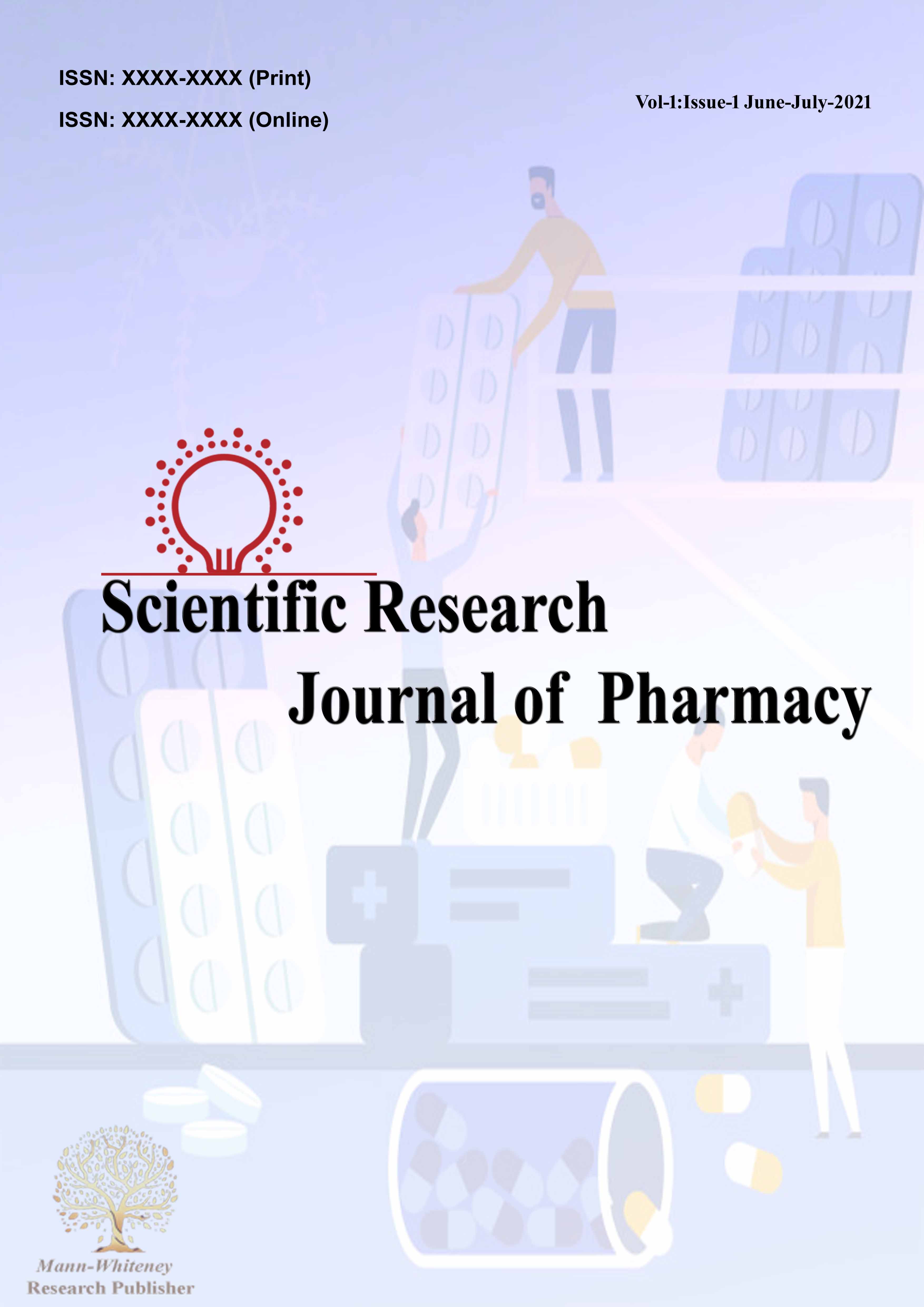
From the antioxidant tests carried out, it can be observed that at concentrations greater than 400 µg/ml, the ethanolic extracts were able to increase availability of nitric oxide under physiological condition. This suggests that the plant extract might be beneficial in the management of sickle cell vaso-occlusive crisis and other nitric-oxide dependent complications of sickle cell disease. However, the n-Hexane extract was observed to be a potential nitric oxide scavenger, therefore can serve as an anti-oxidant in cases of chronic exposure to nitric oxide or nitric oxide toxicity(Figure 1,2).
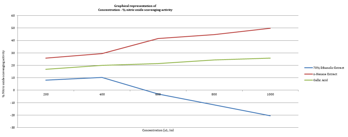
Figure 1: Nitric Oxide Scavenging Activity
Figure 2: Fe2+ Chelation Activity
The results of the Fe2+ chelation assay carried out showed that under normal physiological condition, the n-Hexane extract was able to chelate free ferrous ions, although to a relatively lower extent when compared to the ferrous chelation ability of the EDTA standard used. The ethanolic extract however, was unable to chelate free ferrous ions. Therefore, on further modification, the n-Hexane extract of the T. catappa leaves may be useful as a scavenger for cell free plasma heamoglobin released during intravascular heamolysis and in the treatment of iron overload in patients with sickle cell disease.
The ability of T. catappa leaves extracts to generate and increase nitric oxide bioavailability under physiological condition can be thought to be responsible for its ethnomedicinal use in the management of SCD as a deficiency of nitric oxide is indicated in the pathophysiology in SCD.
Herrick, J.B. “Peculiar elongated and sickle-shaped red blood corpuscles in a case of severe anemia.” Archives of Internal Medicine, vol. 6, 1910, pp. 517–517.
Pauling, L. et al. “Sickle cell anemia, a molecular disease.” Science, vol. 109, 1949, pp. 443–443.
Bunn, H.F. “Pathogenesis and treatment of sickle cell disease.” New England Journal of Medicine, vol. 337, no. 11, 1997, pp. 762–769.
Aslan, M. et al. “Reactive species in sickle cell disease.” Annals of the New York Academy of Sciences, vol. 899, 2000, pp. 375–391.
Nur, E. et al. “Oxidative stress in sickle cell disease; pathophysiology and potential implications for disease management.” American Journal of Hematology, vol. 86, no. 6, 2011, pp. 484–489.
Halliwell, B. and J.M. Gutteridge. Free radicals in biology and medicine. 4th ed., Oxford University Press, 2007.
Furchgott, R.F. and J.V. Zawadzki. “The obligatory role of endothelial cells in the relaxation of arterial smooth muscle by acetylcholine.” Nature, vol. 288, no. 5789, 1980, pp. 373–376.
Stryer, L. Biochemistry. 4th ed., W.H. Freeman and Company, 1995.
Perez, K.M. and M. Laughon. “Sildenafil in term and premature infants: a systematic review.” Clinical Therapeutics, vol. 37, no. 11, 2015, pp. 2598–2607.
Hebbel, R.P. “Auto-oxidation and a membrane-associated ‘Fenton reagent’: a possible explanation for development of membrane lesions in sickle erythrocytes.” Clinical Haematology, vol. 14, no. 1, 1985, pp. 129–140.
Reiter, C.D. et al. “Cell free hemoglobin limits nitric oxide bioavailability in sickle cell disease.” Nature Medicine, vol. 8, no. 12, 2002, pp. 1383–1389.
Voetsch, B. et al. “Nitric oxide insufficiency and atherothrombosis.” Histochemistry and Cell Biology, vol. 122, no. 4, 2004, pp. 353–367.
Rother, R.P. et al. “The clinical sequelae of intravascular hemolysis and extracellular plasma hemoglobin: a novel mechanism of human disease.” Journal of the American Medical Association, vol. 293, no. 13, 2005, pp. 1653–1662.
Aslan, M. et al. “Reactive species in sickle cell disease.” Annals of the New York Academy of Sciences, vol. 899, 2000, pp. 375–391.
Morris, C.R. et al. “Patterns of arginine and nitric oxide in patients with sickle cell disease with vaso-occlusive crisis and acute chest syndrome.” Journal of Pediatric Hematology/Oncology, vol. 22, no. 6, 2000, pp. 515–520.
Azarov, I. et al. “Nitric oxide scavenging by red blood cells as a function of hematocrit and oxygenation.” Journal of Biological Chemistry, vol. 280, no. 47, 2005, pp. 39024–39032.
Minneci, P.C. et al. “Hemolysis-associated endothelial dysfunction mediated by accelerated NO inactivation by decompartmentalized oxyhemoglobin.” Journal of Clinical Investigation, vol. 115, no. 12, 2005, pp. 3409–3417.
Mack, K.A. and G.J. Kato. “Sickle cell disease and nitric oxide: a paradigm shift?” International Journal of Biochemistry and Cell Biology, vol. 38, no. 8, 2006, pp. 1237–1243.
Schrier, S.L. et al. “Diagnosis of hemolytic anemia in adults.” 2017. Accessed June 2021.
Raghupathy, R. et al. “Iron overload in sickle cell disease.” Advances in Hematology, vol. 2010, 2010, pp. 1–9.
Walter, P.B. et al. “Iron metabolism and iron chelation in sickle cell disease.” Acta Haematologica, vol. 122, 2009, pp. 174–183.
Porter, J. and M. Garbowski. “Management of sickle cell disease: consequences and management of iron overload in sickle cell disease.” Hematology American Society of Education Program, vol. 2013, no. 1, 2013, pp. 447–456.
Henn, J.J. et al. “Beach almond (Terminalia catappa) seed production and predation by scarlet macaws and variegated squirrels.” Revista de Biologia Tropical, vol. 62, no. 3, 2014, pp. 929–938.
Manzur, A. et al. “Antimicrobial activity of Terminalia catappa extracts against some pathogenic microbial strains.” Pharmacology & Pharmacy, vol. 2, no. 4, 2011, pp. 299–305.
Bharath, K.G. et al. “Antiulcer activity of ethanolic extract of Terminalia catappa leaves against gastric ulcers.” International Journal of Pharmaceutical Sciences and Drug Research, vol. 6, no. 1, 2014, pp. 38–40.
Gandhi, P.P. et al. “Efficacy of Terminalia catappa wood and bark against some fungal species.” International Journal of Current Microbiology and Applied Sciences, vol. 4, no. 9, 2015, pp. 74–80.
Chukwuma, O.E. “Antioxidative activity of the almond leaves (Terminalia catappa).” International Journal of Nursing, Midwife and Health Related Cases, vol. 1, no. 2, 2015, pp. 29–40.
Arjariya, S. et al. “Antinociceptive potential of Terminalia catappa leaves in Swiss albino rat.” American Journal of Phytochemical and Clinical Therapeutics, vol. 1, no. 1, 2013, pp. 1–9.
Chanda, S. et al. “Antimicrobial activity of Terminalia catappa leaf extracts against clinically important pathogenic microbial strains.” Chinese Medicine, vol. 2, no. 4, 2011, pp. 171–177.
Wen, K.C. et al. “Inhibitory effects of Terminalia catappa on UVB-induced photo-damage in fibroblast cell line.” Evidence-Based Complementary and Alternative Medicine, vol. 2011, 2011, pp. 1–9.
Nagappa, A.N. et al. “Antidiabetic activity of Terminalia catappa fruits.” Journal of Ethnopharmacology, vol. 88, 2003, pp. 45–50.
Lin, C.C. et al. “Evaluation of the antioxidant and hepatoprotective activity of Terminalia catappa.” American Journal of Chinese Medicine, vol. 25, no. 2, 1997, pp. 153–161.
Mgbemene, C.N. and F.C. Ohiri. “Anti-sickling potential of Terminalia catappa leaf extract.” Pharmaceutical Biology, vol. 37, 2008, pp. 152–154.
Samuel, B.B. et al. “Phytochemical and anti-sickling activities of Terminalia catappa.” Journal of Phytomedicine and Therapeutics, vol. 14, 2009, pp. 31–36.
