+91 6002993949
submission@iarconsortium.org
Open Access
ISSN (Print) : 2788-8843
ISSN (Online) : 2788-8851
Caesarean Scar Pregnancy (CSP) is an uncommon form of ectopic pregnancy where embryo implantation occurs within the scar tissue of a previous caesarean section. This case report presents a detailed analysis of a 35-year-old woman with Type-2 CSP, characterized by placental development within a scar niche. Transvaginal ultrasound and magnetic resonance imaging confirmed the diagnosis. The patient underwent surgical management with successful excision of the Caesarean Scar Pregnancy. The report discusses the diagnostic challenges, management options, and potential complications associated with CSP. Timely diagnosis and specialized care are crucial to mitigate life-threatening risks. This case underscores the significance of transvaginal ultrasonography, expert evaluation, and appropriate management for optimal outcomes in Caesarean Scar Ectopic Pregnancies.
Caesarean Scar Pregnancy (CSP) is a distinctive and uncommon form of ectopic pregnancy that involves the implantation of a developing embryo within the scar tissue of a previous caesarean section. This unique condition can be classified into two main types: Type-1 CSP, characterized by implantation on a well-healed scar, and Type-2 CSP, where implantation occurs within a defect or niche of an incompletely healed scar. While CSP can manifest with a range of clinical presentations, some women remain asymptomatic. The primary diagnostic tool for CSP is transvaginal ultrasonography, and the termination of pregnancy is often recommended due to the potential for life-threatening complications [1-4].
Pregnancy termination is recommended after CSP diagnosis since it is associated with several life threatening complications that may arise in late first trimester or in second trimester as well severe maternal morbidity.The literature reports a varying CSP incidence between 1:1800 to 1:2216 pregnancies with a rate of 0.15% in women with previous caesarean sections,& the incidence is rising in parallel with the number of repeat caesarean sections. The incidence of CSP is on the rise in tandem with the increasing rates of repeat caesarean sections [3-6].
Case Report
A 35-year-old woman, G3P1+1 (L1), presented to the antenatal clinic at Kamla Nehru Hospital, Shimla after confirming her pregnancy through a home-based urine pregnancy test. Her obstetrical history included a missed abortion at 8 weeks in 2021, which had been managed with suction and evacuation. Following a detailed evaluation that encompassed ultrasound and magnetic resonance imaging (MRI), the diagnosis of Type-2 Caesarean Scar Pregnancy was established (Figure 1).
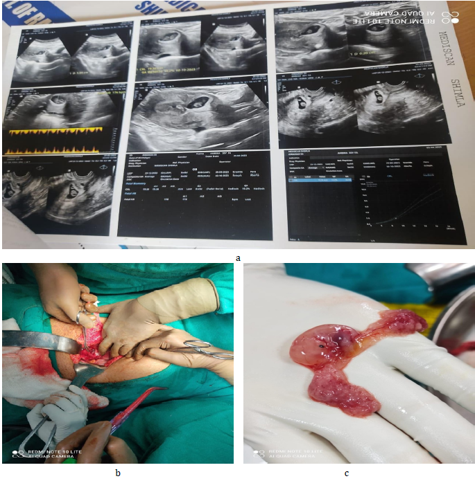
Figure 1: a-c Caesarean Scar Ectopic Pregnancy
Imaging and Investigations
Transvaginal ultrasound displayed a live intrauterine pregnancy within the caesarean scar region with residual myometrial thickness on the outer side of the sac measuring 2mm, with the placenta developing toward the scar tissue (Type-2 CSP). The presence of fetal cardiac activity (FHR-170 BPM) and embryo visualization provided valuable diagnostic information. Her histopathology reports of previous suction and evacuation were evaluated and were found to be normal. A subsequent MRI examination validated the initial diagnosis by revealing a T2 hyperintense sac-like structure measuring 1.4x2.4cm seen in the anterior lower uterine segment beneath the caesarean scar. Notably, the overlying myometrium measuring 2.2mm exhibited thinning, and the placenta's development was discernible. T2 hypointense structure of thickness 2mm was also seen in postero-inferior aspect towards right of the sac-developing placenta. Bilateral adnexa was grossly normal. No free fluid was seen in the pelvis. Uterocervix is 9.3x4.8cm with the endometrial thickness (ET) of 19.6mm. Upon clinical examination,she had normal vitals and cardiovascular and respiratory examination was within normal limits. She was asymptomatic with no abdominal tenderness, cramping or vaginal bleeding.
Management
Given the potential risks associated with CSP, a surgical approach was deemed appropriate for this case. Under spinal anesthesia, a laparotomy was performed to meticulously separate the gestational sac from the scar tissue, followed by the excision of the Caesarean Scar Pregnancy.
The intricate mechanisms driving the development of Caesarean Scar Pregnancy (CSP) remain elusive, despite several theories proposed to elucidate its occurrence. Among these theories, the endogenous migration of the gestational sac through either a wedge defect or microscopic fistula within the scar tissue stands out. This suggests that embryonic tissues could navigate through these imperceptible openings, leading to implantation within the scar. Another plausible explanation involves the invasion of placental villi into weakened or dehiscent areas of the scarred uterine wall, allowing for attachment and subsequent development [2,4].
Moreover, previous interventions such as caesarean sections, dilation and curettage (D&C), hysterotomy, myomectomy, and manual placenta removal may contribute to defects in the scar tissue. These interventions potentially disrupt the natural healing process, creating microtubular tracts that serve as conduits for gestational sac migration. The complex interplay of scar tissue healing and embryo implantation dynamics underscores the multifaceted nature of CSP etiology [3,4,6].
The imperative to detect CSP early is underscored by the recommended protocol for individuals with a history of prior caesarean sections. Early Pregnancy Assessment Clinic (EPAC) evaluations, combining transvaginal and transabdominal ultrasounds, coupled with corroborative MRI, play a pivotal role in achieving accurate diagnosis. The depth of implantation within the scar tissue is a crucial determinant of the risks involved. Cases of deep implantation towards the bladder and abdominal cavity bear an elevated risk of catastrophic outcomes like uterine rupture and severe hemorrhaging, necessitating vigilant monitoring and prompt intervention [6,7].
Management approaches for CSP hinge upon hemodynamic stability and pregnancy progression. Both medical and surgical termination options are considered, with the former presenting a viable choice for stable patients. Expectant management may yield favorable outcomes in Type-1 CSP cases, where implantation is superficial, while the Type-2 cases involving deeper niche implantation necessitate closer monitoring due to their propensity for complications [7,8].
Operative resection methods vary based on factors such as gestational age and patient anatomy. Laparoscopic, hysteroscopic, and laparotomic approaches are employed for operative resection. Suction aspiration guided by ultrasonography is particularly recommended for cases in the early first trimester (5 to 7 weeks). Laparoscopic excision, feasible up to 11 weeks, offers the advantage of complete removal of retained products of conception during surgery, reducing the need for prolonged follow-up. Additionally, retention of the lower segment's anatomy augments positive future fertility outcomes, provided that the procedure is conducted by skilled laparoscopic surgeons. The discussion underscores the imperative of comprehensive care, informed by an evolving understanding of CSP's complex dynamics [5-8].
Caesarean Scar Ectopic Pregnancy presents as a rare obstetrical phenomenon, fraught with the potential for life-threatening situations. Swift and accurate diagnosis, ideally in specialized medical centers, is imperative. Transvaginal ultrasonography serves as a cornerstone in diagnosis, and prompt management decisions involving experienced healthcare professionals are essential for optimal patient outcomes.
Kaelin Agten, A. et al. “The clinical outcome of cesarean scar pregnancies implanted ‘on the scar’ versus ‘in the niche’.” American Journal of Obstetrics and Gynecology vol. 216, no. 5, 2017, pp. 510.e1–510.e6.
Bowman, Z.S. et al. “Cesarean delivery and risk for subsequent ectopic pregnancy.” American Journal of Perinatology vol. 32, no. 9, 2015, pp. 815–820.
Morente, L.S. et al. “Cesarean scar ectopic pregnancy—case series: treatment decision algorithm and success with medical treatment.” Medicina (Kaunas) vol. 57, no. 4, 2021, pp. 362.
Jameel, K. et al. “Cesarean scar ectopic pregnancy: a diagnostic and management challenge.” Cureusvol. 13, no. 4, 2021, pp. e14463.
Valasoulis, G. et al. “Caesarean scar pregnancy: a case report and a literature review.” Medicinavol. 58, no. 6, 2022, pp. 740.
Lin, S.-Y. et al. “New ultrasound grading system for cesarean scar pregnancy and its implications for management strategies: an observational cohort study.” PLOS ONE vol. 13, no. 8, 2018, pp. e0202020.
Deepika et al. “A rare case report of caesarean scar ectopic pregnancy.” Journal of Clinical and Diagnostic Research vol. 11, no. 8, 2017, pp. QD10–QD11.
Onwonga, L. et al. “Causes, assessment and management of cesarean scar pregnancy.” International Journal of Pregnancy & Child Birth vol. 2, no. 5, 2017, pp. 119–124.
