+91 6002993949
submission@iarconsortium.org
Open Access
ISSN (Print) : 2788-8843
ISSN (Online) : 2788-8851
Background: Cutibacterium acnes (formerly Propionibacterium acnes) is a Gram-positive, rod-shaped bacterium that is aerotolerant and grows slowly under anaerobic conditions. Aim: The aim of this study to isolate and identify Cutibacterium acnes in Acne Vulgaris Patients through polymerase chain reaction and its antibiotic sensitivity. Materials and Methods: The study was conducted at the laboratory of the college of Veterinary Medicine for the period from first of October, 2021 to May 2022. The sample of this study consisted of collecting skin swabs from 90 patients who had acne vulgaris and attending dermatology private clinics in the Kirkuk city/ Iraq, for isolating and identifying Cutibacterium acnes. In order to collect the samples from a skin lesion, cotton swabs made of sterile cotton were used. In the clinic, the lesion was opened under aseptic conditions and then placed under the same conditions in a Thioglycolate medium tube. The growing bacteria were diagnosed based on their color, shape and size, edge, height, and whether or not they catalase production. In addition, secondary cultures were done for antibiotic sensitivity tests as well as molecular detection of C.acnes using PA primer and various virulence factors including hemolysis and lipase activity by polymerase chain reaction. Results: In the present study, five antibiotics were chosen to perform the susceptibility tests by disc diffusion assay. The study showed that 91.67% of C. acnes isolates were resistance to erythromycin, resistance to clindamycin (75%) and levofloxacin (33.3%) respectively However, they showed low resistance to tetracycline (8.3%) and (25%) doxycycline was performed. The current results showed that six out the 12 tested isolates that tested were positive for PA form severe acnes . While one strain from mild and moderate groups respectively was positive. The study showed that all C. acnes isolates that positive to PA primer had hemolysis gene as detected by PCR using tly primer. The study showed that 62.5% of C. acnes isolates were succeed with this type of lipase gene as detected by PCR, using lipase primer.
Cutibacterium acnes (formerly Propionibacterium acnes) is a Gram-positive, rod-shaped bacterium that is aerotolerant and grows slowly under anaerobic conditions [1,2]. This bacterium is a major commensal bacterium residing on healthy human skin [3,4] and it is the etiological pathogen for human acne vulgaris [5,6]. It plays an important role in maintaining skin health, but it has also been implicated in the pathogenesis of several diseases and infections, including sarcoidosis, SAPHO (synovitis, acne, pustulosis, hyperostosis, and osteomyelitis) syndrome, endodontic lesions [9], eye infections [1], prosthetic joint infections and acne vulgaris (commonly called acne) [5,6]. Antibiotic-resistant bacteria pose a serious threat to human health and the economy. More than 23,000 deaths result from 2 million infections caused by antibiotic-resistant bacteria every year in the United States [1]. New alternatives to antibiotics are urgently required in the post-antibiotic era. Antibiotic resistance in C. acnes (briefly CA) is a major problem. Treatment of acne vulgaris has failed due to the development of antibiotic resistance [4]. From the 1980s to the 2000s, the antimicrobial resistance of CA increased by approximately 40% worldwide [5]. C. acnes have long been considered to be a commensal bacterium, but its implication in various types of infection have led to its emergence as an opportunistic pathogen of low pathogenicity. This pathogenicity, like that of other skin-related bacteria, may be mediated by several molecular mechanisms, including the production of biofilms and the expression of virulence factors triggering immune responses in the host or promoting the adaptation of C. acnes to its environment. Various putative virulence factor genes have been identified in the C. acnes genome [7,8]. The aim of this study to isolate and identify Cutibacterium acnes in Acne Vulgaris Patients through polymerase chain reaction and its antibiotic sensitivity.
Preparation Cutibacterium Acnes Culture
12 of Cutibacterium acnes isolates from acnes vulgaris patients were used in this study after identification by gram stain, colonies morphology properties and some biochemical tests. The isolates were fisrt refresh by select single colony of Cutibacterium acnes from BHI-blood agar, subcultured onto thioglycolate broth and incubated anaerobically at 37 C0 for 5 days. After centrifuging the culture, it was washed and resuspended in sterile phosphate-buffered saline (PBS, pH 7.2) many times. Calibration of the spectrophotometer was performed at OD600, which is equivalent to 1x10⁸ CFU/mL, and the suspension turbidity was set to a 0.5 McFarland standard.
The Susceptibility Test of Antibiotics
The Kirby-Bauer disk diffusion technique was used to determine the susceptibility of several isolates of Cutibacterium acnes to various antibiotics by using of Levofloxacin (5µg); Doxycycline(10µg); Clindamycin (10µ) Erythromycin (10 µg) and Tetracycline (10µg), tested in duplicate with inoculum (1×10⁸ CFU/mL) on Mueller Hinton agar under anaerobic condition for 5-7 days at 37C, the diameters of the zones of inhibition (IZD) (including the 6-mm disk) were determined. The endpoint was taken as a complete inhibition of growth determine by the naked eye.
Molecular Characterization of Cutibacterium acnes
DNA Extraction: The extraction of genomic DNA of Cutibacterium acnes isolates was accomplished by using the G-spinTM Total DNA Extraction Kit and according to munfactoring iNtRON Biotechnology, Korea
Primers
Three primers including PA, tly (putative cytotoxin /hemolysin) and GehA lipase. used were previously designated by Ishige et al., McDowell et al., and Falcocchio et al., respectively. The primers for the detection of C. acnes associated gene and two virulence factors gene. The primers were prepared according to the information of the manufacturer (Bioneer, Korea) as shown in Table 1
PCR Reactions
In order to diagnose the PA gene of C. acnes and vigilance factors tly and lipase genes conventional polymerase chain reaction were applied. The PCR amplification mixture that was used including, 12.5 μl master mix, 1 μl forward primers,1 µL reverse primers, 5.5 µL PCR water, 5 µL Templet DNA to complete the reaction mixture to 25 µL Table 2.
The PCR tubes containing the amplification mixture are transferred to the Thermocycler and the amplification program is started as shown in Table 3. The purity and concentration of the extracted DNA were measured using a Nanodrop spectrophotometer. the PCR product is analyzed by agarose electrophoresis according to the following steps:10 microliters of the PCR products was electrophoresed through a 2% agarose gel and then stained with ethidium bromide. No band was obtained when DNA sample was replaced by distilled water as a negative control for the PCR procedure.
Table 1: Description of oligonucleotide primers used in the PCR assays
Size of bp | Primer sequence (5- to 3- | Primer |
587 | F : GGCACACCCATCTCTGAGCAC R : GGGTTGTAAACCGCTTTCGCTG | PA |
777 | F:CAGGACGTGATGGCAATGCGA R:TCGTTCACAAGACCACAGTAGC | tly |
510 | F: TCACTGATGAAGATCAACGCAC R: TGCGAATGTCCGACGAAGTCGA | lipase |
Table 2: Components mixture of PCR Master Mix Reaction of PA and virulence factors genes of Cutibacterium acnes
Ingredients | Volume in µl |
Master Mix | 12.5 |
Fowrward Primer | 1 |
Reverse Primer | 1 |
Template DNA | 5 |
Nuclease free water | 5.5 |
Total volume | 25 |
Table 3: PCR program for diagnosing PA and virulence factors( tly and lipase) genes diagnostics.
Primer | PCR Steps | Temp (C0 ) | Time | No. of cycles |
PA of C.acnes
| Initial denaturation | 95 | 5 min | 1x |
Denaturation | 95 | 40s | 35X | |
Annealing | 58 | 1 min | ||
Extension | 72 | 40s | ||
Final extension | 72 | 7min | 1x | |
Lipase
| Initial denaturation | 95 | 5 min | 1x |
Denaturation | 95 | 40s | 35x | |
Annealing | 55 | 1 min | ||
Extension | 72 | 40s | ||
Final extension | 72 | 7min | 1x | |
tly | Initial denaturation | 95 | 5 min | 1x |
Denaturation | 95 | 40s | 35x | |
Annealing | 57 | 1 min |
In the present study, five antibiotics were chosen to perform the susceptibility tests by disc diffusion assay. According to the standard values provided by Clinical and Laboratory Standards Institute guidance(CLSI,2020),the anaerobic culture of Cutibacterium acnes isolates exhibited variety in the resistance to antibiotics. The study showed that 91.67% of C. acnes isolates were resistance to erythromycin, resistance to clindamycin (75%) and levofloxacin (33.3%) respectively However, they showed low resistance to tetracycline (8.3%) and (25%) doxycycline was performed as shown in Table 4.
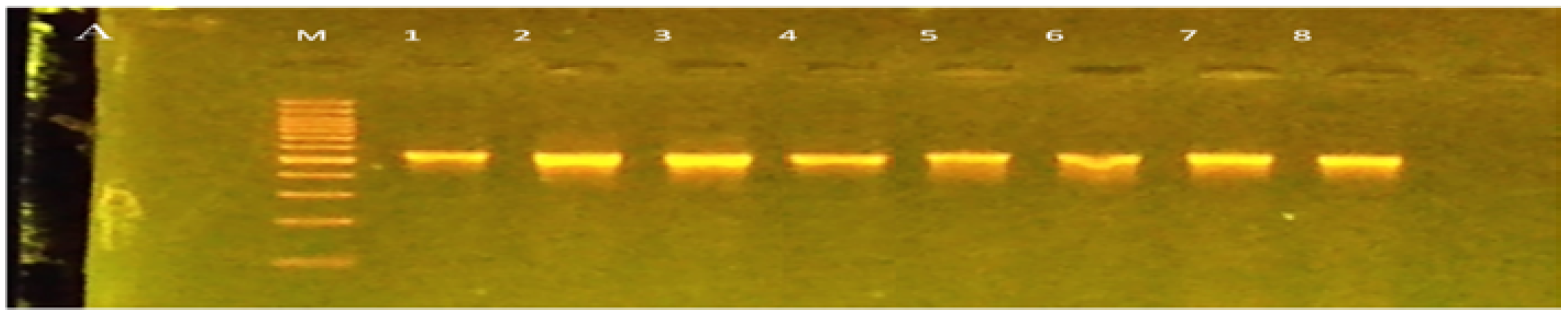
Figure 1: Typical amplification products of conventional PCR with primer PA. PCR products for PA gene of C. acnes were separated on 1.5 % agarose gels containing ethidium bromide and visualized under UV light. M: marker and lanes of eight isolated.
The results of the conventional polymerase chain reaction were performed on DNA extracted from 12 isolates of C.acnes isolates, by migration electrophoresis to estimate the weight of DNA based on the size parameter marker (100-1000 bp). The result of DNA amplification at the size of 587 bp for the PA primer of C.acnes gene was as follows: The eight isolates from severe, moderate and mild groups were detected by specific primer for C. acnes(PA) (Figure1). The current results showed that six out the 12 tested isolates that tested were positive for PA form severe acnes . While one strain from mild and moderate groups respectively was positive. The results of anaerobic culture of acnes isolates and PCR were differed. Potentially PA primer or the specific that used did not identify all types of C. acnes that resembling the observations of Cavalcanti.
The study showed that all C. acnes isolates that positive to PA primer had hemolysis gene as detected by PCR using tly primer, (Figure 2).
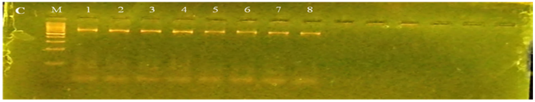
Figure 2: Typical amplification products of conventional PCR with primer tly. PCR products for tly gene of C. acnes were separated on 1.5 % agarose gels containing ethidium bromide and visualized under UV light. M: marker and lanes of eight isolated
The study showed that 62.5% of C. acnes isolates were succeed with this type of lipase gene as detected by PCR, using lipase primer (Figure 3).
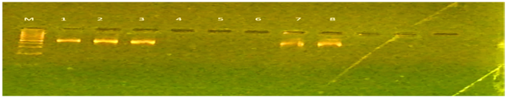
Figure 3: Typical amplification products of conventional PCR with primer lipase. PCR products were separated on 1.5 % agarose gels containing ethidium bromide and visualized under UV light. M: marker and only six showed positive
Table 4: Antibiotic sensitivity of C. acnes isolates
Antibiotics
| Sensitive | Resistance | ||
No. | % | No. | % | |
Levofloxacin | 4 | 66.6 | 8 | 33.3 |
Clindamycin | 3 | 25 | 9 | 75 |
Tetracycline | 1 | 91.67 | 11 | 8.3 |
Doxycycline | 3 | 75 | 9 | 25 |
Erythromycin | 12 | 33.3 | 4 | 66.6 |
Resistance to C. acnes is increasing in Europe and the rest of the world. Untile late 1970s, C. acnes had no resistance to antimicrobials. However, with the widespread use of topical and systemic antibiotics, antibiotic resistance has become a problem [1]. In our isolates the sensitivity rate of tetracycline and Doxycycline against C. acnes isolate was low as found by Mercieca et al., [2].The increasing antibiotic resistance, the use of systemic antibiotics in combination with topical adapalene (with or without benzoyl peroxide) or azelaic acid has a medium strength of recommendation for all severities of acne except comedonal acne. Tetracyclines, such as lymecycline and doxycycline, are the first-line systemic antibiotics recommended in combination with topical treatment. However, they should be limited to a treatment period of three months [3]. Hemolysins can lyse erythrocytes in vitro and are also potentially harmful to other cell types and enzymatic and/or pore-forming processes accomplish hemolysis. Hemolysis is associated with the tly gene and bacteria with this feature is dangerous [4]. In their 2004 study on the first C. acnes genome sequencing, Brüggemann et al. [5] recognized the existence of a hemolysin gene, which has since been validated by several additional laboratories. Many bacterial infections manufacture the hemolysin enzyme as a means of tissue degradation, host cell invasion, nutrition absorption, and dissemination and resistance to host immune response [6,7]. Lee et al., [8] reported that Type 1A and 1B of C. acnes were more β-hemolytic than Type II isolates. Furthermore, the C. acnes isolates at this study are potentially be C. acnes type I. This type of C. acnes strains are more potential to be associated with acne as recommended by Spittaels et al., [9]. They mentioned that of C. acnes strains are belonging to type 1 were more likely to be linked with acne, while strains belonging to type II were linked with healthy skin or deep tissue infections. Lipase is a crucial virulence component for bacteria linked to acne vulgaris. Protease is also present and contributes to the bacterium's pathogenicity. To help bacteria adhere and colonize the sebaceous follicle and induce the development of inflammatory lesions, Coenye et al., [10] reported that the adhesion to the internal surface of a continuous culture vessel in the presence of the free fatty acid, and proposed that triglycerides within nascent sebum, which contains no free fatty acid, were partially converted to free fatty acid by C.acnes lipase. C. acnes releases lipases to degrade sebum lipids, providing energy to the bacteria and skin with hydration. While the lipase serves in the nutrition of C. acnes and maintenance of healthy skin, excessive products (fatty acids) are known as major factors of inflammation. In inflammatory lesions, the quantities of C. acnes were similar to those in healthy skin, but preferential proliferation of type IA C. acnes over type II C. acnes is detected. Although there are only limited genetic studies to reveal the phenotypic and functional differences, virulent and lipase genes showed apparent differences between type IA and type II C. acnes [11,12,13]. Based on the above results, not all C. acnes isolates in which lipase activity was detected lends credence to the theory that lipase activity in C. acnes plays a role in the development of acne. However, each strain of C. acnes produces near to 15 lipases, and many of them are well conserved within types. However, these lipases are not conserved across types. The conserved lipases in type IA are not observed in types IB, IC, or II, and type II conserved lipases are not observed in type IA [14,15,16]. This confirmed the absence of positive PCR products of lipase for all isolates within this study.
The study showed that Cutibacterium acnes isolates were sensitive to tetracycline and doxycycline (91.67 %,75 %) respectively which suggest to use them with topical medicine to treat acnes. Only eight out of 12 isolates were confirmed by convential pcr to detection PA gene of C.acnes the differ between the two ways of identification potentially to fail of culture procedure and the primer that used in this study. The study showed that all C. acnes isolates have hemolysis genes as detected by PCR. While they were variable in lipase production depending on types of C. acnes
Coates, P. et al. “Prevalence of Antibiotic-Resistant Propionibacteria on the Skin of Acne Patients: 10-Year Surveillance Data and Snapshot Distribution Study.” British Journal of Dermatology, vol. 146, no. 5, 2002, pp. 840–848.
Mercieca, L. et al. “The Antibiotic Susceptibility Profile of Cutibacterium acnes in Maltese Patients with Acne.” Journal of Clinical and Aesthetic Dermatology, vol. 13, no. 6, 2020, p. 11.
El Seedawy, M.E.S.H.E. Microbiological and Molecular Studies on Microorganisms Related to Acne Vulgaris in Some Egyptian Hospitals. n.d.
Boyle, K.K. et al. “Pathogenic Genetic Variations of C. acnes Are Associated with Clinically Relevant Orthopedic Shoulder Infections.” Journal of Orthopaedic Research, vol. 38, no. 12, 2020, pp. 2731–2739.
Brüggemann, H. et al. “A Janus-Faced Bacterium: Host-Beneficial and Detrimental Roles of Cutibacterium acnes.” Frontiers in Microbiology, vol. 12, 2021, article 673845.
Ergin, Ç. et al. “Nasal Antibiotic-Resistant Propionibacterium acnes Carriage in Acne Vulgaris Patients in Turkey.” Journal of Dermatology, vol. 33, 2006, pp. 899–901.
Castillo, D.E. et al. “Propionibacterium (Cutibacterium) acnes Bacteriophage Therapy in Acne: Current Evidence and Future Perspectives.” Dermatology and Therapy, vol. 9, no. 1, 2019, pp. 19–31, https://doi.org/10.1007/s13555-018-0275-9.
Lee, J. et al. “Correlation between Hemolytic Profile and Phylotype of Cutibacterium acnes (Formerly Propionibacterium acnes) and Orthopedic Implant Infection.” Shoulder & Elbow, vol. 12, no. 6, 2020, pp. 390–398.
Spittaels, K.J. et al. “Cutibacterium acnes Phylotype I and II Strains Interact Differently with Human Skin Cells.” Frontiers in Cellular and Infection Microbiology, vol. 10, 2020, pp. 1–11, https://doi.org/10.3389/fcimb.2020.575164.
Coenye, T. et al. “The Role of Biofilm Formation in the Pathogenesis and Antimicrobial Susceptibility of Cutibacterium acnes.” Biofilm, vol. 4, 2022, article 100063.
Coley, M.K. et al. “Overview of Treatment Principles for Skin of Color.” Acne Vulgaris, CRC Press, 2011, pp. 82–97.
Cong, T.X. et al. “From Pathogenesis of Acne Vulgaris to Anti-Acne Agents.” Archives of Dermatological Research, vol. 311, no. 5, 2019, pp. 337–349, https://doi.org/10.1007/s00403-019-01908-x.
Corvec, S. “Clinical and Biological Features of Cutibacterium (Formerly Propionibacterium) avidum, an Underrecognized Microorganism.” Clinical Microbiology Reviews, vol. 31, no. 3, 2018, e00064-17.
Coates, P. et al. “Prevalence of Antibiotic-Resistant Propionibacteria on the Skin of Acne Patients: 10-Year Surveillance Data and Snapshot Distribution Study.” British Journal of Dermatology, vol. 146, no. 5, 2002, pp. 840–848.
Coenye, T. et al. “The Role of Biofilm Formation in the Pathogenesis and Antimicrobial Susceptibility of Cutibacterium acnes.” Biofilm, vol. 4, 2022, article 100063.
Corvec, S. et al. “Taxonomy and Phylogeny of Cutibacterium (Formerly Propionibacterium) acnes in Inflammatory Skin Diseases.” Annales de Dermatologie et de Vénéréologie, vol. 146, no. 1, 2019, pp. 26–30.
