Contents
Download PDF
pdf Download XML
716 Views
313 Downloads
Share this article
Research Article | Volume 1 Issue 1 (Jul-Dec, 2020) | Pages 1 - 5
Isolation and Characterization of Chemical Constituents from n-Hexane Extract of Leaf and Flower Part of Catharanthus Roseus, Available in Bangladesh
 ,
1
Chemical Research Division, BCSIR Laboratories Dhaka, Bangladesh Council of Scientific and Industrial Research, Dhamondi, Dhaka-1205, Bangladesh
2
Department of Chemistry, Jahangirnagar University, Savar, Dhaka-1342, Bangladesh
Under a Creative Commons license
Open Access
Received
Sept. 3, 2020
Revised
Oct. 2, 2020
Accepted
Nov. 19, 2020
Published
Dec. 28, 2020
Abstract

Bangladesh is a rich store house of medicinal plants. Catharanthus roseus is administered as a cooling medicine. From this plant, n-hexane extract Compounds: 3-Hydroxy-cyclooctanone (1) of flowers and from leaf compound Lupeol (2), Stigmasterol (3), have been isolated of Catharanthus roseus. The structures of the compounds have been established by different spectroscopic data analysis. From flower and leaf n-hexane extract compounds 3-Hydroxy-cyclooctanone and Lupeol were isolated for the first time from this plant.

Keywords
INTRODUCTION

Medicinal plants are a source of great economic value all over the world. Nature has bestowed on us a very rich botanical wealth and a large number of diverse types of plants grown in different parts of the country [1]. Today, according to world health organization (WHO) as many as 80% of the world’s people depend on traditional medicine for their primary health care needs [2]. High plants are sources of drug which have made important contribution to the welfare and quality of life urban as well as rural communities especially in tropics and sub-tropics [3]. During the early years of human existence, many plants materials by instinct, intuition of trial and error were used to combat different aliments [4]. 

Catharanthus roseus, as a medicinal plant (Common name - Periwinkle, Vinca; Bengali - Nayantara, Synonyms - Vinca rosea; Family - Apocyanaceae) popularly known as madagascar periwinkle is a potential source for anti-leukemic alkaloids. It is cultivated mainly for its alkaloids which are having anticancer activities [5]. It is an evergreen subshrub or harbeceous plant growing up to 1 m tall [6]. This plant is administered as a cooling medicine. It is used for the treatment of diabetes, fever, malaria, throat infection and chest complaints. It is also used for the regulation of menstrual cycles and as a euphoriant [7]. The plant is an important source of indole alkaloids which are present in all plant parts. The physically important and antineoplastic alkaloids namely Vincristine and Vinblastine are mainly present in the leaves whereas antihypertensive alkaloids such as ajmalicine, serpentine and reserpine are reported to be present in the roots [8]. Vincristine and Vinblastine alkaloids are used in the treatment of various types of lymphoma and leukemia [9,10]. 


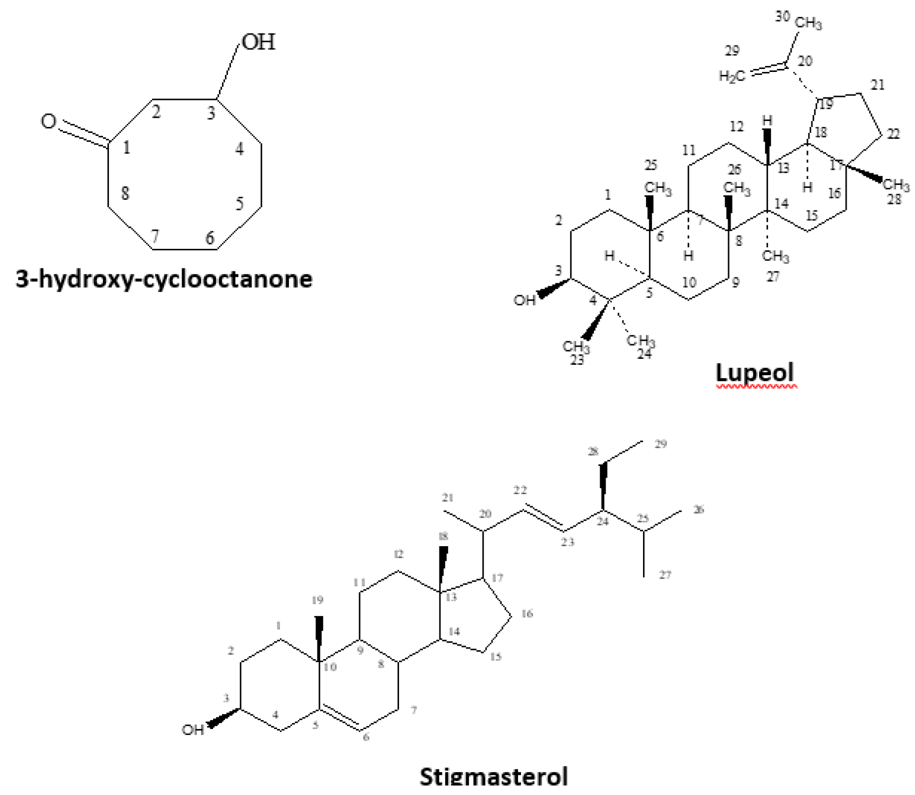
Figure 1: Structures of the Isolated Compounds


These Catharanthus alkaloids are also used for the treatment of both malignant and nonmalignant diseases and in platelet and platelet associated disorder. Previous phytochemical investigations resulted in the isolation of Kaemferol [11], Kaempferol trisaccarides [12], Quercetin [13], Quercetin trisaccarides [11], Syringetin glycosides [14], Malvidin [13] Malvidin 3-0-glucosides, Malvidin 3-0-(6-0-p-coumaroyl) [15], Petunidin [13], Petunidin 3-0-glucosides [15], Petunidin 3-0-(6-0-p-coumaroyl) [13], Hirsutidin [13], Hirsutidin 3-0-glucosides, Hirsutidin 3-0-(6-0-p-coumaroyl) [15]. Catharanthus Plant produce many pharmaceutically important alkaloids. They are antineoplastic medicines and the monoindole alkaloids ajmalicin and serpentine are antihypertension drugs [16-22]. Our recent study on n-hexane part of flower and leaf of this plant has led to the isolation of 3-Hydroxy-cyclooctanone, lupeol and stigmasterol (Figure 1). 3-Hydroxy-cyclooctanone and lupeol were isolated from flower and leaf n-hexane extract for the first time from this plant.

MATERIALS AND METHODS

Melting points were determined by thin disc method on a Fisher-John’s electrothermal melting point apparatus. UV spectra were recorded in methanol on a Shimadzu UV-Visible spectrophotometer. IR spectra were recorded on a Shimadzu FT-IR spectrometer as thin film or KBr disc. NMR spectra were recorded in CDCl3 using Bruker WH 400 MHz NMR spectrometer. Mass spectra of the compounds were measured on Finnigan Mat SSQ 710 spectrometer with ionization induced by electron impact at 70 eV. Separation by column chromatography was carried out using silica gel 40 (70-230 mesh, E. Merck). Thin layer chromatography was carried out on TLC plastic sheets pre-coated with silica gel 60 F254 (E. Merck).


Collection of Plant Material

Fully matured fresh leaves and flowers of this plant were collected from the gardens of Chemistry Department of Dhaka university, Bangladesh in June 2013 and identified by the taxonomist of Bangladesh national Herbarium, Dhaka, where a voucher specimen (No = 39512) has been deposited. The leaves and flowers of C. roseus were separately air dried. These dried samples of leaves and flowers were powdered using 20 mesh screen in Willey mill and then used for subsequent analysis.


Extraction of the flower and leaf parts of C. roseus

Powder of the flowers (200 g) was extracted at room temperature with n-hexane for 5 days and the extract was dried to get a gummy mass (5.0 g) using rotary evaporator. The powder of leaves (510 g) was extracted with methanol at room temperature for 5 days. The filtrate was dried into a gummy mass using rotary evaporator under reduced pressure. The methanol extract (40.0 g) was then triturated by n-hexane (100 ml 3), then these extracts were dried by using a rotary evaporator to get n-hexane extract (11.0 g).
 

Isolation of Compounds from Crude Extracts

The dry mass of n-hexane extract (5.0g) was adsorbed by the column grade silica gel. TLC analysis of the n-hexane extract showed several spots under UV lamp followed by the development by spray reagent on TLC plate. This sample was placed on the top of the column bed packed with TLC grade silica gel in VLC apparatus. It was first eluted with 100% n-hexane and then eluted with mixtures of n-hexane and Ethyl Acetate (EA) and finally with the mixtures of ethyl acetate and methanol with increasing polarity. Twenty-two collections of 200 ml each were collected and combined into eight fractions according to their TLC patterns for further purification. Compound 1 (1.3 mg) was isolated in pure form from fraction 6 using a column eluted with 70% ethyl acetate in chloroform.


The dry mass of leaf n-hexane extract (7.0 g) was dissolved in minimum amount of n-hexane and adsorbed by the column grade silica gel. TLC analysis of the n-hexane extract showed several spots in different solvent systems under UV lamp followed by spray reagent on TLC plate. This adsorbed sample was placed on the top of the silica gel bed (TLC grade) in VLC apparatus. It was first eluted with 100% n-hexane and then mixtures of n-hexane and dichloromethane (DCM) and finally with the mixtures of DCM and methanol with increasing polarity. The eluents were collected in an amount of 200 ml each in 23 conical flasks and were divided into five fractions according to their TLC pattern. Depending on the TLC behavior of VLC fractions, fraction 3 (collection no. 9-13) and fraction 4 (collection no. 14-18) were selected for further investigation due to their good resolution. 


Compound 2 (9.8 mg) was isolated from the VLC fraction 3 (collection no. 9-13) of n-hexane extract using column chromatographic separation eluted wit hexane-ethyl acetate solvent gradient and found as white amorphous solid which was completely soluble in chloroform. Compound 3 (8.5 mg) was found as white crystalline solid by repeated column chromatographic separation of VLC fraction 4 (collection no. 14-18) using hexane-chloroform solvent system in gradient manner as mobile phase. The compound was TLC pure and soluble in chloroform.


Spectroscopic Data of the Isolated Compounds

3-Hydroxy-Cyclooctanone: Yellowish gummy solid; IR (neat) υ 3450 (O-H), 2930, 2870,1710 (C = O), 1065 (C-O) cm-1; 1H NMR (CDCl3) δ 3.77 (1H, unr.s, H-3), 2.61 ( 2H, unr.s, H-2), 2.16 ( 2H, unr. s. H-8), 1.63 (2H, unr.s, H-4), 1.24 ( 6H, unr.s, H-5, H-6 and H-7); 13C NMR (CDCl3) δ 69.5 (C-3), 53.7 ( C-2), 31.7, 29.7, 29.2 ( 3C).


Lupeol

White amorphous solid; mp 214-215 ̊ C; IR (neat) υ 3368 (br. O-H), 2941, 1640 (C = C), 1463, 1380, 1214 (C-O) cm-1; 1H NMR (CDCl3) δ 4.67 (1H, unr. s, Ha-29), 4.55 (1H, unr. s, Hb-29), 3.17 (1H, dd, J = 11.2 and 4.8 Hz, H-3), 2.36 (1H, m, H-19), 1.90 (2H, m, H-2), 1.67 (3H, s, H-30), 1.48-1.65 (3H, m), 1.30-1.47 (14H, m), 1.15-1.29 (6H, m), 1.02 (3H, s), 0.95 (3H, s), 0.93 (3H, s), 0.82 (3H, s), 0.77(3H, s), 0.75 (3H, s); 13C NMR (CDCl3) δ 151.0 (C-20), 109.3 (C-29), 79.0 (C-3), 55.3, 50.4, 48.3, 48.0, 43.0, 42.8, 40.8, 40.0, 38.8, 38.7, 38.0, 37.1, 35.6, 34.3, 29.8, 28.0, 27.5, 27.4, 25.1, 20.9, 19.3, 18.3, 18.0, 16.1, 16.0, 15.4, 14.5; MS m/z 412(M+), 411, 315, 218, 207, 189, 135, 121, 107, 95, 81, 68, 43 (base peak).


Stigmasterol

White amorphous solid; mp 168-170° C; IR (neat) υ 3417 (O-H), 2937, 2865, 1462, 1381, 1051 (C-O) cm-1; 1H NMR (CDCl3) δ 5.34 (1H, unr. s, H-6), 5.14 (1H, dd, J = 15.2 and 8.4 Hz, H-22 or H-23), 5.01 (1H, dd, J = 15.2 and 8.4 Hz, H-22 or H-23), 3.52 (1H, m, H-3), 2.17-2.32 (2H, m), 1.92-2.08 (2H, m), 1.79-1.88 (2H, m), 1.35-1.73 (14H, m), 1.08-1.28 (6H, m), 1.01 (3H, d, J = 7.2 Hz, H-21), 0.99 (3H, s, H-19), 0.83 (6H, d, J = 6.0 Hz, H-26 and H-27), 0.79 (3H, t, J = 6.8 Hz, H-29), 0.68 (3H, s, H-18); 13C NMR (CDCl3) δ 140.7, 138.3, 129.3, 121.7, 71.8 (C-3), 56.8, 55.9, 51.2, 50.1, 42.3, 42.2, 40.5, 39.7, 37.2, 36.5, 31.9 (3C), 31.6, 28.9, 25.4, 24.3, 21.2, 21.1 (2C), 19.4, 19.0, 12.2, 12.0; MS m/z 412, 397, 394, 351, 271, 255, 213, 159, 133, 83, 69, 55 (base peak).

RESULTS AND DISCUSSION

n-hexane extract of flower of this plant has yielded to the isolation of 3-Hydroxy-cyclooctanone 1 (1.3 mg) from flowers and from leaf has yielded lupeol (9.8mg) and stigmasterol (8.5mg) (3). Fractionation and isolation works on the n-hexane by repeated column chromatography have afforded to 3-Hydroxy-cyclooctanone 1 (1.3 mg) from flowers and from leaf has yielded lupeol (9.8mg) and stigmasterol (8.5mg) (3) (Figure 1). The structures of the compounds were elucidated using spectroscopic techniques. The compound 1 (1.3 mg) was found as yellow gummy solid and was soluble in chloroform. The IR spectrum of the compound showed a broad absorption band at 3450 cm-1 due to the O-H stretching vibrations. The bands at 1710 and 1065 cm-1 were found due to the carbonyl (C = O) and C-O stretching vibrations in the molecule.


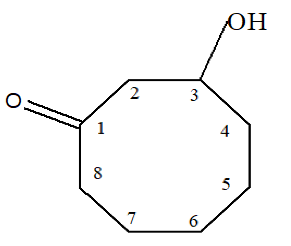
In the 1H NMR spectrum, the 1H unresolved singlet at δ 3.77 indicated the presence of >CHOH group in the structure. The methylene protons at C-2 which is attached to the carbonyl carbon and >CHOH group was assigned by the peak at δ 2.61. The two-proton peak at δ 2.16 clearly showed the methylene protons at C-8 which is attached to the carbonyl group. Two methylene protons at C-4 indicated by the poeak at δ 1.63. The other three methylene groups were ascertained by the peak at δ 1.24. The 13C NMR spectrum showed five signals for seven carbons but the carbonyl carbon was not shown in this spectrum due to the very small amount of the compound. Two peaks at δ 69.5 and 53.7 clearly indicated the carbons at position-3 and -2, respectively. Due to the lack of mass spectrum, the compound 1 was only suggested as 3- hydroxyl-cyclooctanone on the basis of above spectral analysis. This compound is isolated for the first time from the flower as well as from the plant C. roseus. The structure of the compound 1 is given in Figure 2.


Compound 2 (9.8 mg) was found as white amorphous solid and was soluble in chloroform. Melting point of the compound was found as 214-215 ̊C. It showed single spot on TLC plate with Rf value 0.51 in 100% CHCl3. The mass spectrum of the compound showed a molecular ion peak at m/z 426 corresponding to the molecular formula C30H50O. The IR spectrum of the compound (2) showed a broad absorption band at 3368 cm-1 due to the O-H stretching vibration and band at 2941 cm-1 due to the C-H stretching vibration. The absorption band found at 1640 cm-1 due to the C = C stretching and bands at 1463 and 1380 cm-1 due to the C-H bending vibrations and band at 1214 cm-1 due to the C-O stretching vibration in the molecule.


Figure 2: Structure of 3-hydroxy-cyclooctanone


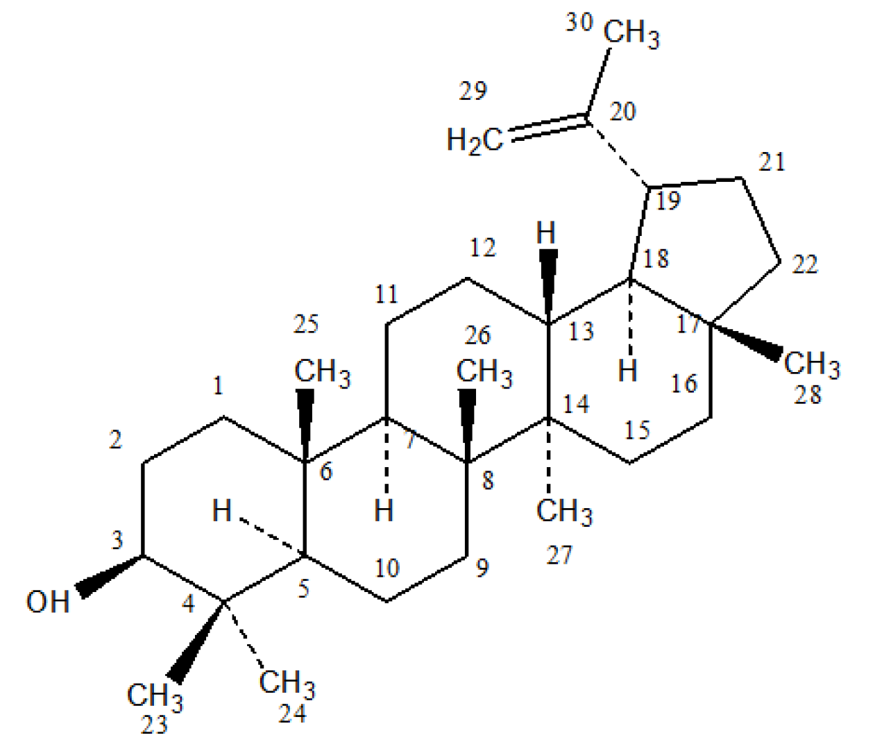
Figure 3: Structure of Lupeol


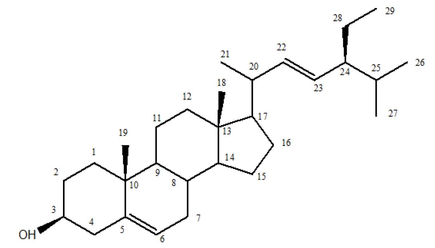
Figure 4: Structure of Stigmasterol


In the 1H NMR spectrum, the two unresolved singlets integrated for one proton each indicated the two olefinic protons attached to C-29. The one proton doublet of doublet at δ 3.17 showed the presence of >CHOH group in the molecule. The olefinic carbons (C-20 and C-29) and the carbon attached with oxygen were also indicated by the peaks at δ 151.0, 109.3 and 79.0 in the 13C NMR spectrum, respectively. The seven methyl groups in the molecule are clearly showed by the seven 3H-singlets at δ 1.67, 1.02, 0.95, 0.93, 0.82, 0.77 and 0.75.


The presence of 30 carbons in the molecule was clearly indicated by the 30 peaks in the 13C NMR spectrum. The analysis of DEPT-135 along with the 1H NMR spectrum undoubted showed that the molecule contains seven methyl, eleven methylene, six methine and six quaternary carbons in its structure. The molecular formula and all of the above spectral data analysis clearly informed that the compound is a pentacyclic triterpenoid containing one hydroxyl and a double bond. Finally, the structure of the compound was confirmed by the fragment ions present in the mass spectrum.


Based on all spectroscopic data, literature values [23] and melting point of the compound, it was confirmed that the compound 2 is Lupeol. The compound was found from the leaves as well as from the plant C. roseus for the first time. The structure of the compound 2 is given in Figure 3.


Lupeol

Compound 3 (8.5 mg) was found as white amorphous solid and was soluble in chloroform. Melting point of the compound was found as 168-170°C. It showed single spot on TLC plate with Rf value 0.55 in 100% CHCl3. The mass spectrum of the compound showed a molecular ion peak at m/z 412 which is corresponding to the molecular formula C29H48O. The IR spectrum of the compound showed a broad absorption band at 3417 cm-1 due to the O-H stretching vibration and bands at 2937 and 2865 cm-1 due to the saturated C-H stretching vibrations. The absorption band at 1051 cm-1 indicated the presence of C-O stretching vibration in the molecule.


In the 1H NMR spectrum, the three one-proton peaks in the olefinic region at δ 5.34, 5.14 and 5.01 indicated the presence of three olefinic protons at C-6, C-22 and C-23, respectively. This was strongly supported by the four signals at δ 140.7, 138.3, 129.3 and 121.7 in the 13C NMR spectrum which clearly indicated the presence of two double bonds in the molecule. The one-proton multiplet at δ 3.52 was found in the 1H NMR spectrum due to proton at C-3 which is attached to a hydroxyl group and this was also supported by the peak at δ 71.8 in the 13C NMR spectrum. The presence of six methyl groups in the structure was clearly showed by the two singlets at δ 0.99 (3H, H-19), 0.68 (3H, H-18), two doublets at δ 1.01 (3H, H-21) and 0.83 (6H, H-26 and H-27) and one triplet at δ 0.79 (3H, H-29) in the 1H NMR spectrum.


The presence of 29 carbons in the molecule clearly indicated by the 26 signals in the 13C NMR spectrum. The intensified peaks at δ 31.9 and 21.1 were found due to three carbons and two carbons, respectively. All the above data suggested that the compound 3 is a steroidal molecule containing one hydroxyl, two double bonds and six methyl groups. Finally, the structure of the compound was confirmed by the fragment ions present in the mass spectrum.


Based on all spectroscopic data, literature values [24] and melting point of the compound 3, it was confirmed that the compound is Stigmasterol. The structure of the compound 3 is given in Figure 4.

CONCLUSION

Literature survey showed that very little phytochemical studies have been done on n-hexane extract on flower and leaves of the plant Catharanthus roseus. The isolation and characterization of three compounds from leaf and flower part of the plant have been reported here. We believe, there is a scope to do more detailed phytochemical and biological study on this plant in future.


Acknowledgment

We are grateful to Division in charge, Chemical Research Division, BCSIR Laboratories, Dhaka and Director, BCSIR Laboratories, Dhaka, for providing necessary facilities to carry out this research work.

REFERENCES
  1. Nair, L.D. et al. “Comparative study on proximate analysis conducted on medicinal plants of Chhattisgarh, CG, India.” Research Journal of Chemical Sciences, vol. 2, no. 9, 2012, pp. 18–21.

  2. Uduak, E. and A. Emana. “Proximate composition and phytochemical constituents of Aspillia Africana (Pers) C.D. Adams and Tithonia diversifolia (Hemsl) A. gray stems (Asteraceae).” Indian Journal of Pharmaceutical and Biological Research, vol. 1, no. 1, 2013, pp. 23–30.

  3. Sofowora, A.A. Medicinal Plants and Traditional Medicine in West Africa. 2nd ed., John Wiley and Sons, 1993, p. 289.

  4. Ogundare, A.O. “Antimicrobial effect of Tithonia diversifolia and Jatropha gossypifolia Leaf Extracts.” Trends in Applied Science and Research, vol. 2, no. 2, 2007, pp. 145–150.

  5. Jaleel, C.A. et al. “Alterations in non-enzymatic antioxidant components of Catharanthus roseus exposed to Paclobutrazol, gibberellic acid and Pseudomonas fluorescens.” Plant Omics Journal, vol. 2, no. 1, 2009, pp. 30–40.

  6. Huxley, A. New RHS Dictionary of Gardening. Macmillan, 1992.

  7. Ambusta, C.S. The Wealth of India: Raw Materials. Revised ed., Publication and Information Directorate, CSIR, 1992, p. 117.

  8. Mishra, P. et al. “Pattern of diversity for morphological and alkaloid yield-related traits among the periwinkle catharanthus roseus accessions collected from in and around the indian subcontinent.” Genetic Resources and Crop Evolution, vol. 48, 2001, pp. 273–286.

  9. Farnsworth, N.R. et al. “Antiviral activity of selected catharanthus alkaloids.” Journal of Pharmaceutical Sciences, vol. 57, 1968, pp. 2174–2175.

  10. Gajalakshmi, S. et al. “Pharmacological activities of catharanthus roseus: A prospective review.” International Journal of Pharmaceutical and Biological Sciences, vol. 4, no. 2, 2013, pp. 431–439.

  11. Forsyth W.G.C. and N.W. Simmonds. “Anthocyanidins of Lochnera rosea.” Nature, vol. 180, 1957, p. 247.

  12. Nishibe S. et al. “Bioactive compounds from Catharanthus roseus and Vinca minor.” Natural Medicines (Tokyo), vol. 50, 1995, pp. 378–383.

  13. Bruneton J. Pharmacognosy: Phytochemistry, Medicinal Plants. 2nd ed., Intercept Ltd., 1999, pp. 227–231.

  14. Filippini R. et al. “Production of Anthocyanins by Catharanthus roseus.” Fitoterapia, vol. 74, 2003, pp. 62–67.

  15. Srinivasa R.A. and M.A. Fazil. “Simultaneous estimation of quercetin and Rutin in Ethanolic Extract of Catharanthus roseus Linn. Leaves by HPLC Method.” International Global Research Analysis, vol. 2, no. 7, 2013.

  16. Atta-Ur-Rahman et al. “16-Epi-19-S-Vindlinine, an Indole alkaloid from C. roseus.” Planta Medica, vol. 47, 1983, pp. 246–247.

  17. Atta-Ur-Rahman and J. Fatima. “Isolation and structure of rosicine from C. roseus.” Tetrahedron Letters, vol. 25, 1984, pp. 6051–6054.

  18. Atta-Ur-Rahman and I. Ali. “Isolation of Rhazinal from the leaves of C. roseus.” Journal of Natural Products, vol. 47, no. 2, 1984, p. 389.

  19. Atta-Ur-Rahman and I. Ali. “Isolation and 13C-NMR studies on Cathovaline, an alkaloid from leaves of C. roseus.” Planta Medica, vol. 5, 1985, pp. 447–448.

  20. Atta-Ur-Rahman et al. “Leurosinone: A new binary indole alkaloid from C. roseus.” Perkin Transactions I, no. 8, 1988, pp. 2175–2178.

  21. Auriola S. et al. “Identification of indole alkaloids from c. roseus with liquid chromatography/mass spectrophotometry using collision-induced dissociation with Thermospray Ion Repeller.” Biomed Environ Mass Spectrum, vol. 19, no. 7, July 1990, pp. 400–404.

  22. Nikita V. et al. “Studies in the proximate analysis of leaves of murraya koenigi l. from amravati, Maharashtra state.” International Journal of Natural Products Research, vol. 1, no. 3, 2012, pp. 40–41.

  23. Abdullahi S.M. et al. “Isolation of lupeol from the stem bark of Lonchocarpus sericeus (Papilionaceae).” Scholars Academic Journal of Biosciences, vol. 1, no. 1, 2013, pp. 18–19.

  24. Rajput A.P. “Isolation of Stigmastrol and β-Sitosterol from chloroform extract of leaves of Corchorus fascicularis Lam.” International Journal of Biological Chemistry, vol. 6, no. 4, 2012, pp. 130–135.
Recommended Articles
Research Article
Amit Jain’s System of Practice: From Modern to Super Modern Diabetic Foot Surgery
Published: 11/12/2020
Download PDF
Research Article
Curcumin: A Possible Treatment for Coronavirus Infection
Published: 10/10/2021
Download PDF
Research Article
Awareness Regarding Danger Sign and Symptoms During Pregnancy among Antenatal Mothers Attending Outpatient Department of Rural Hospital in Himachal Pradesh
Published: 26/05/2023
Download PDF
Review Article
Role of Nanotechnology in Pharmaceutical Science: A Review
...
Published: 10/09/2021
Download PDF
Chat on WhatsApp
Flowbite Logo
PO Box 101, Nakuru
Kenya.
Email: office@iarconsortium.org

Editorial Office:
J.L Bhavan, Near Radison Blu Hotel,
Jalukbari, Guwahati-India
Useful Links
Order Hard Copy
Privacy policy
Terms and Conditions
Refund Policy
Shipping Policy
Others
About Us
Team Members
Contact Us
Online Payments
Join as Editor
Join as Reviewer
Subscribe to our Newsletter
+91 60029-93949
Follow us
MOST SEARCHED KEYWORDS
Copyright © iARCON International LLP . All Rights Reserved.