
+91 6002993949
submission@iarconsortium.org
Open Access
ISSN (Print) : XXXX-XXXX
ISSN (Online) : 2708-3594
Blue sclera is a bluish discolouration of the white sclera of eye. The blue colour is caused by thinness of collagen layers of the sclera that allows the veins of the choroid layers to be seen. Blue sclerae are characteristic of a number of conditions, particularly connective tissue disorders and musculoskeletal disorders. In this article we have presented a few clinical cases which were diagnosed on the basis of this ‘blue sclera’.
Osteogenesis imperfecta (ICD 10 code Q78.0) is most frequently associated with blue sclera. It is a severe disease in bone mass that makes the bones brittle [1]. The other clinical features of OI are loose joints, frequent fractures, dental abnormalities, progressive hearing loss and a positive family history. The underlying mechanism is usually a problem with connective tissue due to lack of type 1 collagen [2]. This deficiency arises from an amino acid substitution of glycine to bulkier amino acids in the collagen triple helix structure. The larger amino acid side-chains create steric hindrance that creates a bulge in the collagen complex, which in turn influences both the molecular nano mechanics and the interaction between molecules, which are both compromised [3]. As a result, the body may respond by hydrolysing the improper collagen structure. If the body does not destroy the improper collagen, the relationship between the collagen fibrils and hydroxyapatite crystals to form bone is altered, causing brittleness [4]. It occurs due to mutation in COL1A1 and COL1A2 genes in more than 90% of the cases. It can be either a new mutation or genetically inherited as autosomal dominant trait. Blue sclera is rarely found normally as an inherited trait. There are other few diseases where blue sclera is a rare occurrence or accidental finding [5]. These are as follows:
Ehlers: Danlos Syndrome [5]: A group of rare autosomal dominant disorder of connective tissue, which commonly presents with joints hypermobility
loose, unstable joints that dislocate easily, joint pain, fatigue, easy bruises on skin and digestive problems, such as heartburn and constipation [6]
Marfan Syndrome [5]: A genetic disorder of autosomal dominant inheritance caused by mutations in the FBN1 gene, clinically presenting with tall and thin, with long arms, legs, finger & amp; toes, overly-flexible joints, serious complication of heart, lungs & amp; aorta and tendency to develop mitral valve prolapse [7]
Pseudoxanthoma elasticum [5]
Williems De Vries Syndrome [5]
Diamond Blackfan Anaemia [5]: A congenital erythroid aplasia, where abnormally low levels of RBC with normal platelet & WBC count clinically presents with normocytic or macrocytic anaemia, craniofacial & amp; urogenital malformations, cardiac defects & amp; cleft palate. The patients have tendency to develop myelodysplastic syndrome [8]
Severe iron deficiency anaemia [5]
Juvenile Paget’s disease [5]
Acid phosphatase deficiency [5]
Presentation of Few Clinical Cases
Case 1: A 13-year-old girl visited the OPD with nausea, loss of appetite, weakness and light headedness. She had a history of fainting from over exertion. On examination, the colour of her skin was pale and she had blue sclera (Figure 1).
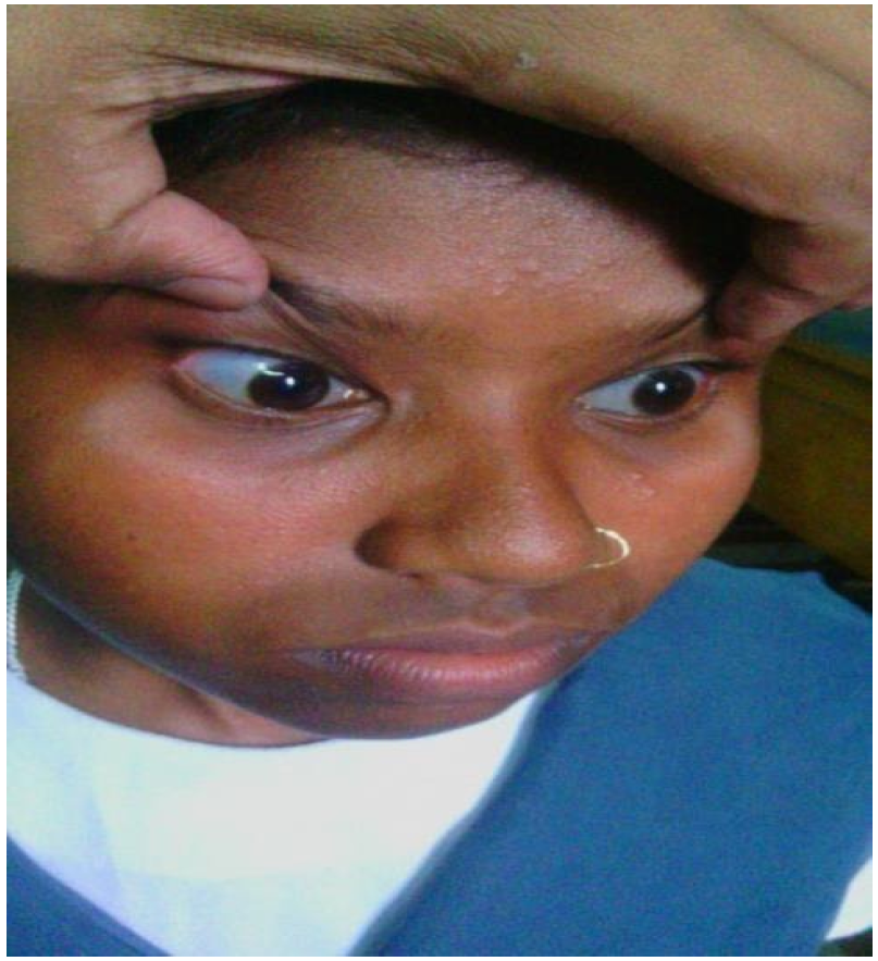
Figure 1: A 13-Year-Old Girl Visited the OPD with Nausea, Loss of Appetite, Weakness and Light Headedness, she Colour of Her Skin Was Pale and She Had Blue Sclera
Laboratory tests were done which revealed her RBC count was 3.5 million cells/mcL, Haemoglobin level was 6.8 µg/dl, ferritin level was 9.2 ng/ml and TIBC was 400 mcg/dL. The blue sclera was thus due to severe iron deficiency anaemia.
Case 2
An 8-year-old school going boy visited the OPD with complaint of blueness of his eyes (Figure 2) since early childhood. His vision was normal (6/6) with no other discomfort in the eye.
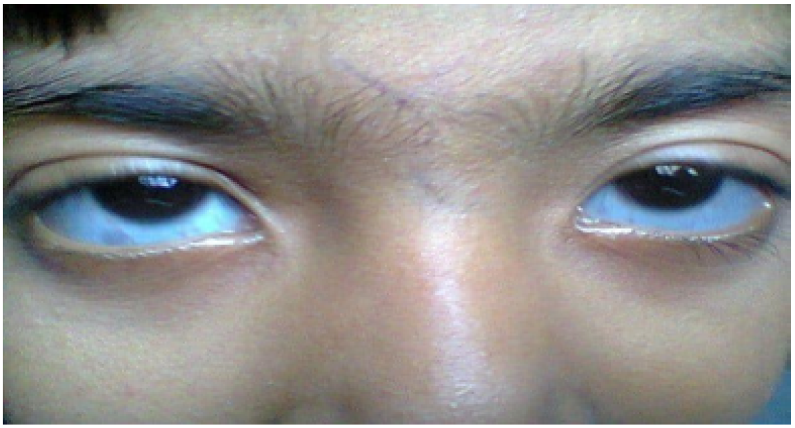
Figure 2: An 8-Year-Old School Going Boy Visited the OPD with Complaint of Blueness of his Eyes
His past history revealed that he had suffered from recurrent fractures of left tibia, left ankle and right femur since early childhood.
His milestones were timely and there were no other illnesses. This presentation is typical of Osteogenesis imperfecta, which is a genetic disease with a defect in Type I collagen
Case 3
A 38 years old woman came to the OPD with complaint of pain in lower back, both knee joints and wrist joints for last 6 years. During case taking and examination, it was seen that her sclera was bluish (Figure 3).
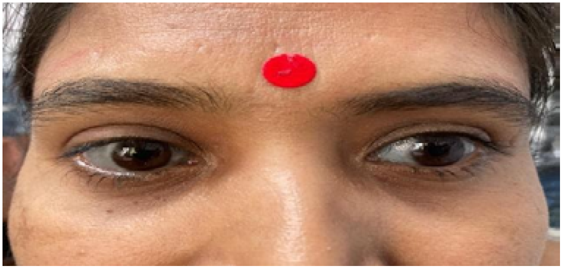
Figure 3: A 38 Years Old Woman Came to the OPD with Complaint of Pain in Lower Back, Both Knee Joints and Wrist Joints for Last 6 Years. During Case Taking and Examination, It Was Seen That Her Sclera Was Bluish
She was sent to an ophthalmologist for evaluation of any pathology in her eyes. Her visual acuity was normal with no other pathology in the eye. On detailed history taking, she gave a history of recurrent falls and fracture of different bones in her body. This fracture started from the age of 5 years. Her stature was also very short with short limbs. There was no evidence of osteoporosis. This was another case of Osteogenesis Imperfecta.
Case 4
A 5-year-old male child was brought to the OPD with complaint of frequent and easy fractures of bones for last 2 and half years. He fractured his ribs and elbow from minor injuries, He had no other complaints. His past history signified that he had suffered from breathing distress during birth. He was very lean and thin. His family history also revealed of no such history. The sclera of his eyes had a bluish tinge (Figure 4).
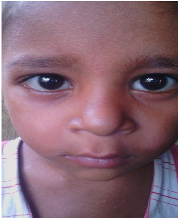
Figure 4: The Sclera of His Eyes had a Bluish Tinge
Thus, it was suspected as a case of OI.
Repertorial Study
For Blue Sclera:
Boenninghausen’s characteristics and repertory-EYE -Blue sclerotic–Ars, Ver.A [9]
Murphy’s repertory-EYE-Discolouration–Bluish, sclera-Ars, Calc, carc, Puls, Tub [10]
Murphy’s repertory–EYE–Blue, Discolouration-Sclera–Children–Carc [10]
Murphy’s repertory–EYE–Blue, Discolouration-Sclera–Porcelain like–Carc [10]
Complete Repertory–EYES–Blueness, Sclera–Ars, Bell, Calc, Carc, Cupr, Ferr-i, Plb, Tub, Verat [11]
Complete Repertory–EYES–Blueness, Sclera, Children–Carc, Cupr, Tub [11]
Complete Repertory–EYES–Blueness, Porcelain like, Sclera–Carc [11]
Knerr’s Repertory of Hering’s Guiding Symptoms–EYES–Sclerotica, Bluish–bell. [11]
For Osteogenesis Imperfecta
Kent’s repertory – GENERALITIES – Softening of bones – Bell, Calc, Merc, Puls [12].
Boenninghausen’s characteristics and repertory – SENSATION
& COMPLAINTS- Bones – brittle as if – CALC, LYC, MERC, Phos.ac, SIL, SUL. [9].
Murphy’s repertory – Bones – BRITTLE, bones – Bufo., Calc., Calc-f., CALC- P.,
Carc., Lac-ac., Sil., Symph [10].
Murphy’s repertory – Bones – OSTEOGENESIS, imperfect – Calc-f, Calc-p., Sil [10].
For Paget’s Disease
Murphy’s repertory – LIMBS – Paget’s Disease – Calc-p., Sil [10]
Murphy’s repertory – CLINICAL – Osteitis deformans, Paget’s Disease – Calc-p[10]
For Ehlers- Danlos Syndrome
Boenninghausen’s characteristics and repertory – UPPER EXTREMITIES – Loose, joints, shoulder – Croc, Kali-bi, Staph [9]
Murphy’s repertory – KNEES – Looseness, knee, joints – Phos, Ruta [10]
Complete Repertory – GENERALITIES – Loose, as, if, Joints – Aesc, Apis, Arg-n, Bry, Calc, Calen, Coca, Croc, Kali-bi, Lac-h, Laur, Lyss, M-p-a, Mez, Mobil-ph, Ozone, Phos, Sabad, Staph, Stram, Sumb, Thuj [11]
Phatak Repertory – Joints, Loose – Bov, Chel, Med, Ph-ac, Psor, Thuj [11]
Symptoms from Materia Medica
Arsenicum Album [13]: Blue colour around the eyes
Veratrum Album: Blueness of eyes [13], Eyes surrounded by blue or black rings [13], Blueness of the left eye with frequent eructation [14], Dull appearance of the eyes with blue rings around them [14]
Calcarea Carb: Bluish cornea or bluish spots on it [15].
Belladonna: Sclerotia bluish; livid lead-coloured spots on eyelids [15]
Cuprum Met: Eyes: sunken, with blue rings [15]
Plumbum Met: Disc of eye was prominent, it outlines hazy, its colour of an opaque bluish-white [15]. Deep bluish redness of sclerotia, most marked at corneal margin [13] (For bones related symptoms)
Calc Carb: Sensation as if right hip and thigh were brittle as well as short and small [15]. Bones soft, develop very slowly [16]
Mercurius: Softening of the bones, so that they can bend [13]
Lycopodium: Softening of bones; caries [15]
Silicea: Softening and ulceration of femur [13]
Sulphur: Curvature of spine, vertebrae softened [15]. Looseness in the knees, as if they would give way [17]
Calc Phos: Defective bone growth, bones thin and brittle [13]
Staphysagria: Dislocation pain in right shoulder joint, only on moving [13]
Crocus Sat: Pain on moving upper arm, as if head of humerus was loose and easily dislocated, cracking [15]
Declaration of Patient Consent
Appropriate patient consent forms have been signed by the patients and guardians in case of minors. They have given their consent for their images and clinical information to be used in the article. The patients understand that their names and initials won’t be published and their identity will be tried to be concealed, but anonymity cannot be guaranteed.
Prockop, D.J. and J.F. Bateman. “Heritable disorders of connective tissues.” Harrison’s Principles of Internal Medicine, edited by D.L. Kasper, et al., vol. 2, 19th ed., McGraw–Hill, 2015, p. 2507.
Rosenberg, A.E. “Bones, joints & soft tissue tumours.” Robbins Basic Pathology, edited by V. Kumar, et al., 9th ed., Elsevier, 2013, p. 767.
Gautieri, A. et al. “Molecular and mesoscale disease mechanisms of osteogenesis imperfecta.” Biophysical Journal, vol. 97, no. 3, 2009, pp. 857–865. https://doi.org/10.1016/j.bpj.2009.04.059
“Osteogenesis imperfecta foundation: Fast facts.” Osteogenesis Imperfecta Foundation, archived 2007.
NHS UK. “Ehlers-danlos syndrome.” May 2021, www.nhs.uk/conditions/ehlers-danlos-syndromes/
NHS UK. “Marfan Syndrome.” May 2021, www.nhs.uk/conditions/marfan-syndrome/
DBA UK. “The diamond blackfan anaemia charity.” May 2021, www.diamondblackfan.org.uk
Glorieux, F.H. et al. “Cyclic administration of pamidronate in children with severe osteogenesis imperfecta.” New England Journal of Medicine, vol. 339, 1998, pp. 947–952.
Boger, C.M. Boenninghausen’s Characteristics and Repertory. Reprint ed., B. Jain Publishers, 2002, p. 310.
Murphy, R. Homoeopathic Medical Repertory. 2nd rev. ed., IBPS, 2006, p. 539.
Hompath Zomeo 3.0. Repertory section.
Kent, J.T. Repertory of the Homoeopathic Materia Medica. Reprint of 6th American ed., B. Jain Publishers, 2006.
Clarke, J.H. A Dictionary of Practical Materia Medica. 34th impression, B. Jain Publishers, 2017.
Hahnemann, S. Materia Medica Pura. Vols. 1–2, 1st ed., April 2007, B. Jain Publishers.
Hering, C. The Guiding Symptoms of Our Materia Medica. B. Jain Publishers, November 2016.
Allen, H.C. Keynotes and Characteristics with Comparisons of Some of the Leading Remedies of the Materia Medica with Bowel Nosodes. Low price ed., 16th impression, B. Jain Publishers, 2017.
Hahnemann, S. The Chronic Diseases: Their Peculiar Nature and Their Homoeopathic Cure. 1st ed., April 2008, B. Jain Publishers.
