
+91 6002993949
submission@iarconsortium.org
Open Access
ISSN (Print) : 2709-3239
ISSN (Online) : 2709-3220
Ophthalmomyiasis is a rare ophthalmic condition characterized by larval infestation of eye and ocular adnexa due to accidental contact with gravid female flies. Based on infestation of ocular tissue by larva, ophthalmomyiasis is described as ophthalmomyiasis externa, ophthalmomyiasis interna and orbital myiasis. In this article we present the case study of a 25 year old male presenting to the eye OPD with foreign body sensation in right eye. We discuss the management of the cause of foreign body sensation.
Ophthalmomyiasis is a rare ophthalmic condition characterized by larval infestation of eye and ocular adnexa due to accidental contact with gravid female flies. [1]. It is mainly reported amongst people who have close association with animals such as shepherds, farmers and workers in animal husbandry [2]. Based on infestation of ocular tissue by larva, ophthalmomyiasis is described as ophthalmomyiasis externa, ophthalmomyiasis interna and orbital myiasis. When larvae are found on ocular surface and eyelids it is termed as ophthalmomyiasis externa. If there is involvement of anterior segment and posterior segment of eye due to globe penetration by larvae, it is described as ophthalmomyiasis interna and when larvae infestation involves the orbital structures it is called orbital myiasis [3]. Treatment of ophthalmomyiasis depends on the ocular tissue involved. Early diagnosis of ophthalmomyiasis and initiation of treatment helps in effective management and avoidance of complications.
A 25 year old patient presented to ophthalmology out-patient department of Civil Hospital Sarahan with the complaints of profuse watering and foreign body sensation in his right eye. Patient noticed these symptoms suddenly after waking up in morning. He gave history of working on farm with cattles and sheep. On examination, visual acuity was 6/6 (Snellens chart) for both eyes. Right upper eyelid was mildly odematous and mild conjunctival congestion was noted in right eye. Small, translucent larvae approx. 0.75 mm to 1 mm long with blackish cephalic ends were seen over the cornea, bulbar conjuctiva and some in the upper and lower fornix (Figure 1 and 2).
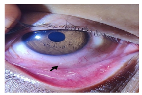
Figure 1: Larva Present Over Inferior Bulbar Conjunctiva at 6 O’clock Position
Figure 2: Larva Moving from Cornea Towards Bulbar Conjunctiva
On exposure to light larvae started moving towards fornix and burrowing into conjunctiva sac to escape. Larvae were immobilized by putting topical anesthetic drops (0.5% proparacaine) in right eye and a total of 9 larvae were mechanically removed with plain forceps. Patients was then put on moxifloxacin eyedrops. He was examined again next day to look for any residual larvae. No larva was found on examination and patient was asymptomatic. A larva removed from right eye was sent for microbiological examination and the report indicated first-stage larvae of Oestrous ovis.
The most common cause of external ophthalmomyiasis globally is Oestrus ovis and it affects human eye either by direct contact of gravid flies or by hands contaminated with larvae [4].
Ophthalmomyiasis is more common in rural areas of tropical and subtropical regions. Other factors that have been associated with ophthalmomyiasis are poor hygiene, immunocompromised patients and open wounds [5].
Patients of ophthalmomyiasis externa usually mimic catarrhal and adenoviral conjuntivitis and present as foreign body sensation, pain, watering, mucoid discharge, conjunctival congestion, eyelid edema, photophobia and sometimes punctate subconjunctival hemorrhages [6].
Detailed slit lamp examination is important for diagnosis giving special attention to fornices in all suspected cases because larvae of oestrus ovis hide in fornices as they are photophobic [7].
Mainstay of treatment in cases of ophthalmomyiasis externa is manual removal of larvae with plain forceps after instillation of topical anesthetic agent. Saline irrigation of conjunctival sac does not help in removal of larvae, as they are attached firmly to conjunctiva with their oral hooks. Topical antibiotic is given after manual removal of larvae to prevent secondary infections. Topical steroid treatment can be given in patients to suppress inflammation if necessary [8]. As these larvae tend to hide in conjunctival sac when exposed to light, an early follow-up examination is important to ensure there are no residual larvae and no risk of ophthalmomyiasis internal [9].
Sreejith R. et al. “Oestrus Ovis Ophthalmomyiasis with Keratitis.” Indian Journal of Medical Microbiology, vol. 28, no. 4, 2010, p. 399.
Pather S. et al. “Ophthalmomyiasis Externa: Case Report of the Clinicopathologic Features.” International Journal of Ophthalmic Pathology, vol. 2, no. 2, 2013.
Al-Amry M. et al. “External Ophthalmomyiasis: A Case Report.” Saudi Journal of Ophthalmology, vol. 28, no. 4, 2014, pp. 322-324.
Vogt R. et al. “Ophthalmomyiasis Externa.” Der Ophthalmologe, vol. 112, 2015, pp. 61-63.
Singh P. and K. Tripathy. Ophthalmomyiasis. StatPearls, StatPearls Publishing, 2021. https://www.ncbi.nlm.nih. gov/books/NBK560884/.
Istek Ş. “Ophthalmomyiasis Externa from Hakkari, the South East Border of Turkey.” Case Reports, 2014, bcr2013201226. https://doi.org/10.1136/bcr-2013-2012 26.
Wong D. “External Ophthalmomyiasis Caused by the Sheep Bot Oestrus Ovis L.” The British Journal of Ophthalmology, vol. 66, no. 12, 1982, p. 786.
Sundu C. et al. “Ophthalmomyiasis Externa: A Report of Three Cases.” Turkish Journal of Ophthalmology, vol. 45, no. 5, 2015, p. 220.
Choudhary P. et al. “Red Eye: Rule Out Ophthalmomyiasis Too.” Indian Journal of Ophthalmology, vol. 61, no. 6, 2013, p. 293.
