
+91 6002993949
submission@iarconsortium.org
Open Access
ISSN (Print) : 2709-3239
ISSN (Online) : 2709-3220
Poland syndrome is a rare congenital disorder typically characterized by unilateral absence or hypoplasia of the pectoralis major muscle, often accompanied by ipsilateral hand anomalies such as brachydactyly or syndactyly. While its clinical presentation is relatively well defined, associations with other congenital defects are uncommon. We report the case of an 18-year-old male who presented with left-sided chest wall asymmetry and brachydactyly, consistent with Poland syndrome, along with the novel finding of ipsilateral congenital ptosis—a feature not previously documented in the medical literature. Detailed physical, neurological, and ophthalmologic examinations, along with imaging studies, confirmed the diagnosis and ruled out other neurological etiologies. The absence of functional impairment led to conservative management with clinical monitoring. This case expands the known phenotypic spectrum of Poland syndrome and underscores the importance of thorough clinical evaluation in detecting atypical manifestations of rare congenital anomalies.
Poland syndrome is an uncommon congenital disorder marked by the partial or complete absence of the pectoralis major muscle on one side of the body, frequently accompanied by ipsilateral upper limb anomalies such as syndactyly (webbed fingers) or brachydactyly (shortened fingers). The condition predominantly affects males, with a male-to-female ratio of approximately 3:1, and commonly involves the right side of the body in sporadic cases. In contrast, familial cases tend to show no gender bias or consistent laterality. The estimated incidence is approximately 1 in 30,000 live births, placing it among the rarer congenital syndromes encountered in clinical practice [1-5].
The exact etiology of Poland syndrome remains unclear, but the most widely accepted theory involves a vascular disruption sequence during embryogenesis. Specifically, an interruption in the embryonic blood supply—most likely affecting the subclavian artery—may impair the development of the chest wall and adjacent musculoskeletal structures during the sixth week of gestation. Despite its distinct physical manifestations, the syndrome often goes undiagnosed until adolescence or adulthood when asymmetry of the chest wall becomes more apparent [2,5,6,7].
While the hallmark features of Poland syndrome are well characterized, its association with other congenital anomalies remains infrequently reported. This case report presents an 18-year-old male with typical features of Poland syndrome affecting the left side of his body, uniquely accompanied by congenital ptosis (drooping of the eyelid) on the same side. To the best of our knowledge, this is the first documented case reporting ipsilateral congenital ptosis as a possible component of the syndrome. Such atypical associations not only deepen our understanding of the clinical variability of Poland syndrome but also emphasize the importance of comprehensive physical examinations and multidisciplinary evaluation in cases presenting with congenital asymmetries.
An 18-year-old male presented to the Department of Pulmonary Medicine at Indira Gandhi Medical College, Shimla, with a primary complaint of visible chest wall asymmetry that he had noticed becoming more apparent during adolescence. The patient reported no pain, respiratory difficulty, or functional limitations associated with this asymmetry. There was no history of trauma, prior surgical procedures, or significant comorbidities. Notably, the patient’s family history was unremarkable, with no known congenital anomalies or similar deformities in first-degree relatives, suggesting a sporadic presentation.
On physical examination, a distinct flattening of the left anterior chest wall was observed, specifically involving the left infraclavicular and mammary regions. Palpation revealed an absence of muscle bulk in the area typically occupied by the sternal head of the pectoralis major muscle. The skin overlying the defect was normal in appearance, with no signs of scarring or prior intervention.
Further inspection of the left upper limb revealed ipsilateral brachydactyly, characterized by shortened phalanges in the fingers of the left hand, without syndactyly. The affected hand exhibited a mild degree of hypoplasia, although the patient retained full range of motion and normal grip strength. The right upper limb and chest were entirely normal in appearance and function.
An additional and noteworthy finding was the presence of left-sided congenital ptosis. The patient’s left upper eyelid exhibited significant drooping, partially covering the superior portion of the iris in the primary gaze position. The patient and his parents confirmed that this eyelid droop had been present since birth and had remained unchanged over time. There was no history of trauma or neurological illness associated with the ptosis. Importantly, the levator function was preserved, and there were no signs suggestive of third nerve palsy or Horner’s syndrome.
A comprehensive ophthalmologic examination was conducted and revealed no visual impairment. Pupillary size and reactivity were normal in both eyes, and ocular movements were full and symmetrical. A concurrent neurological examination was also normal, with no evidence of focal deficits, cranial nerve involvement, or muscle weakness elsewhere in the body.
Radiographic evaluation was performed to further assess the chest wall abnormality. A posteroanterior (PA) chest radiograph demonstrated increased translucency over the left upper thoracic region, suggestive of reduced soft tissue mass. The underlying bronchovascular markings and bony structures were otherwise unremarkable. No rib anomalies or scapular abnormalities were identified.
To more precisely delineate the anatomical defect, magnetic resonance imaging (MRI) of the thorax was obtained. The MRI confirmed the absence of the sternal head of the left pectoralis major muscle, while the clavicular head appeared to be intact. There was no evidence of any associated rib hypoplasia, scoliosis, or internal organ displacement.
Given the constellation of findings—including unilateral absence of the sternal head of the pectoralis major, ipsilateral brachydactyly, and congenital ptosis—the patient was diagnosed with Poland syndrome with an atypical presentation. The association of ipsilateral congenital ptosis has not been previously described in the literature, thus representing a novel phenotypic variant of the syndrome.
The patient was counseled extensively about the benign nature of the condition. As he was asymptomatic with no functional impairment and expressed no cosmetic concerns requiring correction, no surgical intervention was recommended. He and his family were reassured about the non-progressive nature of the syndrome and advised periodic clinical follow-up.
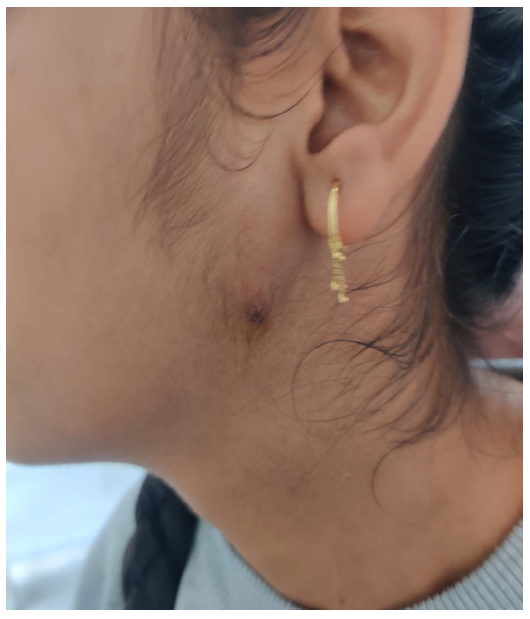
Figure 1: (a) Flattening of left infraclavicular and mammary area b) Brachydactyly (Left sided) c) Ptosis (congenital) of left eye
Figure 2: (a) Chest radiograph (PA) view showing translucency on left side with normal bronchovascular markings and (b) MRI chest showing absent sternal head of left pectoralis major muscle (Arrow)
Poland syndrome is an uncommon congenital anomaly first described by British surgeon Sir Alfred Poland in 1841, characterized by unilateral absence or underdevelopment of the pectoralis major muscle, often accompanied by ipsilateral upper limb deformities. Though its exact pathogenesis remains unclear, the prevailing hypothesis attributes the condition to a disruption in the embryonic blood supply—most notably involving the subclavian artery or its branches—during the sixth week of gestation. This interruption likely results in hypoplasia or aplasia of tissues dependent on this vascular territory, including the pectoral muscles, ribs, breast tissue, and digits of the upper limb.
In the present case, the patient demonstrated classic features of Poland syndrome, including the absence of the sternal head of the pectoralis major muscle and ipsilateral brachydactyly. These findings alone substantiate the diagnosis. However, the case is unique in its association with ipsilateral congenital ptosis—an anomaly not previously reported in the context of Poland syndrome to our knowledge. This expands the phenotypic spectrum of the condition and raises intriguing questions about its embryologic underpinnings.
The presence of ptosis, defined as drooping of the upper eyelid due to levator muscle dysfunction or innervation issues, introduces new complexity to the understanding of Poland syndrome. In this case, the ptosis was congenital, isolated, and ipsilateral to the musculoskeletal defects. Neurological and ophthalmologic evaluations ruled out common differential diagnoses such as third cranial nerve palsy, Horner’s syndrome, or myasthenia gravis. Notably, the levator function and ocular motility were preserved, supporting a primary structural or developmental etiology.
It is conceivable that the ptosis observed in this patient may share a common vascular or neurovascular disruption with the other manifestations of Poland syndrome. While traditional vascular disruption theory explains the musculoskeletal defects, it does not typically extend to craniofacial anomalies. However, some studies have linked Poland syndrome with broader vascular disruption sequences, such as the "subclavian artery supply disruption sequence" (SASDS), which includes Klippel-Feil and Möbius syndromes. These conditions may involve ocular or cranial nerve abnormalities due to early embryonic vascular compromise, suggesting a broader field of embryologic vulnerability. The occurrence of ptosis in our patient may thus represent an extension of the SASDS concept, implicating a more widespread disturbance in regional development than previously appreciated [3,5,6].
From a clinical management perspective, Poland syndrome is often diagnosed late due to the subtlety of physical signs in early childhood and the absence of functional impairment in many cases. Imaging, particularly MRI, plays a vital role in confirming muscle aplasia and evaluating the extent of associated anomalies [7,8,9]. In our case, the absence of significant symptoms, normal pulmonary and limb function, and lack of cosmetic concern meant that no surgical intervention was necessary. However, surgical options such as muscle transposition or implant-based reconstruction may be considered in patients with functional deficits or significant asymmetry affecting quality of life.
The psychosocial impact of such congenital anomalies, even when mild, must not be underestimated. While our patient demonstrated no functional limitations or psychological distress, other individuals may experience body image concerns, particularly during adolescence. Comprehensive care should include psychological counseling when indicated, and interdisciplinary coordination among pulmonologists, plastic surgeons, neurologists, and ophthalmologists is essential for holistic management [7-10].
Importantly, the inclusion of ptosis in this case underscores the value of thorough systemic examination and documentation in all congenital presentations. It also highlights the potential for underrecognized associations in rare syndromes, which may only come to light through careful observation and case reporting. This case therefore not only contributes a novel clinical insight but also serves as a reminder of the variability and complexity inherent in congenital disorders like Poland syndrome.
This case highlights a rare and atypical presentation of Poland syndrome in an 18-year-old male, marked not only by the classical features of unilateral pectoralis major aplasia and ipsilateral brachydactyly, but also by the novel association of ipsilateral congenital ptosis—a finding not previously reported in the literature. This unusual constellation broadens the phenotypic spectrum of Poland syndrome and suggests the possibility of a more extensive embryological disruption than traditionally recognized. Through detailed clinical examination and imaging, the diagnosis was established, and unnecessary interventions were avoided due to the absence of functional impairment. This case underscores the importance of vigilant physical assessment and comprehensive documentation in congenital syndromes, and it contributes to the evolving understanding of the variable manifestations of Poland syndrome.
Rajawat, G.S., N. Khippal, and R.P. Takhar. "Poland Syndrome with Associated Synbrachydactyly and Dextrocardia: A Rare Case." International Journal of Research in Medical Sciences, vol. 6, 2018, pp. 2860–2862.
Bavinck, J.N., and D.D. Weaver. "Subclavian Artery Supply Disruption Sequence: Hypothesis of a Vascular Etiology of Poland, Klippel-Fiel, and Mobius Anomalies." American Journal of Medical Genetics, vol. 23, 1986, pp. 903–918.
Poudel, S., et al. "Right-Sided Poland Syndrome with No Classical Hand Deformity: A Case Report." Radiology Case Reports, vol. 20, no. 1, Jan. 2025, pp. 515–520.
Baldelli, I., A. Baccarani, C. Barone, et al. "Consensus Based Recommendations for Diagnosis and Medical Management of Poland Syndrome (Sequence)." Orphanet Journal of Rare Diseases, vol. 15, 2020, Article ID 201.
Hashim, E.A.A., B.H. Quek, and S. Chandran. "A Narrative Review of Poland’s Syndrome: Theories of Its Genesis, Evolution and Its Diagnosis and Treatment." Translational Pediatrics, vol. 10, no. 4, 2021, pp. 1008–1019.
Dustagheer, S., M.H. Basheer, A. Collins, and C. Hill. "Further Support for the Vascular Aetiology of Poland Syndrome—A Case Report." Journal of Plastic, Reconstructive & Aesthetic Surgery, vol. 62, no. 10, Oct. 2009, pp. e360–e361.
Hashim, E.A.A., B.H. Quek, and S. Chandran. "A Narrative Review of Poland’s Syndrome: Theories of Its Genesis, Evolution and Its Diagnosis and Treatment." Translational Pediatrics, vol. 10, no. 4, Apr. 2021, pp. 1008–1019.
Geeroms, B., L. Breysem, and M. Aertsen. "An Atypical Case of Poland Syndrome with Bilateral Features and Dextroposition of the Heart." Journal of the Belgian Society of Radiology, vol. 103, no. 1, 2019, Article ID 45.
Haider Khan, Zainab, and Suma Kaza. "Poland Syndrome Presenting with Failure to Thrive: A Case Report." Journal of Pediatrics, Perinatology and Child Health, vol. 8, 2024, pp. 95–97.
Ramakrishnan, K.K., S. Sharmeela, B.R. Govindarajan, and S.G. Subramonian. "Understanding Poland Syndrome: A Collaborative Approach to Patient Care." Romanian Journal of Pediatrics, vol. 73, no. 3, Jan. 2025, pp. 235–241.
