+91 6002993949
submission@iarconsortium.org
Open Access
ISSN (Print) : 2789-6056
ISSN (Online) : 2789-6064
Antimicrobial susceptibility testing (AST) is a laboratory procedure performed by medical technologists to identify which antimicrobial regimen is specifically effective for individual patients. On a larger scale, it aids in the evaluation of treatment services provided by hospitals, clinics, and national programs for the control and prevention of infectious diseases. Clinical laboratories currently employ several methods depending on the laboratory test menu that they provide. These approaches include the disk diffusion and minimum inhibitory concentration (MIC) methods. Commercial systems also became available across health centers and hospital facilities, utilizing both phenotypic and genotypic characterization of bacterial resistance. While routine antimicrobial susceptibility testing for gram-positive (e.g., Staphylococcus aureus) and gram-negative bacteria (e.g., Pseudomonas aeruginosa) are commonly available in peripheral laboratories, drug susceptibility testing (DST) for Mycobacterium tuberculosis is usually carried out within more complex facilities like reference laboratories. Despite the differences in the techniques for susceptibility tests, all laboratories must be critical on each step of the sampling and testing process so that test results are obtainable with consistently high levels of accuracy and reliability. In this article, we elaborate the importance of AST, Standardization of Antimicrobial Sensitivity Testing (AST) Methods, Various types of AST including Disk Diffusion Method (Kirby Bauer method and Stokes method), Dilution methods (Broth dilution method and Agar dilution method), Epsilometer test and Automated Antimicrobial Susceptibility Test Systems (Vitek 2 systems, The MicroScan WalkAway system and The Phoenix system).
Antimicrobial sensitivity testing is done to provide testing of bacterial pathogen to set of available antibiotics to determine its susceptibility profile and so predict the effectiveness of the antibiotic regimen and secondly to assess the effectiveness of the treatment regimen [1].
Purposes
To guide the clinician in selecting the best antibiotic agent for an individual patient
To control the use of antibiotics in clinical practice
To accumulate epidemiological information on the resistance of microorganisms of public health importance within the community
To reveal the changing trends in the local isolates
Improved patient outcomes
Lower mortality rates
Less diagnostic procedures ordered including Laboratory tests and Diagnostic imaging procedures
Shortened ICU stays
Decreased ventilator days
Fewer prescriptions for inadequate antibiotic therapy
Shortened length of stay
Lower total cost of hospitalization
Testing Methods
Three general methods are available to detect and evaluate antimicrobial susceptibility
Methods that directly measure the activity of one or more antimicrobial agents against a bacterial isolate
Methods that directly detect the presence of a specific resistance mechanism in a bacterial isolate
Special methods that measure complex antimicrobial organism interactions
The method used depends on factors such as clinical need, accuracy, and convenience. Given the complexities of antimicrobial resistance patterns, a laboratory may commonly use methods from more than one category
Definitions of Susceptibility Testing Interpretive Categories
Susceptible (S) Indicates that the antimicrobial agent in question may be an appropriate choice for treating the infection caused by the organism. Bacterial resistance is absent or at a clinically insignificant level.
Resistant (R) Indicates that the antimicrobial agent in question may not be an appropriate choice for treatment, either because the organism is not inhibited with serum-achievable levels of the drug or because the test result highly correlates with a resistance mechanism that indicates questionable successful treatment
Intermediate (I) Indicates a number of possibilities, including:
The potential utility of the antimicrobial agent in body sites where it may be concentrated (e.g., the urinary tract) or if high concentrations of the drug are used
Possible effectiveness of the antimicrobial agent against the isolate, but possibly less so than against a susceptible isolate
Use as an interpretive safety margin to prevent relatively small changes in test results from leading to major swings in interpretive category (e.g., resistant to susceptible or vice versa)
Standardization of Antimicrobial Sensitivity Testing (AST) Methods
Growth medium (typically a Mueller-Hinton base)
Bacterial inoculum size
pH
Cation concentration
Blood and serum supplements
Thymidine content
Incubation atmosphere
Incubation temperature
Incubation duration
Antimicrobial concentrations
Media
Best Medium – MHA (Mueller Hinton Agar)
Shows acceptable batch to batch reproducibility for susceptibility testing
Low in sulphonamide, trimethoprim and tetracycline inhibitors
Gives satisfactory growth of most non fastidious pathogens
A large body of data and experience has been collected concerning susceptibility tests performed with this medium
MHA (Mueller Hinton Agar)
Cool the medium to 45–50 ⁰C and pour into the plates. Allow to a depth of approximately 4 mm
A 9-cm plate requires approximately 25 ml of medium
When the agar has solidified, for 10–30 minutes at 35 ⁰C by placing them in the upright position in the incubator with the lids tilted
If it is not to be used immediately, the agar medium can be stored in a refrigerator (2 to 8degree C) for 2 weeks
A supply of cotton wool swabs on wooden applicator sticks should be prepared [2]
They can be sterilized in tins, culture tubes, or on paper, either in the autoclave or by dry heat
Inoculum
Inoculum is usually maded from broth culture that has been incubated for 4 to 6 hours
The density of the suspension is adjusted to approximately 108 colony-forming units(CFUs) per milliliter by comparing its turbidity to a McFarland 0.5 BaSO4 standard
Commonly, the degree of cloudiness in the broth is compared with the standard, visualizing the two against a white background on which black lines have been drawn
Nephelometers may be used to determine turbidity
Turbidity standard is checked by spectrophotometer – 625nm – absorbance should be 0.008 to 0.10
Seal the tubes containing the Mcfarland standards and store in dark at RT
The standard turbidity should be mixed throroughly every time before use
Check the density monthly and replace it monthly
Ph
Tests are performed using media for which the pH has been standardized at physiologic levels.
Media pH should be between 7.2 and 7.4 at room temperature.
The pH of broth media may be tested directly with a pH electrode, and agar media may be tested by macerating enough of the agar so that the tip of the electrode can be submerged, by allowing a portion of agar to solidify around the electrode, or by using a properly calibrated surface electrode
Cation Concentration
Agar and broth media varygreatly in the concentration of divalent cations [3].
By convention, antibiotic susceptibility testing is done under physiologic conditions.
MHA has very low concentrations of divalent cations, but is adjusted to physiologic concentrations (20 to 35 mg/L Mg2+ and 50 to 100 mg/L Ca2+) during production
Serum
Antibiotics differ greatly in the degree to which they bind to proteins. In the bloodstream, free antibiotic is in equilibrium with serum protein-bound antibiotic
Free and protein-bound antibiotic can be measured, but it is not clear which is the more useful result
The CLSI method does not include added serum because of the difficulty in standardization of the product and uncertainty about how to interpret the results
Environmental Conditions
Antibiotic susceptibility tests are routinely incubated in ambient air at 35°C. Agar or broth is incubated in an ambient air incubator.
Incubation times vary depending on the test system used. The recommended incubation time for conventional disk diffusion systems is 16 to 18 hours, while that for dilution tests is longer at 16 to 20 hours
However, tests to determine the MICs of staphylococci for oxacillin and vancomycin, and the MIC of enterococci for vancomycin should be incubated a full 24 hours [4]
Selection of Antimicrobial Agents for Testing
The antimicrobial agents chosen for testing against a particular bacterial isolate are referred to as the antimicrobial battery or panel
A laboratory may use different testing batteries, but the content and application of each battery are based on specific criteria
Final decision should not be made independently by the laboratory; input from the medical staff (particularly infectious diseases specialists) and pharmacists (e.g., a Pharmacy and Therapeutics committee) is imperative
Specific Criteria
Organism Identification or Group: certain antimicrobials were developed specifically for use against particular organisms, but not against others (e.g., ceftazidime for use against Pseudomonas aeruginosa but not against Staphylococcus aureus); such agents should be included only in the appropriate battery.
Acquired Resistance Patterns Common to Local Microbial Flora.
Depending on the testing method, some agents do not reliably detect resistance and should not be included in the battery.
Site of Infection.
Availability of Antimicrobial Agents in the Formulary
CLSI Grouping of Recommended Antimicrobial Agents for Testing and Reporting
Antibiotic Stock Solutions
Buy commercial pure source of antibiotics
Don’t use injectable solutions
Accurate weighing of powders is must
Standard strains of stock cultures should be used to evaluate the stock solution After preparing the stock solution, make 5 ml aliquots and should be frozen.
Calculation of stock solution
1000 * V * C = W
P
Dried Filter Paper Discs
Whatmann no.1 filter paper is made to form a disc size of 6mm
Keep in petridish and sterilize in a hot air oven
With the help of antibiotic delivery loop which has a 20G wire with diameter of 2mm
The antibiotic is delivered (0.005ml)
Storage of discs
Refrigerate at 2 to 8 deg C
Beta lactam class drugs should be frozen
The drugs should be kept outside at Room Temperature 1 to 2 hours before work
The dispensing apparatus which is used to deliver the drugs should also be refrigerated
Check the expiry of drugs
AST Types (figure-1)
Qualitative: For the testing of isolates from “healthy” patients with intact immune defenses. – For such as uncomplicated urinary tract infections
Quantitaive: In the treatment of serious infections such as endocarditis or osteomyelitis. – For infections in high-risk patient groups such as immune compromised patients (e.g. transplant patients). Those who are critically ill
Disk Diffusion Method
It is the Simplest and most convenient method widely used everywhere and Developed by Kirby, Sherrris, Bauer and Turk in 1966. Types are Kirby Bauer method and Stokes method

Figure 1: Types of Antimicrobial Sensitivity Testing

Figure 2: Kirby Bauer Method
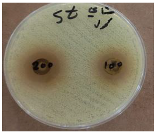
Figure 3: Broth Dilution Method
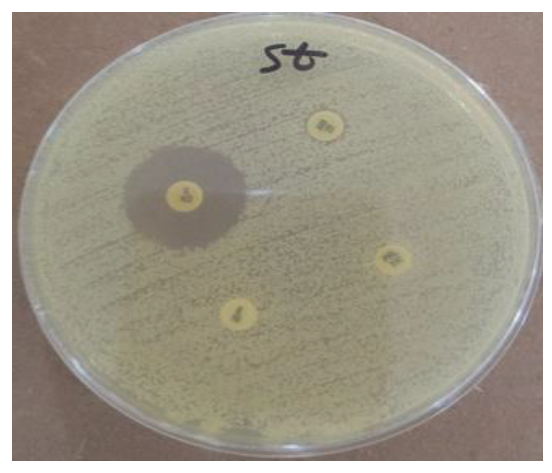
Figure 4: Broth Dilution Method
Figure 5: Agar Dilution Method
Figure 6: Epsilometer Test

Figure 7: Vitek 2
Kirby Bauer Method
Application of Discs
150 mm plate - 12 discs
100 mm plate - 6 discs
Drug should not be relocated
Distance from the lid edge - 15mm
Distance between two drug from center to center - 24mm
Inoculum - disc placement - incubation (only 15 minutes delay is acceptable each) [5]
Procedure
To prepare the inoculum from the primary culture plate, touch the top of the colony 3 to 5, of similar appearance, of the organism to be tested
Transfer this growth to a tube of saline. Compare the tube with the turbidity standard and of the test suspension to that of the standard by adding more bacteria or more sterile saline
Inoculate the plates by dipping a sterile swab into the inoculum. Remove excess inoculum by pressing and rotating the swab firmly against the side of the tube above the level of the liquid
Streak the swab all over the surface of the medium three times, rotating the plate through an angle of 60⁰ after each application.Finally, pass the swab round the edge of the agar surface
Leave the inoculum to for a few minutes at room temperature with the lid closed. They may be placed on the inoculated plates using sterile forceps, a template, a sterile needle tip and antibiotic disc dispenser
The plates should be placed in an incubator within 30 minutes of preparation. Temperatures above 35 degree celsius invalidate results for oxacillin/methicillin. Disks should not be moved after diffusion
Interpretation/Results
A dark background and reflected light are used to examine disk diffusion plates. The plate is situated so that a ruler or caliper can be used to measure the inhibition zone diameters for each antimicrobial agent.In instances not involving swarming organisms or the testing of sulfonamides and trimethoprim, hazes of growth that occur in more obvious inhibition zones should not be ignored. Interpretation should be done after 16 to 18 hours, Confluent lawn of growth, Zones of inhibition - uniformly circular and read with reflected light but For MRSA - read in transmitted light. When proteus is tested the thin veil of swarming growth after the zone of inhibition should be ignored.
Stokes Method
Built in controls against many variables and provide dependable results
A standard sensitive strain of the bacterium is inoculated in the middle third of the culture plate
The test bacterium is inoculated in the upper and lower third of the plate
Antibiotic discs are placed between the standard and test inocula so that zones of inhibition formed around each disc are composed of standard and test bacteria
The results are reported as Susceptible, Intermediate , Resistant
Dilution Methods
In this, the minimum concentration of antimicrobial to inhibit or to kill the microorganism is determined. MIC is the lowest concentration of antimicrobial that will inhibit the visible growth of an organism in an ideal growth condition while MBC is the least concentration of the test antibiotic which will completely kill the bacteria tested. It is of two types, Broth dilution method and Agar dilution method
Broth Dilution Method
The Mueller-Hinton preparation is the standard medium used for most broth dilution testing, and conditions in the medium (e.g., pH, cation concentration, thymidine content) are well controlled by commercial manufacturers. Broth dilution testing is divided into two general categories: microdilution and macrodilution. The principle of each test is the same; the only difference is the volume of broth in which the test is performed. For microdilution testing, the total broth volume is 0.05 to 0.1 mL; for macrodilution testing, the broth volumes are usually 1 mL or greater. Because most susceptibility test batteries require testing of several antibiotics at several different concentrations, the smaller volume used in microdilution allows this to be conveniently accomplished in a single microtiter tray
Broth dilution
Media: cation adjusted MH broth with a pH of 7.2 to 7.4
For H.influenzae – HTM
For TMP-SMX – Thymidine free medium
For oxacillin Resistance – MH broth with 2% NaCl
Working Antibiotic Solution
Take original stock solution
Prepare stock dilutions of the antibiotic of concentrations 1000 and 100ug/ml
Arrange two rows of 12 sterile 7.5*1.3cm capped tubes in the rack
Take a 30ml universal screw capped bottle
Add 8ml broth and required antibiotic concentration and mix the contents
Take 2ml + 2ml and put it in first tube in both rows Remaining 4ml broth - add 4ml fresh broth
Transfer 2ml+2ml and put in second tube
Continue this dilution upto 11 tubes
12th tube acts as control.
Inoculation
Inoculation of 1st row with one drop of overnight broth culture 1 in 1000 dilutions
2nd row with control with known sensitivity organisms
Final inoculum = 5 * 105 cfu/ml
Incubate at 37degC for 16 to 18 hours
Inoculate another tube with 2ml broth and keep at 4degC in a refrigerator overnight to be used as standard for determinattion of complete inhibition
Interpretation
Positive control tube – turbidity
Negative control tube – clear
MIC end point is read
Agar Dilution Method
1ml concentration of the drug + 24 ml of MHA
Inoculum – make 1:10 dilution of 0.5Mcfarland standard inoculum which delivers 107 cfu/ml
Using pipette or calibrated loop , deliver 0.001 ml on the surface of the agar giving the final inoculum of 104 cfu /spot
Inoculate a control plate
Interpretation
Examine the drug free control growth of the test organism for viability and purity
Place the plate on a dark background and examine them for the lowest concentration that inhibits visible growth
A single colony of a faint haze is not recorded as growth
Epsilometer Test
An exponential gradient testing methodology
A predefined stable antimicrobial gradient is present on a thin inert carrier strip
Following incubation, the E strip releases drug and a symmetrical inhibition ellipse is produced
MIC = intersection of the inhibitory zone edge and the calibrated carrier strip
Automated Antimicrobial Susceptibility Test Systems
Vitek 2 systems (bioMérieux, Inc., Durham, NC)
The MicroScan WalkAway system (Beckman Coulter, Inc., Brea, CA)
The Phoenix system (BD Microbiology Systems, Sparks, MD)
Vitek 2
AST inoculum is automatically introduced by a filling tube into a miniaturized, plastic, 64-well, closed card containing specified concentrations of antibiotics.
Cards are incubated in a temperature-controlled compartment.
Optical readings are performed every 15 minutes to measure the light transmitted through each well, including a growth control well.
Algorithmic analysis of the growth kinetics in each well is performed by the system’s software to derive the MIC data.
The MIC results are validated with the Advanced Expert System (AES) software, a category interpretation is assigned, and the organism’s antimicrobial resistance patterns are reported.
Resistance detection is enhanced with the sophisticated AES software, which can recognize and report resistance patterns using MICs. In summary, this system facilitates standardized susceptibility testing in a closed environment with validated results and recognition of an organism’s antimicrobial resistance mechanism in 6 to 8 hours for most clinically relevant bacteria
MicroScan WalkAway System
It uses the broth microdilution panel format manually inoculated with a multiprong device. Inoculated panels are placed in an incubator-reader unit, where they are incubated for the required time, and then the growth patterns are automatically read and interpreted. Depending on the microdilution tray used, bacterial growth may be detected using spectrophotometry or fluorometry
Spectrophotometric analyzed panels require overnight incubation, and the growth patterns may be read manually
Fluorometric analysis is based on the degradation of fluorogenic substrates by viable bacteria. The fluorogenic approach can provide susceptibility results in 3.5 to 5.5 hours. Either full dilution schemes or breakpoint panels are available
In addition to speed and facilitation of workflow, the automated systems provide increasingly powerful computer-based data management that can be used to evaluate the accuracy of results, manage larger databases, and interface with the pharmacy to improve and advance the utility of antimicrobial susceptibility testing data
Phoenix System
The Phoenix system provides a convenient, albeit manual, gravity-based inoculation process. Growth is monitored in an automated fashion based on a redox indicator system, with results available in 8 to 12 hours. Supplemental testing (e.g., confirmatory extended-spectrum beta-lactamase [ESBL] test for E. coli) is included in each panel, reducing the need for additional or repeat testing. Interpretation of results is augmented by a rules-based data management expert system.
Wikipedia. "Antibiotic sensitivity testing." Available at: https://en.wikipedia.org/wiki/Antibiotic_sensitivity_testing (Accessed on 16 June 2022).
MedlinePlus. "Antibiotic sensitivity test." Available at: https://medlineplus.gov/lab-tests/antibiotic-sensitivity-test/ (Accessed on 17 June 2022).
Healthline. "Sensitivity analysis." Available at: https://www.healthline.com/health/sensitivity-analysis (Accessed on 18 June 2022).
Testing.com. "Antibiotic susceptibility testing." Available at:https://www.testing.com/tests/antibiotic-susceptibi lity-testing/ (Accessed on 19 June 2022).
TMCC. "Antimicrobial susceptibility testing." Available at: https://www.tmcc.edu/microbiology-resource-center /lab-protocols/antimicrobial-susceptibility-testing (Accessed on 17 June 2022).
