Breast Cancer (BC) is a major health issue that affects millions of people around the world. The NF-κB gene signaling plays a critical action in the processes of initiation, progression and metastasis. In addition, miR-34a gene plays a major role in the CD4+ T cell activity of the Triple-Negative BC (TNBC), an unusual type of BC. The current study was conducted to evaluate the levels of mRNA Gene expression of NF-κB and miR-34a genes in human patients with BC. The study was performed on 16 blood samples from confirmed cases of BCs. Some other 16 blood samples from healthy individuals also were collected. The samples were subjected to a quantitative real-time PCR (qRT-PCR) method that targeted both NF-κB and miR-34a genes to evaluate levels of produced mRNA of these genes. The results demonstrated that the mRNA levels for the NF-κB gene significantly (p<0.01) decreased (0.04-fold change) in patients in a comparison with those from control group (0.23-fold change). For the miR-34a gene, the findings revealed that the mRNA levels significantly (p<0.01) increased (0.74-fold change) in patients compared to those from the control group (0.37-fold change). These results suggest important roles of the NF-κB and miR-34a genes in human patients with BC as it may refer to increases in resistance to chemotherapy, especially in the case of miR-34a gene, which needs further future investigation of their roles in the survivability and resistance to chemotherapy.
Carcinogenesis, defined by six primary hallmarks, has the potential to manifest in any cellular, tissue, or organ context, thereby inducing pathological changes that give rise to a wide range of cancer types [1]. The primary mechanisms facilitating its progression encompass evasion of programmed cell death, unrestricted proliferative potential, augmented formation of new blood vessels, insensitivity to inhibitory signals for growth, self-stimulation of growth signals and the ability to spread to distant sites [2]. The process of carcinogenesis is complex and influenced by multiple factors, with genetic susceptibility and environmental causes playing significant roles. The incidence of cancer-related mortality exhibits a concerning upward trend, positioning it among the prominent contributors to global mortality rates. While it is true that a considerable proportion of cancer cases may not inevitably lead to mortality, they do have a substantial negative impact on the overall quality of life and necessitate substantial financial resources [1].
In addition to its prevalence, BC stands as the primary contributor to cancer-related mortality among women on a global scale. On a global scale, BC accounted for a total of 684,996 deaths, with a 95% uncertainty interval ranging from 675,493 to 694,633. The age-adjusted rate of BC mortality was estimated to be 13.6 per 100,000 individuals. This information is supported by reference. While developed regions exhibited the highest incidence rates, it is noteworthy that Asian and African countries collectively accounted for 63% of the total deaths in the year 2020. The survival rates for women diagnosed with BC differ significantly between high- and low-income countries [3,4].
The global BC Mortality-to-Incidence Ratio (MIR) in 2020, which serves as an appropriate measure of 5-year survival rates, was reported as 0.30. When considering the clinical scope of BC, it has been observed that in regions with well-established healthcare systems such as Hong Kong and Turkey, the five-year survival rates for restricted BC were found to be 89.6%, while for geographic region cancer, the survival rates were 75.4% [4–6].
The nuclear factor kappa-light-chain-enhancer of activated B cells (NF-κB) is a collection of transcription factors that have a significant impact on the regulation of inflammatory signaling pathways [7–9]. The irregularities of NF-κB signaling have been found to be associated with immune disorders and cancer, making this pathway crucial in the processes of cancer formation, oncologic expansion and the establishment of therapeutic interventions [10–12]. The function of CD4+ T cells in TNBC, an unusual type of BC. can potentially be regulated by miR-34a, as indicated by recent research. This regulatory mechanism involves the regulation of gene expression, specifically targeting genes that have an impact on T-cell infiltration into cancers [13,14].
BC is a major health issue that affects millions of people around the world. The NF-κB gene signaling plays a critical action in the processes of initiation, progression and metastasis. In addition, miR-34a gene plays a major role in the CD4+ T cell activity of the TNBC [13,15–18]. The current study was conducted to evaluate the levels of mRNA Gene expression of NF-κB and miR-34a genes in human patients with BC.
Samples
The current study was conducted to evaluate the levels of mRNA Gene expression of NF-κB and miR-34a genes in human patients with BC. The study was performed on 16 blood samples from confirmed cases of BCs. Some other 16 blood samples from healthy individuals also were collected.
Level of mRNA
Total RNA Extraction: The total RNA was extracted using the AddBIO kit (Korea) and following the its protocol. Briefly, 200μl blood was used in 1ml Lysis Buffer. The resulted RNA was evaluated using a Quantus™ Fluorometer (Promega, USA). The RNA was stored under -80°C for later Lab work.
Synthesis of cDNA
The cDNA was synthetized by using the Add BIO kit (Korea) and following its procedure as follows: in a 20µl-total volume, 2μl, 10μl, 2μl, 1μl and 5μl (100ng) of H2O, 2X add script cDNA, dNTPs, random oligos hexamer and RNA, respectively, were mixed in a reaction tube. The thermal cycler conditions included priming, Reverse Transcriptase (RT) and RT inactivation at 25°C, 50°C and 80°C for 10mins, 60mins and 5mins, respectively.
QRT-PCR
The samples were subjected to a qRT-PCR method by using the AddScript RT-qPCR Syber master kit (AddBio, Korea) and following its instructions. In a 20µl-total reaction, H2O, AddScript RT-qPCR, each primer sequence (direction) (0.05pmol/20μl) and cDNA at 3μl, 10μl, 2μl and 3μl, respectively. The thermal cycler included the use of the following conditions: one-repeat activation, one-repeat initial denaturation, 40x-repeat denaturation, 40x-repeat annealing, 40x-repeat extension, one-repeat melting analysis, one-repeat melting analysis and melting analysis at 50°C, 95°C, 95°C, 60°C, 60°C, 95°C, 60°C, +0.3°C of 95°C for 2mins, 5mins, 15s, 60s, 35s, 15s, 60s and 15s, respectively.
These methods targeted both NF-κB and miR-34a genes to evaluate levels of produced mRNA of these genes and GABDH gene as a housekeeping gene. The primer sequences were F: AAGACCCACCCCACCATCAA and R: AAACTGTGGATGCAGCAGCGGTC [19], F: AGGGTGGCAGTGTCTTAGC and R: GAGCAGGGTCCGAGGT [20] and F: GAAGGTGAAGGTCGGAGTCA and R: TTGAGGTCAATGAAGGGGTC, respectively. For the normalization process, the 2-∆∆CT method was employed as described by Schmittgen and Livak [21].
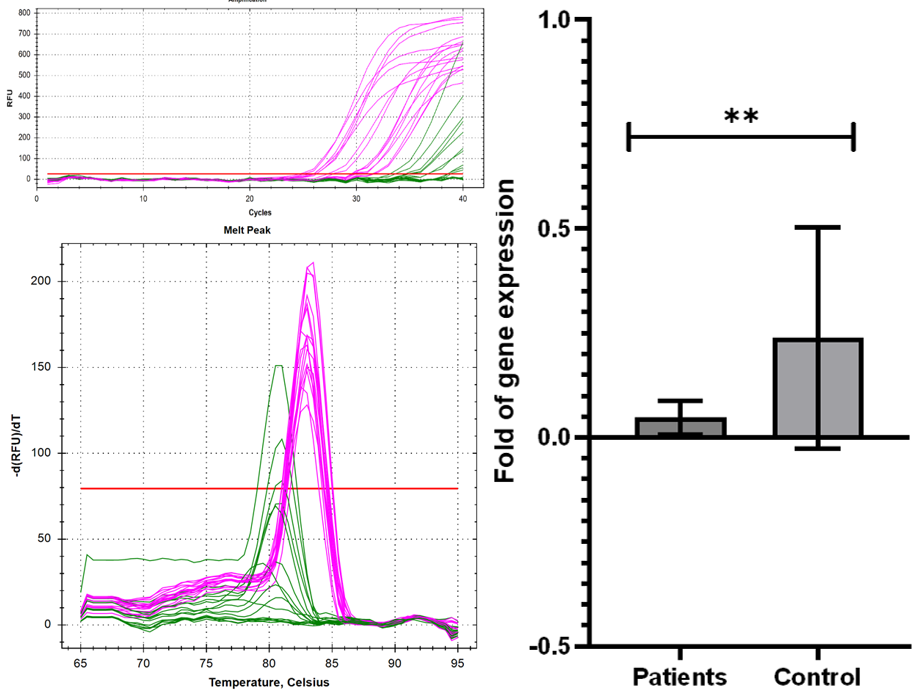
The results demonstrated that the mRNA levels for the NF-κB gene significantly (p<0.01) decreased (0.04-fold change) in patients in a comparison with those from control group (0.23-fold change) (Figure 1).

Figure 1: NF-κB Gene mRNA Expression in Blood Samples of Breast Cancer Patients. A: Amplification Curve. B: Melting Curve. C: Fold Change of NF-κB Expression
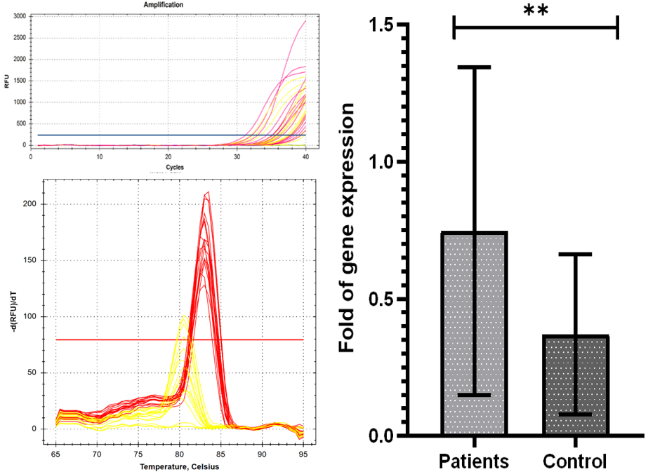
For the miR-34a gene, the findings revealed that the mRNA levels significantly (p<0.01) increased (0.74-fold change) in patients compared to those from the control group (0.37-fold change) (Figure 2).

Figure 2: miR-34a Gene mRNA Expression in Blood Samples of Breast Cancer Patients. A: Amplification Curve. B: Melting Curve. C: Fold Change of NF-κB Expression
The current study found declines in the levels of NF-κB mRNA in BC patients. This could be as a result to the ongoing treatment in these patients. In addition, the suppression of NF-κB/p65 using dehydro-xymethylepoxy-quinomicin (DHMEQ) resulted in a reduction in the migratory and invasive capabilities of MDA-MB-231 and HCC-1954 human BC cell lines, as reported in a previous study [22]. Furthermore, these findings were corroborated by another study, which demonstrated that DHMEQ effectively suppressed the three-dimensional invasion of BC. This inhibitory effect was attributed to the suppression of matrix metalloproteinase, an essential peptidase involved in the breakdown of the extracellular structure within the cancer microenvironment. Additionally, DHMEQ was found to inhibit interleukin-6 (IL-6) activity [23].
The NF-κB signaling pathway is known to exert significant influence in the processes of cancer beginning, development and metastasis. The activation of NF-κB by RANKL in breast cancer leads to cellular proliferation through the targeting of the cyclin D1 gene. The NF-κB pathway plays a crucial role in promoting cell survival by upregulating inhibitors of apoptosis and Bcl-xL. Regarding ovarian cancer, the activation of NF-κB leads to the activation of the oncogene PI3K-110α, which subsequently results in an increase in p65RelA. This rise in p65RelA relates to heightened chemoresistance and unfavorable prognoses in individuals diagnosed with epithelial ovarian cancer [24,25]. Moreover, it has been observed that individuals with BRCA1 mutations who are also impacted by NF-κB signaling exhibit elevated incidences of breast and ovarian cancer. This line of reasoning justifies the need for further exploration of immunotherapy approaches, such as Vigil and PD-L1 inhibition, which have the potential to counteract the impact of NF-κB in the development of chemoresistance in BC. Further investigation into the inhibition of NF-κB, with a particular focus on contemporary immunotherapy, is warranted in order to enhance the efficacy of cancer therapeutics [26,27].
BC ranks as the fourth most prominent contributor to cancer-related mortality in the United States. Projections for the year 2019 estimate the occurrence of around 268,600 instances of invasive BC. BC is a complex condition that exhibits heterogeneity, characterized by variations in hormone receptor and human epidermal growth factor (ErbB2/(Her2/neu)) receptor situation. Breast tumors lacking expression of both hormone receptors and epidermal growth factor receptors are commonly referred to as TNBC and frequently occur in women harboring a BRCA1 mutation [28–32].
Although these tumor types exhibit differences, most BC tumors, along with other types of cancer, commonly display constitutive activation of NF-κB. In the context of BC, the expression of NF-κB is significantly elevated in grade III tumors, with a prevalence of 86.9%, compared to grade I tumors, where it is observed in only 37.5% of cases (p = 0.002). The transcription factor NF-κB is crucial for the proper growth of the mammary gland, a process that is regulated by the interaction between the receptor activator of NF-κB ligand (RANKL), its corresponding receptor RANK and the decoy receptor osteoprotegerin (OPG) [33–36]. The activation of NF-κB by RANKL leads to cellular proliferation through the targeting of cyclin D1. Additionally, RANKL plays a role in cell survival by safeguarding cells from apoptosis and facilitating the renewal of tumor cells. A study conducted by Kiechl et al. [28] determined that serum levels of RANKL/OPG may serve as potential indicators for the susceptibility and prognosis of BC, particularly among post-menopausal women [37–43].
The current study revealed increases in the mRNA level of the miR-34a in BC patients. This could indicate increases in resistance to chemotherapy. In a study conducted by Kastl et al. [44], it was observed that the drug resistance of in BC is associated with the direct interaction between miR-34a. This correlation is likely attributed to the distinct mechanisms of action exhibited by the drugs. The miR-34a exhibits a ubiquitous presence in all normal tissues, except for the pulmonary organs. According to reports, miR-34a plays a role in regulating various cellular processes, such as tumor cell growth, dissemination and apoptosis. It has been observed that the expression of miR-34a is notably reduced in various tumor tissues, such as colon cancer [45].
These results suggest important roles of the NF-κB and miR-34a genes in human patients with BC as it may refer to increases in resistance to chemotherapy, especially in the case of miR-34a gene, which needs further future investigation of their roles in the survivability and resistance to chemotherapy.
Łukasiewicz, S. et al. “Breast cancer—Epidemiology, risk factors, classification, prognostic markers and current treatment strategies—An updated review.” Cancers, vol. 13, no. 17, August 2021, pp. 4287. https://doi.org/10.3390/cancers13174287
DeSantis, C.E. et al. “Breast cancer statistics, 2015: Convergence of incidence rates between black and white women.” CA: A Cancer Journal for Clinicians, vol. 66, no. 1, 2016, pp. 31–42. https://doi.org/10.3322/caac.21320
Sung, H. et al. “Global cancer statistics 2020: Globocan estimates of incidence and mortality worldwide for 36 cancers in 185 countries.” CA: A Cancer Journal for Clinicians, vol. 71, no. 3, May 2021, pp. 209–249. https://doi.org/10.3322/caac.21660
Sharma, R. “Global, Regional, national burden of breast cancer in 185 countries: Evidence from GLOBOCAN 2018.” Breast Cancer Research and Treatment, vol. 187, no. 2, June 2021, pp. 557–567. https://doi.org/10.1007/s10549-020-05916-1
Duggan, C. et al. “The BHGI 2018 global summit on improving breast healthcare through resource-stratified phased implementation: methods and overview.” Cancer, vol. 126, suppl. 10, May 2020, pp. 2339–2352. https://doi.org/10.1002/cncr.32899
Sankaranarayanan, R. et al. “Cancer survival in africa, asia and central america: a population-based study.” The Lancet Oncology, vol. 11, no. 2, February 2010, pp. 165–173. https://doi.org/10.1016/S1470-2045(09)70335-3
Lawrence, T. “The nuclear factor NF-κB pathway in inflammation.” Cold Spring Harbor Perspectives in Biology, vol. 1, no. 6, December 2009, pp. a001651. https://doi.org/10.1101/cshperspect.a001651
Mitrakas, L. et al. “Primary high-grade non-muscle-invasive bladder cancer: High NFκB expression in tumor specimens distinguishes patients who are at risk for disease progression.” Pathology & Oncology Research, vol. 25, no. 1, January 2019, pp. 225–231. https://doi.org/10.1007/s12253-017-0341-3
Ms, H. et al. “NF-κB in Immunobiology.” Cell Research, vol. 21, no. 2, February 2011. https://doi.org/10.1038/cr.2011.13
Sartori, T. Efeitos da Glutamina e Taurina sobre a Via de Sinalização do NFκB em Células Raw 264.7 Estimuladas com LPS. Universidade de São Paulo, 2016. https://doi.org/10.11606/T.9.2016.tde-28042016-104534
Meads, M.B. et al. “A NOVEL TNF receptor-associated factor 6 binding domain mediates Nf-kappa b signaling by the common cytokine receptor beta subunit.” Journal of Immunology, vol. 185, no. 3, August 2010, pp. 1606–1615. https://doi.org/10.4049/jimmunol.0903646
White, K.L. et al. “Genomics of the NF-κB signaling pathway: Hypothesized role in ovarian cancer.” Cancer Causes & Control, vol. 22, no. 5, May 2011, pp. 785–801. https://doi.org/10.1007/s10552-011-9753-7
Yin, M. et al. “Anti-tumor effects of miR-34a by regulating immune cells in the tumor microenvironment.” Cancer Medicine, vol. 12, no. 10, 2023, pp. 11602–11610. https://doi.org/10.1002/cam4.5827
Jhan, J.R. et al. “Triple-negative breast cancer and the potential for targeted therapy.” Pharmacogenomics, vol. 18, no. 17, November 2017, pp. 1595–1609. https://doi.org/10.2217/pgs-2017-0117
Gupta, S.C. et al. “Regulation of survival, proliferation, invasion, angiogenesis and metastasis of tumor cells through modulation of inflammatory pathways by nutraceuticals.” Cancer and Metastasis Reviews, vol. 29, no. 3, September 2010, pp. 405–434. https://doi.org/10.1007/s10555-010-9235-2
Chaturvedi, M. et al. “NF-κB addiction and its role in cancer: ‘one size does not fit all.’” Oncogene, vol. 30, no. 14, April 2011, pp. 1615–1630. https://doi.org/10.1038/onc.2010.566
“Multiple myeloma: Lusting for NF-KappaB.” PubMed, https://doi.org/10.1038/nrc2085
Ling, J. et al. “Crosstalk between NFkB and glucocorticoid signaling: A potential target of breast cancer therapy.” Cancer Letters, vol. 322, no. 2, September 2012, pp. 119–126. https://doi.org/10.1016/j.canlet.2012.02.015
Alamro, R.A. et al. “Inflammatory gene mRNA expression in human peripheral blood and its association with colorectal cancer.” Journal of Inflammation Research, vol. 11, September 2018, pp. 351–357. https://doi.org/10.2147/JIR.S176546
Irani, S. et al. “Evaluation of miR-34a Effect on CCND1 mRNA Level and Sensitization of Breast Cancer Cell Lines to Paclitaxel.” Iranian Biomedical Journal, vol. 24, no. 6, November 2020, pp. 361–369. https://doi.org/10.29252/ibj.24.6.361
Schmittgen, T.D. and K.J. Livak. “Analyzing real-time pcr data by the comparative CT method.” Nature Protocols, vol. 3, no. 6, June 2008, pp. 1101–1108.
Pires, B.R.B. et al. “NF-kappaB Is involved in the regulation of EMT genes in breast cancer cells.” PLOS One, vol. 12, no. 1, 2017, e0169622. https://doi.org/10.1371/journal.pone.0169622
Ukaji, T. et al. “Inhibition of MMP-2-mediated cellular invasion by NF-κB inhibitor DHMEQ in 3D culture of breast carcinoma MDA-MB-231 Cells: A model for early phase of metastasis.” Biochemical and Biophysical Research Communications, vol. 485, no. 1, March 2017, pp. 76–81.
Yang, N. et al. “Transcriptional regulation of PIK3CA oncogene by NF-kappaB in ovarian cancer microenvironment.” PLOS One, vol. 3, no. 3, March 2008, e1758. https://doi.org/10.1371/journal.pone.0001758
Buckley, N.E. et al. “A BRCA1 deficient, NFΚB driven immune signal predicts good outcome in triple negative breast cancer.” Oncotarget, vol. 7, no. 15, April 2016, pp. 19884–19896. https://doi.org/10.18632/oncotarget.7663
Pruthi, S. et al. “Identification and management of women with brca mutations or hereditary predisposition for breast and ovarian cancer.” Mayo Clinic Proceedings, vol. 85, no. 12, December 2010, pp. 1111–1120.
Kiechl, S. et al. “Aberrant regulation of RANKL/OPG in women at high risk of developing breast cancer.” Oncotarget, vol. 8, no. 3, January 2017, pp. 3811–3825. https://doi.org/10.18632/oncotarget.13949
Tsubaki, M. et al. “Activation of NF-κB by the RANKL/RANK system up-regulates snail and twist expressions and induces epithelial-to-mesenchymal transition in mammary tumor cell lines.” Journal of Experimental & Clinical Cancer Research, vol. 32, no. 1, September 2013, p. 62. https://doi.org/10.1186/1756-9966-32-62
DeSantis, C.E. et al. “Breast cancer statistics, 2019.” CA: A Cancer Journal for Clinicians, vol. 69, no. 6, 2019, pp. 438–451. https://doi.org/10.3322/caac.21583
H., S. et al. “EMT and tumor metastasis.” Clinical and Translational Medicine, vol. 4, February 2015. https://pubmed.ncbi.nlm.nih.gov/25852822/
Gooding, A.J. and W.P. Schiemann. “Epithelial-mesenchymal transition programs and cancer stem cell phenotypes: Mediators of breast cancer therapy resistance.” Molecular Cancer Research, vol. 18, no. 9, September 2020, pp. 1257–1270. https://doi.org/10.1158/1541-7786.MCR-20-0115
Infante, M. et al. “RANKL/RANK/OPG system beyond bone remodeling: Involvement in breast cancer and clinical perspectives.” Journal of Experimental & Clinical Cancer Research, vol. 38, no. 1, January 2019, pP. 12. https://doi.org/10.1186/s13046-018-1015-2
Sarkar, D.K. et al. “Role of NF-κB as a prognostic marker in breast cancer: A pilot study in Indian patients.” Indian Journal of Surgical Oncology, vol. 4, no. 3, September 2013, pp. 242–247.
W., W. et al. “Targeting the NFκB signaling pathways for breast cancer prevention and therapy.” Current Medicinal Chemistry, vol. 22, no. 2, 2015. https://pubmed.ncbi.nlm.nih.gov/25386819/
Jhan, J.R. and E.R. Andrechek. “Triple-negative breast cancer and the potential for targeted therapy.” Pharmacogenomics, vol. 18, no. 17, November 2017, pp. 1595–1609. https://doi.org/10.2217/pgs-2017-0117
Hunter, J.E. et al. “c-Rel and its many roles in cancer: An old story with new twists.” British Journal of Cancer, vol. 114, no. 1, January 2016, pp. 1–6. https://doi.org/10.1038/bjc.2015.409
Son, D.S. and K.F. Roby. “Interleukin-1alpha-induced chemokines in mouse granulosa cells: Impact on keratinocyte chemoattractant chemokine, a cxc subfamily.” Molecular Endocrinology, vol. 20, no. 11, November 2006, pp. 2999–3013.
Fujii, A. et al. “Interleukin-8 gene and protein expression are up-regulated by interleukin-1β in normal human ovarian cells and a granulosa tumor cell line.” Fertility and Sterility, vol. 79, no. 1, January 2003, pp. 151–157.
“Interleukin-8 gene and protein expression are up-regulated by interleukin-1β in normal human ovarian cells and a granulosa tumor cell line.” ScienceDirect. https://www.sciencedirect.com/science/article/pii/S0015028202044084
Watson, M.A. et al. “Isolation and molecular profiling of bone marrow micrometastases identifies TWIST1 as a marker of early tumor relapse in breast cancer patients.” Clinical Cancer Research, vol. 13, no. 17, September 2007, pp. 5001–5009. https://doi.org/10.1158/1078-0432.CCR-07-0887
Devanaboyina, M. et al. “NF-κB signaling in tumor pathways focusing on breast and ovarian cancer.” Oncology Reviews, vol. 16, 2022, pp. 10568. https://doi.org/10.4081/oncol.2022.10568
Bassères, D.S. and A.S. Baldwin. “Nuclear factor-kappab and inhibitor of kappaB kinase pathways in oncogenic initiation and progression.” Oncogene, vol. 25, no. 51, October 2006, pp. 6817–6830. https://doi.org/10.1038/sj.onc.1209942
Kastl, L. et al. “miRNA-34a is associated with docetaxel resistance in human breast cancer cells.” Breast Cancer Research and Treatment, vol. 131, no. 2, January 2012, pp. 445–454. https://doi.org/10.1007/s10549-011-1424-3
Fang, C. et al. “Long non-coding RNA HNF1A-AS1 mediated repression of mir-34a/sirt1/p53 feedback loop promotes the metastatic progression of colon cancer by functioning as a competing endogenous RNA.” Cancer Letters, vol. 410, December 2017, pp. 50–62. https://doi.org/10.1016/j.canlet.2017.09.012