Contents
Download PDF
pdf Download XML
905 Views
113 Downloads
Share this article
Case Report | Volume 1 Issue 1 (Jul-Dec, 2020) | Pages 1 - 7
Accessory Breast Cancer Patient: Follow-Up Case Report
 ,
1
King Abdulaziz Medical City, AlHasa, Saudi Arabia
2
Mohammed Dossary Hospital, AL Khobar, Saudi Arabia
Under a Creative Commons license
Open Access
Received
July 2, 2020
Revised
Aug. 23, 2020
Accepted
Sept. 19, 2020
Published
Oct. 10, 2020
Abstract

Accessory breast is a congenital atavism ‎condition. Accessory breast tissue may be arising anywhere along the mammary line because of the failure ‎of complete maturation during embryogenesis. The malignancy in accessory breast tissue is considered as ‎primary breast cancer. Axillary breast cancer is not well recognized site of primary breast cancer.‎‏ ‏ This case report for a 55 year-old premenopausal female who presented with axillary immobile mass in her left axilla that diagnosed after extensive investigations as stage II B, ER, PR positive and HER neu positive poorly differentiated ‎accessory breast adenocarcinoma.‎ The patient was staged as stage II B and we followed NCCN guidelines 2013 for breast cancer in management, so our patient was surgically treated, followed by postoperative adjuvant chemotherapy in the form of 4 cycles doxorubicin and cyclophosphamide followed by 4 cycles of paclitaxel and 17 cycles trastuzumab. Subsequently radiotherapy was given followed by hormone therapy. We followed up the patient for 6 years and she is doing well. Accessory breast cancer is a rare disease and misdiagnosis of these cases is very immense and lead to extensive unnecessary investigations. Physicians have to be aware about these cases. Management of accessory breast cancer is according to the same guidelines for management of breast cancer. Follow up data should extensively encourage determining the prognosis of accessory breast cancer in comparison to usual breast cancer.

Keywords
INTRODUCTION

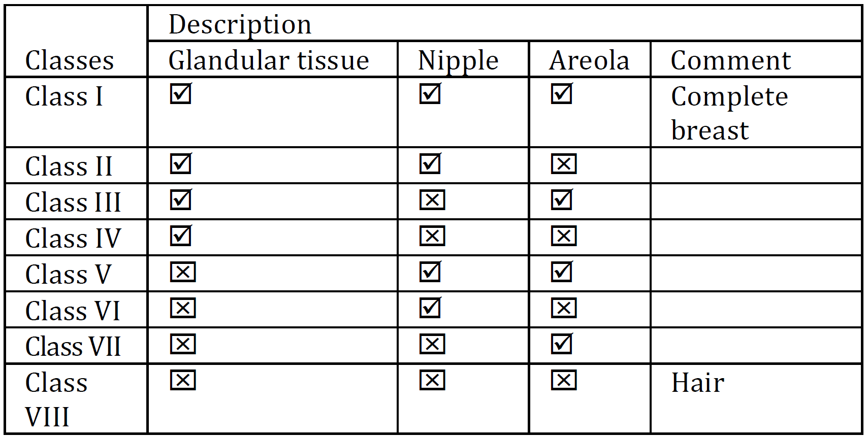
Accessory breast cancer is a rare disease developed from accessory breast tissue. Accessory breast tissue can be found along any point of the mammary lines, including in the thoracic and abdominal region (67%) and down to the groin. Ectopic breast tissue can also be found in locations such as the face, back, and thighs, but the predominant site is the axilla. The incidence of supernumerary breast and ectopic breast tissue around the world is 1–6% [1]. It affects 2–6% of females and 1–3% of males ‎but there is no definite incidence number for accessory breast malignancy; furthermore, reports for accessory breast cancer only include case reports and case series. Occurrence rates differ extensively according to ethnicity and gender, ranging from as low as 0.6% in Caucasians (relatively common among Asian women) to as high as 5% in Japanese females and Native ‎American ‎populations [2,3]. The 1915 Kajava classification system classified accessory breasts as Class I–VIII according to anatomical structure, and is ‎still ‎used today (Table 1) [4,5].


Accessory breast tissue can be located along the chest wall, vulva, axilla, knee, lateral thigh, buttocks, face, ear, and neck. Changes or symptoms may be noticed during puberty, at different times of the menstrual cycle, or during pregnancy and breastfeeding in women. Of note, the accessory breast tissue is often not detected until puberty because it is activated by hormones [6].


Table 1: Anatomical Structure

CASE REPORT

A 55-year-old is premenopausal female with diabetes, a blood pressure of 130/70 mmHg, a weight of 76 kg, and a height of 167 cm and no family history of breast cancer. She was presented to the general surgery outpatient department on the 4th of December 2011. The patient had a history of left axillary immobile mass of around 3.0 x 2.0 cm (length/width) which had increased in size within the prior six months, but had no history of a lump in the breast or discharge from the nipple. The patient mentioned that this axillary mass had been present since puberty but had been gradually increasing in size. The patient had a negative family history of malignancy and ultrasound (USG) breast screening was performed which showed no mass in the breast. Multiple lymph nodes were seen, however, in the left axilla, with the largest lymph node being 4.4 x 3.5 cm (Figure 4, 5, 6). 


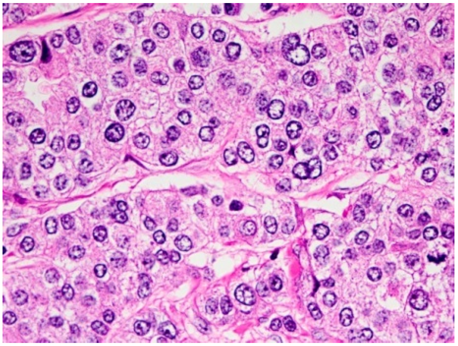
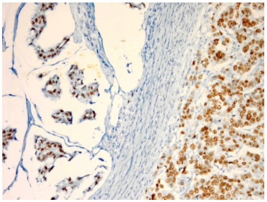
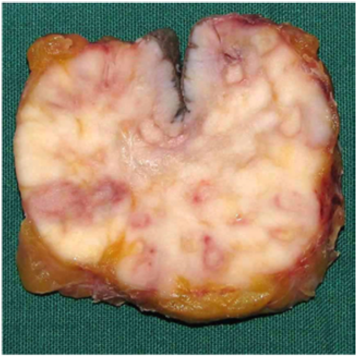
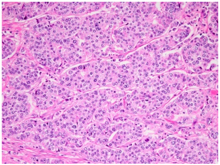
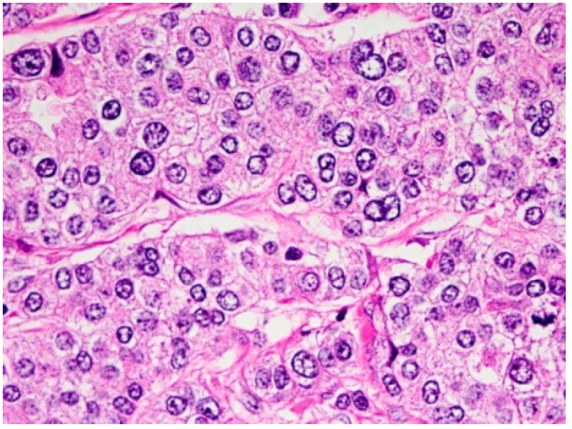
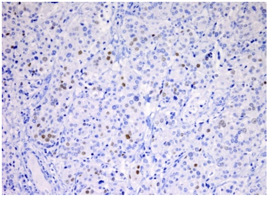
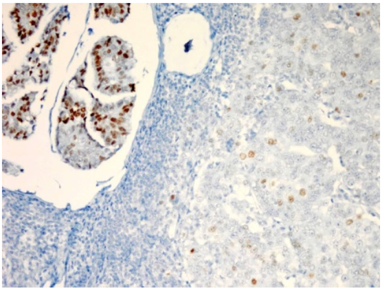
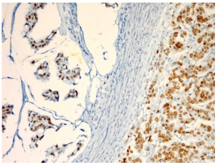
On the 18th of January 2012, tissue biopsy was taken from the left axillary mass which measured 4.5 cm in maximum diameter, weighed 30 g, and to which bio-section showed a greyish-‎‎‎white solid cut surface with pinkish and yellowish areas (Figure 3). Four blocks ‎representing ‎the ‎whole ‎cross-section of the tumour were embedded in three ‎cassettes.‎‏‎ Microscopically the sections showed lobules of a malignant epithelial tumour ‎involving ‎the ‎subcutaneous ‎adipose ‎tissue, dermis, and reaching up to the overlying epidermis. ‎It showed ‎large ‎areas of ‎partial or ‎complete ischaemic necrosis, scattered mitotic figures, and tumour cells arranged in solid sheets or lobules without glandular differentiation (Figure 1). Immunohistochemistry (IHC) methodology reported that the tumour ‎cells ‎stained ‎positively for pan-cytokeratins (CK) (AE1/AE3), suggestive of lymph node micrometastases [7]; CK7; epithelial membrane antigen; ER, that formed 50% of tumour cells; and PR, ‎‎that formed 5% of tumour cells (Figure 2,7,8,9,10).


IHC results were exclusively positive; HER2, BerEP4 (histologic stain used to aid in the diagnosis of basal cell carcinoma), mammoglobulin (highly specific for most breast cancers), CK5/6 (aid to differentiate between mesothelioma and other forms of cancer), thyroid transcription factor-1 (sensitive marker for pulmonary and thyroid adenocarcinomas), CD10 (sensitive immunohistochemical marker of normal endometrial stroma), smooth muscle actin (aid in diagnosis of ovarian carcinoma), S100 (common marker of neural tissue/lesions and melanoma), ‎chromogranin (aid in diagnosis of carcinoid tumours), synaptophysin (neuroendocrine marker), CK20, P63 (rules out invasion in breast tumours by determining presence of myoepithelial cells), and P53 all ‎showed ‎a negative ‎reaction.‎ Therefore, the ‎possibilities of neuroendocrine, primary lung, primary renal tumours‎, among ‎others ‎were not ‎supported by the IHC results‎‏. Reports showed moderate to poorly differentiated adenocarcinoma in favour of primary breast cancer (Table 2).


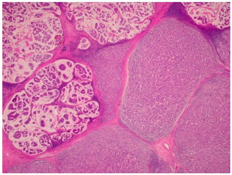
Figure 1: Histological examination of specimen. Sections show large deposits of carcinoma replacing most of the lymph node structure


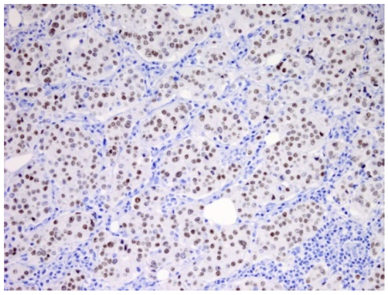
Figure 2: The tumour cells in both mucinous and nonmucinous areas show a positive staining for oestrogen recept


Figure 3: Left axillary mass


Figure 4:The Tumor Cells are Arrange in Solid Sheets or Lobules without Glandular Differentiation


Figure 5: Sections show large deposits of carcinoma replacing most of the lymph node structure


On the first ‏March‎2013, the patient was referred to medical oncologists for further management and ‏workup. Chest, abdomen, and pelvis CT scans were performed to rule out any primary sites, which all came back negative, and only the left axillary lymph node was seen to be involved. Bone scan results, as well as upper gastrointestinal endoscopy and colonoscopy results investigating for primary tumours, were also negative, and there were no signs of any malignancy.‏ MRI of the breast revealed no pathology apart from multiple axillary lymph nodes.‏‏ Tumour markers; cancer antigen-breast (CA 15-3) was high at 107.7 and ‏CA 125 was normal (Table 2).


Figure 6: A Large Component of Mucinous Carcinoma Is Present in one of these Lymph Node


Figure 7: The Tumor Cells Stain Positively for CK7. ER (50%of Tumor Cells)


Figure 8: The tumor cells stain positively for CK7,PR (5% of tumor cells)


In April ‏‎2013‎‏, ‏she ‎underwent left axillary clearance and the histopathology report confirmed primary breast adenocarcinoma. 


On the ‎‏‎19th of ‎May ‎‏‎2013, the patient received chemotherapy; AC protocol ‏doxorubicin 60 mg/m‎and cyclophosphamide 600 mg/m2 every 21 days for 4 cycles, followed by 4 ‎cycles of ‎paclitaxel 175 mg/m2 and 17 cycles of Trastuzumab (first cycle 8mg /kg as loading dose then 6mg/kg for subsequent doses). Every cycle was 21 days and the patient tolerated it well. The patient then underwent radiotherapy and hormonal therapy (tamoxifen 20 mg oral daily). She underwent follow-up ‎every 6 months until present. To assess the prognosis for the case, the Nottingham prognostic index for breast cancer was used. It gave an index value of 4.6, placing it in the moderate group, with 5 year survival [8]. The NHS Predict tool was applied as well for prognostic assessment, including hormonal status, and showed a result of 10 years survival [9]. In 2020, after 7 years of starting the treatment, the patient is doing well, free from any ‎signs of malignancy, recurrence, or complications. The mammography (MMG) results were negative.


Figure 9: The Tumor Cells in Both Mucinous and Non Mucinous Areas show a Positive Staining for ER


Figure 10: The Tumor Cells in Both Mucinous and Non Mucinous Areas show a Positive Staining for PR


Table 2: Immunohistochemistry test results

Test 

Reaction

Comment 

CK AE /1/3

Suggestive of lymph node micrometastases.

CK7

Oestrogen receptor (that formed 50% of tumour cells) and

 progesterone receptor (‎‎that formed 5% of tumour cells).

 EMA

Epithelial membrane antigen.

 HER-‎‎2

Human epidermal growth factor receptor 2.

BerEP4 


Negative

Histologic stain used to aid in the diagnosis of basal cell carcinoma.

Mammoglobulin 


Negative

Highly specific for most breast cancers.

CK5/6 


Negative

Cytokeratin 5/6: help to differentiate between mesothelioma and other forms of cancer.

TTF-‎‎


Negative

Thyroid transcription factor-1: sensitive marker for pulmonary and thyroid adenocarcinomas.

CD10 

Negative

Sensitive immunohistochemically marker of normal endometrial stroma.


SMA 

Negative

Smooth Muscle Actin aid in diagnosis of ovarian carcinoma.

 S100 

Negative

Common marker of neural tissue/lesions and melanoma.

 CgA 

Negative

Chromogranin help diagnose carcinoid tumours.

Synaptophysin

Negative  

Common neuroendocrine marker. 


 CK20


Negative  

Cytokeratin 20.

p63 


Negative  

(Rule out invasion in breast tumours by determining presence of myoepithelial cells).


p53  

Negative

Tumour marker in early stages of lung, skin, head and neck, and oesophageal cancer.

(CA 15-3) 

High 107.7

Tumour marker for breast cancer.

CA 125 


Normal

Tumour marker for breast cancer.

DISCUSSION

Accessory breast is a congenital condition when accessory normal breast tissue is present at abnormal sites [10]. ‎Accessory breast is also known as polymastia, supernumerary breasts, auxiliary breast, ectopic breast, adnexal, or mammae erraticae. These types of breasts sometimes appear to have/lack nipples or areolae, making them ambiguous [11].


In Caucasians the incidence of ectopic breast tissue is 1–4%, while it is more frequent in the Far East, especially Japan, among both genders. Its incidence rate was evaluated as 5.19% in Japanese women, and as 1.68% in Japanese men with hereditary factor [12]. These abnormal tissues are most frequently seen in the axilla, followed by the area just lower and in half of the cases this abnormality is shown on both sides [12]. In rare cases it was reported in some other areas including the acromial or scapular region, vulva, and in the midline of the thorax and abdomen [13]. Ectopic breasts enlarge during pregnancy and lactation and may lactate if they have a working ductal system. Breastfeeding from ectopic breasts was reported [12]. Disorders of breast tissue such as adenofibroma, cysts, and carcinomas have also been reported in ectopic breasts similar to normal breast tissue and carcinomas are rare [14,15,16]. The types of carcinoma seen within the ectopic breast tissue include ductal, lobular, mucinous, medullary, papillary, and invasive secretory (juvenile) carcinomas [13,15,16].


Ectopic breast tissue develops embryologically because of failed resolution of the mammary ridge or milk line. An ectodermal tissue extends from the axilla to the inguinal folds and shows in the 6th week of gestation. The axilla is the most frequent site followed by the area inferior to the normal breast and may appear anywhere along the milk line [17].


Hormonal influences affect ectopic breast tissue, just as they would in a healthy breast, and can develop similar types of benign and malignant disorders. Fibroadenomas, cysts, duct hyperplasia, and infrequently carcinoma can arise [18,19]. The incidence of accessory breast cancer as per reports is between 0.3% and 0.6% of all breast cancers [19].


Ectopic primary breast cancer in the axilla comprises 60–70% of all cases reported [20]. Differential diagnosis of the accessory breast cancer includes many disorders. In the axillary area, it can be confused with lipoma, lymphoma, lymphadenitis, metastatic lymphadenopathy, sebaceous cyst, and hidradenitis suppurativa. MMG and USG of the breast can aid the exclusion of other breast pathologies. USG can also detect ectopic breast tissue as an echogenic area resembling normal glandular tissue, as well as detect characteristics of the mass. MMG cannot capture ectopic breasts because of their peculiar location, but it can be visualised in the axilla by oblique and exaggerated craniocaudal views [21].


Cancer of ectopic breasts is depicted as a typical malignant mass with the same characteristics of those of metastatic axillary lymph nodes associated with malignant tumour. No specific findings for accessory breast cancer are detected [21]. Using MRI, the signal intensity of ectopic breast tissue is similar to that of the adjacent breast tissue, but with variability of the amount of interspersed fat. Pathological confirmation through fine needle aspiration cytology or Tru-cut® biopsy of the mass should be performed to harvest suspect cells or tissue. Invasive ductal carcinoma, like in traditional breast cancer, is the most common histological type with 79% of all accessory breast cancer [22].


Lobular, mucinous, medullary, apocrine, and papillary carcinomas are detected in these cases and cystosarcoma phyllodes are also described. In 2009, Nihon-Yanagi et al.,[20] found that medullary, mucinous, and apocrine carcinomas were more common among accessory breast cancer for unknown reasons. 


Regarding the management, surgical interference of accessory breast cancer combines wide resection of the tumour with surrounding tissue, skin, and axillary lymph nodes dissection.18 Ipsilateral mastectomy has no additional benefit regarding survival considering that MMG and USG of the anatomic breast are normal, as was in the present case, but should be performed when differential diagnosis is challenging in some situations.18


In 2015, Zhang, S. et al., [18] recommended mastectomy if the accessory breast is closely connected to normal breast tissue, otherwise it is unnecessary. 


The same regimens and protocols for postoperative treatment are used for anatomic breast carcinoma. Radiotherapy of the tumour site is recommended to control local spread, and radiation of the ipsilateral anatomic breast is not usually performed. Adjuvant therapy is mostly required because lymph node disease is usually also present. The prognosis of accessory breast cancer is difficult to assess due to limited follow-up and staging data as well as small sample size. Some authors have reported worse outcomes of accessory breast cancer than other anatomic breast cancer as the tumour is near the axillary lymph nodes and is therefore exposed to early metastasis to these nodes [18].


94 Japanese cases were reviewed in a report in 2009 and indicated that accessory breast cancer has a higher risk of lymph node metastasis than usual breast cancer [20].


Accessory breast tissue is more disposed to malignant change than normal breast tissue. No definite number is reported for the incidence of accessory breast cancer among population [23]. Accessory breast cancer patients experience clinical presentations as swelling, thickening, tenderness, irritation, and sometimes limited motion of shoulder. These symptoms are commonly exacerbated at the onset of puberty and pregnancy [24]. Accessory breast cancer is a rare entity and has a substantial chance for misdiagnosis especially when there is an absence of anatomical breast structure such as the nipple and areola [25]. The accessory breast may also not show up; therefore, MRI can be used to differentiate it from the normal breast tissue [3]. 


The diagnosis as well as symptoms of accessory breast carcinoma are the same as forbreast carcinoma [16], except for some differences including excess axillary fat, lymphadenitis, lymphoma, ‎metastatic carcinoma, and hidradenitis suppurativa [6]. Accessory breast cancer is ‎diagnosed as breast carcinoma using MMG and ‎USG, followed by pathologic diagnosis by fine needle aspiration cytology or core biopsy [26].


The mammogram is an effective tool for the assessment of breast carcinoma but not for accessory breast cancer assessment. Core tissue biopsy especially when accompanied by IHC is a very effective tool for early diagnosis and treatment of accessory breast carcinoma [2].


The presented case was very perplexing because it was a rare condition for primary axillary breast cancer. The lack of areola and nipple (according to Kajava classification 1915‎‏ this case classified as class IV) made the clinical diagnosis very difficult, confounded by the IHC results not being conclusive. This leads to extensive and unnecessary lab tests and investigations. The possibility of the normal breast being the primary site was not supported by negative staining of mammoglobulin, gross cystic disease fluid protein. Furthermore, IHC did not support the possibilities of a neuroendocrine tumour or the lung, and the kidney as the primary sites.


In the beginning the case was suspected to be a metastatic adenocarcinoma with small components of mucinous carcinoma; however, the lymph nodes were positive (13/15) with large deposits of carcinoma replacing most of the lymph node structure favouring primary breast including the possibility of axillary (accessory) primary breast. The possibilities included breast, and to a lesser extent, primary adnexal tumour. Oncologists were not satisfied about histopathology results, and so all thoughts were redirected towards it being a secondary tumour from the breast or other sites. As such, more investigations were requested including CT, MRI, CA 125, and upper gastrointestinal endoscopy, but all results were negative for the detection of unknown primary tumours.


A Tumour board committee was held to discuss this case and it was agreed that it is a primary accessory breast tumour because no original tumour could be detected in the breast, adnexa, colon, or lung. The histopathology was in favour of primary breast since CA 15-3 high and ER and PR were positive as well as previous case reports and literature review for similar cases. 


The patient was staged as Stage IIB, ER/PR positive and HER2/neu negative poorly differentiated adenocarcinoma.


The patient was treated with common breast carcinoma methods following NCCN guideline 2012 [27] by surgery followed by chemotherapy. This included four cycles of AC and 4 cycles of docetaxel followed by a 50 g radiotherapy and tamoxifen for 5 years, and cancer antigen (CA 15-3) showed a significant decline after treatment (Figure 1).


Prognosis of accessory breast cancer is difficult to institute because of the absence or limited follow-up data as well as the small sample size [28], in our follow-up case the patient is stable with very good performance status for 7 years and without any complications.


CONCLUSION

Accessory breast cancer is an uncommon type of cancer and the incidence of it has no definite number. The diagnosis of these cases is challenging, and misdiagnosis will lead to extensive and unnecessary investigations. Despite the fact that carcinoma arising in axilla as a primary site is a rare condition, still the possibility of accessory breast cancer should still be considered. ‎Management of accessory breast cancer should follow the same guidelines for breast cancer. It is important to encourage follow up data for these cases to establish the prognosis of accessory breast cancer comparing breast carcinoma. 

REFERENCE
  1. Rizvi, G. et al. “Fibroadenoma of Ectopic Breast Tissue in Axilla.” Journal of Case Reports, vol. 2, no. 2, 2012, pp. 36–38.

  2. Sookar, N. et al. “The Dilemma of Metastatic Axillary Cancer versus Primary Ectopic Breast Cancer.” J Surg Sci Oper Care, vol. 1, 2018, p. 102.

  3. Laor, T. et al. “MRI Appearance of Accessory Breast Tissue: A Diagnostic Consideration for an Axillary Mass in a Peripubertal or Pubertal Girl.” American Journal of Roentgenology, vol. 183, no. 6, 2004, pp. 1779–1781.

  4. Famá, F. et al. “Prevalence of Ectopic Breast Tissue and Tumor: A 20-Year Single Center Experience.” Clinical Breast Cancer, vol. 16, no. 4, 2016, pp. e107–e112.

  5. Amaranathan, A. et al. “An Ectopic Breast Tissue Presenting with Fibroadenoma in Axilla.” Case Reports in Surgery, 2013, article 947295.

  6. Jatoi, I., and A. Rody. “Breast Cancer Screening.” Management of Breast Disease, 2nd ed., Springer Publishing, 2016, pp. 131–145.

  7. Lerwill, M.F. “Current Practical Applications of Diagnostic Immunohistochemistry in Breast Pathology.” The American Journal of Surgical Pathology, vol. 28, no. 8, 2004, pp. 1076–1091.

  8. Todd, J.H. et al. “Confirmation of a Prognostic Index in Primary Breast Cancer.” British Journal of Cancer, vol. 56, no. 4, 1987, pp. 489–492.

  9. National Health Service. “Predict Breast Cancer Tool.” 2019, breast.predict.nhs.uk/tool. Accessed October 2019.

  10. Patel, P.P. et al. “Accessory Breast Tissue.” Eplasty, 2012.

  11. Zhang, R.R. et al. “Unusual Presentation of Multiple Fibroadenomas in Bilateral Breasts and Axillary Accessory Breast.” Breast Cancer: Basic and Clinical Research, vol. 6, 2012.

  12. Önel, S. et al. “Ectopic Breast Cancer: A Case Report.” Turkish Journal of Surgery, vol. 29, no. 2, 2013, p. 96.

  13. Chung-Park, M. et al. “Mucinous Adenocarcinoma of Ectopic Breast Tissue of the Vulva.” Archives of Pathology & Laboratory Medicine, vol. 126, no. 10, 2002, pp. 1216–1218.

  14. Avilés Izquierdo, J. et al. “Pigmented Axillary Nodule: Carcinoma of an Ectopic Axillary Breast.” Dermatologic Surgery, vol. 31, no. 2, 2005, pp. 237–239.

  15. Shin, S.J. et al. “Invasive Secretory (Juvenile) Carcinoma Arising in Ectopic Breast Tissue of the Axilla.” Archives of Pathology & Laboratory Medicine, vol. 125, no. 10, 2001, pp. 1372–1374.

  16. Pardo, M. et al. “Mammary Carcinoma in Ectopic Breast Tissue: A Case Report.” Revista Medica de Chile, vol. 129, no. 6, 2001, pp. 663–665.

  17. Cherrabi, F. et al. “Ectopic Breast Carcinoma: A Case Report.” Tropical Journal of Obstetrics and Gynaecology, vol. 35, no. 1, 2018, pp. 90–92.

  18. Zhang, S. et al. “Diagnosis and Treatment of Accessory Breast Cancer in 11 Patients.” Oncology Letters, vol. 10, no. 3, 2015, pp. 1783–1788.

  19. Abisowo, O.Y. et al. “Feto Maternal Outcome of Induced versus Spontaneous Labour in a Nigerian Tertiary Maternity Unit.” Tropical Journal of Obstetrics and Gynaecology, vol. 34, no. 1, 2017, pp. 21–27.

  20. Nihon-Yanagi, Y. et al. “A Case of Ectopic Breast Cancer with a Literature Review.” Surgical Oncology, vol. 20, no. 1, 2011, pp. 35–42.

  21. Adler, D.D. et al.“Accessory Breast Tissue in the Axilla: Mammographic Appearance.” Radiology, vol. 163, no. 3, 1987, pp. 709–711.

  22. Francone, E. et al. “Ectopic Breast Cancer: Case Report and Review of the Literature.” Aesthetic Plastic Surgery, vol. 37, no. 4, 2013, pp. 746–749.

  23. Gilmore, H.T. et al.“Supernumerary Nipples and Accessory Breast Tissue.” South Dakota Journal of Medicine, vol. 49, no. 5, 1996, pp. 149–151.

  24. Lesavoy, M.A. et al.“Axillary Breast Tissue: Clinical Presentation and Surgical Treatment.” Annals of Plastic Surgery, vol. 35, no. 4, 1995, pp. 356–360.

  25. Loukas, M. et al. “Accessory Breasts: A Historical and Current Perspective.” The American Surgeon, vol. 73, no. 5, 2007, pp. 525–528.

  26. Roorda, A.K. et al.“Ectopic Breast Cancer: Special Treatment Considerations in the Postmenopausal Patient.” The Breast Journal, vol. 8, no. 5, 2002, pp. 286–289.

  27. National Comprehensive Cancer Network. NCCN Breast Cancer Guidelines 2012. 2012, www.nccn.org/professionals/physician_gls/pdf/breast.pdf. Accessed October 2019.

  28. Evans, D.M., and D.P. Guyton. “Carcinoma of the Axillary Breast.” Journal of Surgical Oncology, vol. 59, no. 3, 1995, pp. 190–195.

Recommended Articles
Research Article
A study on the feasibility of online classes for medical education
Published: 15/02/2021
Download PDF
Research Article
Amyand’s Hernia: A Rare form of Common Entity
Download PDF
Research Article
Prevalence of Typhoid Fever among Patients Attending General Sani Abatcha Specialist Hospital Damaturu, Nigeria
...
Published: 10/10/2020
Download PDF
Research Article
A case report of management of a Case of Hyperalgesia by Ultrasound guided Fluoroscopically Verified Transforaminal Nerve Root Block
...
Published: 29/06/2024
Download PDF
Chat on WhatsApp
Flowbite Logo
PO Box 101, Nakuru
Kenya.
Email: office@iarconsortium.org

Editorial Office:
J.L Bhavan, Near Radison Blu Hotel,
Jalukbari, Guwahati-India
Useful Links
Order Hard Copy
Privacy policy
Terms and Conditions
Refund Policy
Shipping Policy
Others
About Us
Team Members
Contact Us
Online Payments
Join as Editor
Join as Reviewer
Subscribe to our Newsletter
+91 60029-93949
Follow us
MOST SEARCHED KEYWORDS
Copyright © iARCON International LLP . All Rights Reserved.